Review
. 2025 May:106:101360.
doi: 10.1016/j.preteyeres.2025.101360. Epub 2025 Apr 23.

Advances and therapeutic opportunities in visual cycle modulation

Affiliations
Review

Advances and therapeutic opportunities in visual cycle modulation

Jordan Zaluski et al. Prog Retin Eye Res. 2025 May.

Abstract

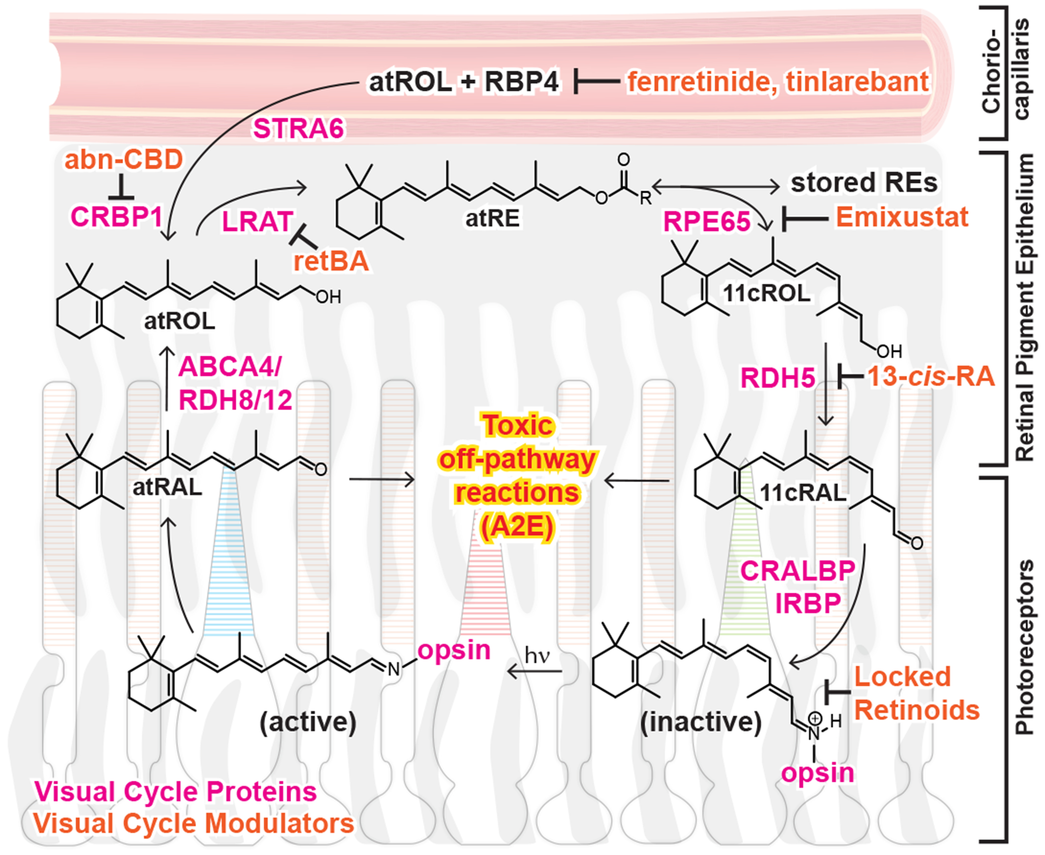
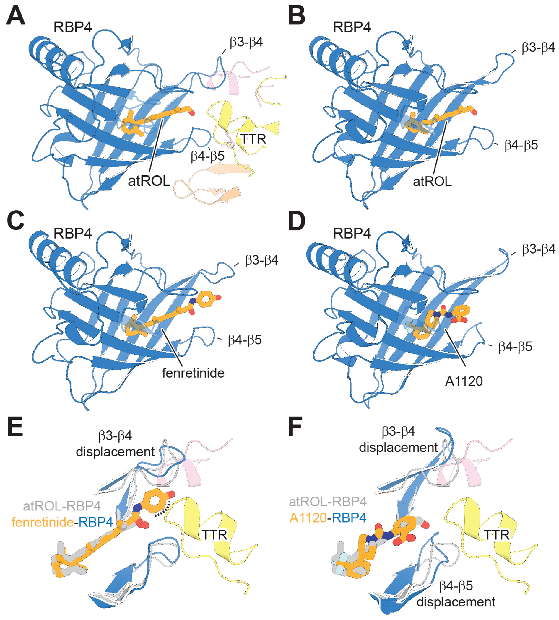
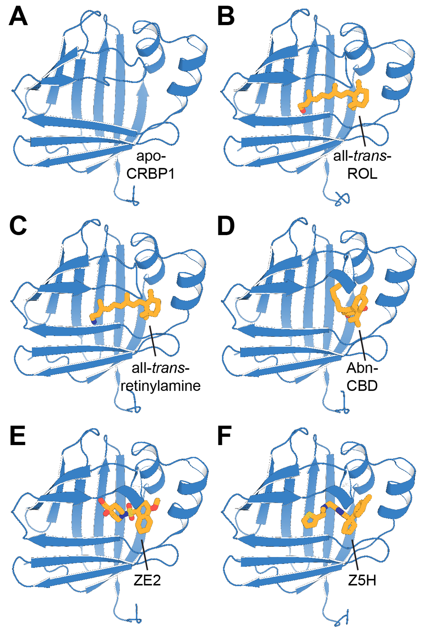
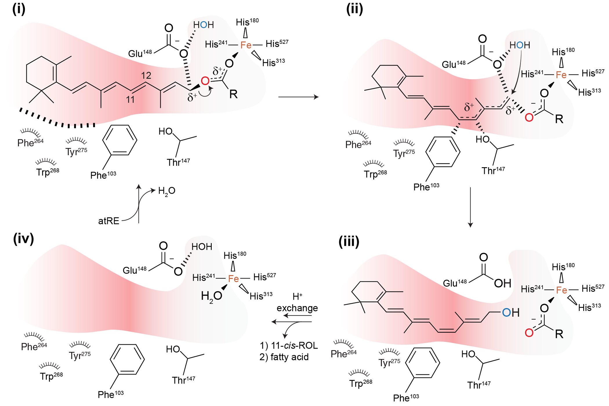
The visual cycle is a metabolic pathway that enables continuous vision by regenerating the 11-cis-retinal chromophore for photoreceptors opsins. Although integral to normal visual function, the flux of retinoids through this cycle can contribute to a range of retinal pathologies, including Stargardt disease, age-related macular degeneration, and diabetic retinopathy. In such conditions, intermediates and byproducts of the visual cycle, such as bisretinoid components of lipofuscin, can accumulate, concomitant with cellular damage and eventual photoreceptor loss. This has inspired efforts to modulate the visual cycle, aiming to slow or prevent the formation of these toxic intermediates and thus preserve retinal structure and function. Over the past two decades, multiple strategies to modulate the visual cycle have emerged. These include both intrinsic approaches, targeting key enzymes, retinoid-binding proteins, or receptors within the pigment epithelium or photoreceptors (e.g., RPE65, CRBP1, and rhodopsin inhibitors/antagonists) and extrinsic strategies that indirectly alter retinoid availability within the retina (e.g., RBP4 antagonists). Many of these agents have shown promise in animal models of visual cycle-associated retinal diseases, reducing pathological changes, and improving retinal survival. Several have advanced into clinical studies, although none are currently FDA-approved. Challenges remain in optimizing drug specificity and duration of action while minimizing side effects such as nyctalopia. In this review, we comprehensively examine current and emerging visual cycle modulators, discuss their medicinal chemistry, mechanisms of action, efficacy in preclinical and clinical studies, and highlight future opportunities for drug discovery aimed at safely and effectively preserving vision through modulation of this biochemical pathway.

Keywords: Drug development; Inhibitors; Ophthalmology; RPE65; Retinol-binding protein; Retinopathy; Stargardt disease; Visual cycle; Vitamin A.


Figures

Figure 1:
Figure 1:. Modulation of the visual cycle by small-molecule inhibitors.
Light activation of visual pigments triggers a G protein-mediated signaling cascade and membrane hyperpolarization causing changes in photoreceptor signaling. This process involves “bleaching” of the visual pigment, where the bound retinal molecule is hydrolytically released. The visual cycle is a series of chemical reactions that regenerates 11-cis-retinal for rod and cone visual pigments. Retinal is subject to off-pathway toxic reactions, which are exacerbated due to certain environmental and genetic perturbations. VCMs that act directly on proteins within the RPE or photoreceptors or indirectly by lowering the concentrations of visual cycle retinoids can favorably modify retinoid flux through the cycle thereby ameliorating the toxic off-pathway reactions. Abbreviations: atRAL, all-trans-retinal; 11cRAL, 11-cis-retinal; 11cROL, 11-cis-retinol; RE, retinyl ester A2E, N-retinyl-N-retinylidene ethanolamine; LRAT, lecithin:retinol acyl transferase; RBP4, serum retinol-binding protein; RDH, retinol dehydrogenase; Rho, rhodopsin; RPE65, retinoid isomerohydrolase; STRA6 stimulated by retinoic acid 6.
Figure 2:
Figure 2:. Fenretinide and A1120 disrupt the RBP-TTR complex.
A) holo-RBP4 in complex with the TTR tetramer (PDB accession code 3BSZ). B) holo-RBP4 alone (PDB accession code 5NU7). C) RBP4 in complex with fenretinide (PDB accession code 1FEL). D) RBP4 in complex with A1120 (PDB accession code 3FMZ). E) Comparison of the holo-RBP4-TTR complex with the RBP4-fenretinide complex, showing steric clashes (dashed line) and structural alterations in key loop regions that prevent the RBP4-fenretinide complex from binding to the TTR tetramer. F) Comparison of the holo-RBP4-TTR complex with the RBP4-A1120 complex, showing structural alterations in key loop regions that prevent the RBP4-A1120 complex from binding to the TTR tetramer.
Figure 3:
Figure 3:. Crystal structures of CRBP1-ligand complexes.
A) apo-CRBP1 (PDB accession code 5H9A). B) holo-CRBP1 (PDB accession code 5HBS). C) CRBP1-retinylamine complex (PDB accession code 5HA1). D) CRBP1-Abn-CBD complex (PDB accession code 6E5L). E) CRBP1-ZE2 complex (PDB accession code 8GEY). F) CRBP1-Z5H complex (PDB accession code 8GD2).
Figure 4:
Figure 4:. Proposed catalytic mechanism of RPE65.
The figure is reused with permission (Kiser, 2022).
Figure 5:
Figure 5:. Crystal structure of the RPE65 in complex with emixustat and palmitate (PDB accession code 4RSC).
The emixustat and palmitate ligands are shown as orange and blue sticks, respectively. The iron cofactor is shown as a brown sphere.
Figure 6:
Figure 6:. Catalytic mechanism of LRAT and mode of action of various serine protease and papain protease inhibitors.
A) The catalytic ping-pong bi-bi mechanism of LRAT is depicted. B) Schematics of small molecule inhibitors of papain and protease inhibitors. Catalytic Cys161 residue of LRAT is depicted in green while the inactivated Cys161-small molecule adduct is depicted in red. In the case of LRAT, serine is not present in the active site, so the free serine and serine adduct are shown in blue. C) Early studies utilized hydrolase inhibitors to determine that the key catalytic residue in LRAT was cysteine. Specifically, the thiol-specific inhibitors, NEM and pAPAO, inhibited LRAT activity, whereas serine hydrolase inhibitors displayed minimal inhibitory activity.
Figure 7:
Figure 7:. Mechanism of action of ALK-001 is based on the biosynthesis of A2E.
A) all-trans-retinal can form Schiff base Intermediate I with phosphatidylethanolamine (PE, shown embedded in a lipid bilayer). This species can undergo an imine to enamine tautomerization to form Intermediate II. A second molecule of all-trans-retinal can form a subsequent Schiff base to give Intermediate III. This molecule can then undergo spontaneous electrocyclic ring closure followed by oxidation to the pyridinium species followed by hydrolysis from PE to give A2E (highlighted in yellow). B) Gildeuretinol acetate (ALK-001) is metabolized, enters the visual cycle through the canonical vitamin A pathways, and can be converted to 2H3-retinal through these pathways. Presumably 2H3-retinal can form Schiff bases with PE to afford 2H3-Intermediate I (highlighted in blue). This intermediate will undergo imine to enamine tautomerization at a dramatically reduced rate as this process likely involves a 1,5-hydride shift. This process will be substantially inhibited due to the primary isotope effect. This effectively renders 2H3-Intermediate I unable to form A2E.

Similar articles

References

    1. Abbas F, Becker S, Jones BW, Mure LS, Panda S, Hanneken A, Vinberg F, 2022. Revival of light signalling in the postmortem mouse and human retina. Nature. 606, 351–357. 10.1038/s41586-022-04709-x - DOI - PMC - PubMed
    1. Abi-Mosleh L, Infante RE, Radhakrishnan A, Goldstein JL, Brown MS, 2009. Cyclodextrin overcomes deficient lysosome-to-endoplasmic reticulum transport of cholesterol in Niemann-Pick type C cells. Proc. Natl. Acad. Sci. U. S. A 106, 19316–19321. 10.1073/pnas.0910916106 - DOI - PMC - PubMed
    1. Ahmed CM, Dwyer BT, Romashko A, Van Adestine S, Park EH, Lou Z, Welty D, Josiah S, Savinainen A, Zhang B, Lewin AS, 2019. SRD005825 Acts as a Pharmacologic Chaperone of Opsin and Promotes Survival of Photoreceptors in an Animal Model of Autosomal Dominant Retinitis Pigmentosa. Transl. Vis. Sci. Technol 8, 30. 10.1167/tvst.8.6.30 - DOI - PMC - PubMed
    1. Akita H, Tanis SP, Adams M, Balogh-Nair V, Nakanishi K, 1980. Nonbleachable rhodopsins retaining the full natural chromophore. J. Am. Chem. Soc 102, 6370–6372. 10.1021/ja00540a047 - DOI
    1. Akula JD, Hansen RM, Tzekov R, Favazza TL, Vyhovsky TC, Benador IY, Mocko JA, McGee D, Kubota R, Fulton AB, 2010. Visual cycle modulation in neurovascular retinopathy. Exp. Eye Res 91, 153–161. 10.1016/j.exer.2010.04.008 - DOI - PubMed