. 2025 May 12;12(5):ENEURO.0054-25.2025.
doi: 10.1523/ENEURO.0054-25.2025. Print 2025 May.

Investigating Mechanically Activated Currents from Trigeminal Neurons of Nonhuman Primates

Affiliations

Investigating Mechanically Activated Currents from Trigeminal Neurons of Nonhuman Primates

Karen A Lindquist et al. eNeuro. .

Abstract

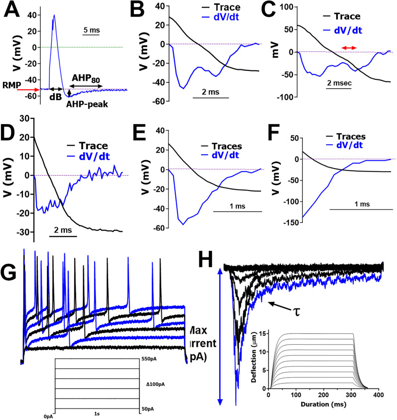
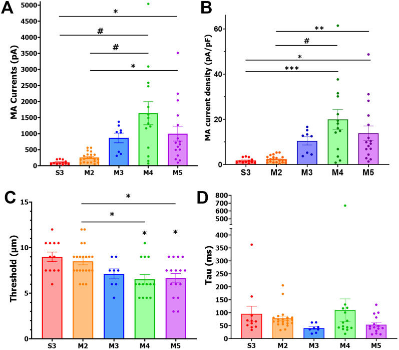
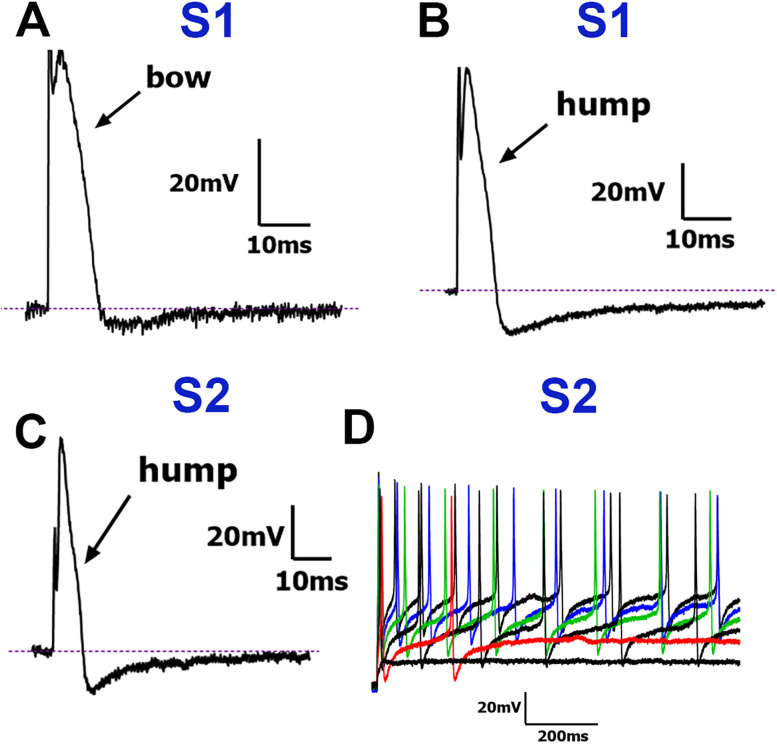
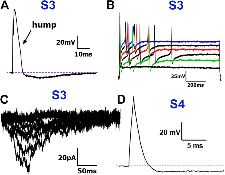
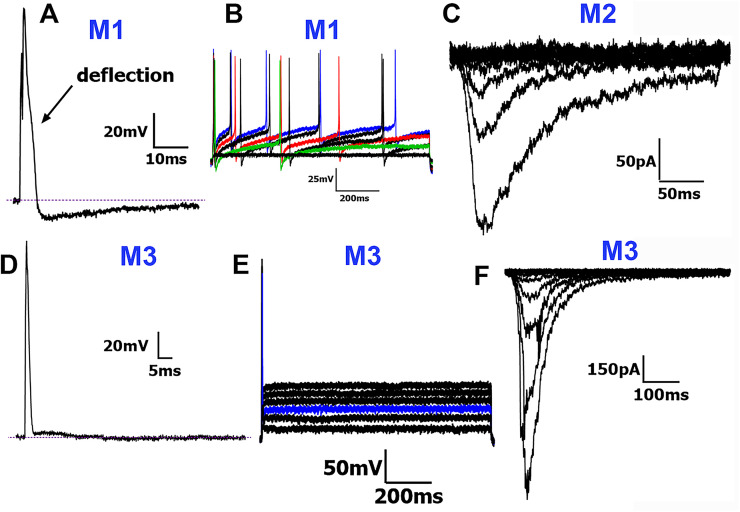
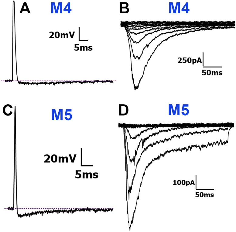
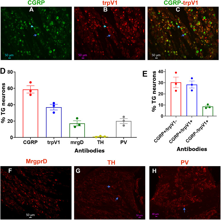
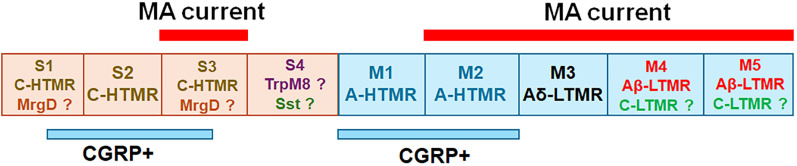
Pain sensation often involves mechanical modalities. Mechanically activated (MA) ion channels on sensory neurons underly responsiveness to mechanical stimuli. MA current properties have mainly been derived from rodent sensory neurons. This study aimed to address gaps in knowledge regarding MA current properties in trigeminal (TG) neurons of a higher-order species, common marmoset nonhuman primates (NHP). MA currents triggered by a piezoactuator were recorded in patch-clamp configuration. MA responses were associated with action potential (AP) properties, such as width, dV/dt on the falling phase, and presence/absence of AP firing in NHP TG neurons. According to responsiveness to mechanical stimuli and AP properties, marmoset TG neurons were clustered into four S-type and five M-type groups. S-type TG neurons had broader AP with two dV/dt peaks on the AP falling phase. Only one S-type group of NHP TG neurons produced small MA currents. M-type TG neurons had narrow AP without two dV/dt peaks on the AP falling phase. M-type NHP TG neurons, except for one group, showed MA currents. We additionally used immunohistochemistry to confirm the presence of known sensory neuronal types such as unmyelinated peptidergic CGRP+/trpV1+, unmyelinated nonpeptidergic MrgprD+ and CGRP-/trpV1+, and myelinated peptidergic CGRP+/trpV1- and nonpeptidergic CGRP- and PV+ NHP TG neurons. Overall, marmoset TG neurons and associated MA currents have many similarities compared with reported data from mouse sensory neurons. However, there are notable differences such as lower percentage of small NHP TG neurons responding to mechanical stimuli and absence of fast inactivating MA currents.

Keywords: mechnoactivated current; nonhuman primates; sensory neurons; trigeminal ganglia.


Conflict of interest statement

The authors declare no competing financial interests.

Figures

Figure 1.
Figure 1.
Electrophysiology protocols. A, A single action potential (AP) is elicited in an NHP TG neuron by a brief current injection of 1 nA for 0.5 ms. We analyzed indicated AP and afterhyperpolarization (AHP) parameters, including resting membrane potential (RMP), duration at base (dB), magnitude of AHP peak, and the time required for the AHP (measured in ms) to decay by 80% (AHP80). B, C, dV/dt for the falling phase of broad (dB > 5 ms) AP with a characteristic “hump” or “bow.” D, dV/dt for the falling phase of AP (dB < 5 ms) with a characteristic slight “deflection.” E, F, dV/dt for the falling phase of fast AP (dB < 4 ms) with a “straight” falling phase. G, AP trains are triggered by applying steps of increasing current injections, 50–550 pA with 100 pA increments. A schematic of the protocol used, and a sample recording is shown. H, MA currents are activated by a piezoactuator controlled by a piezoelectric device. A graphical representation of actuator movement is shown. Each poke extends by 1.5 µm increments and is held for 300 ms before returning to the starting position for 10 s of relaxation. A total of 10 progressively deeper pokes are administered, each increasing by an additional 15 µm with the final poke going in 15 µm deep. Sample recording from an NHP TG neuron is shown; τ is indicated.
Figure 2.
Figure 2.
MA currents from NHP TG neurons. Five NHP neuronal subgroups (labeled under the x-axis) responded to mechanical stimulation. These neuronal groups showed different characteristics. A, Max MA current amplitudes (pA) from these five MA groups of NHP TG neurons. B, Max MA current density (pA/pF) from these MA groups of NHP TG neurons. C, Activation threshold in actuator distance (μm) traveled for these NHP TG neuronal subgroups. D, Decay kinetics (ms) of MA currents for these NHP TG neuronal subgroups. Data were analyzed by one-way ANOVA in each column compared with others followed by Bonferroni's post hoc tests; *p < 0.05; **p < 0.01; ***p < 0.001; #p < 0.0001.
Figure 3.
Figure 3.
Traces representing current signatures from S1 and S2 NHP TG neuronal groups. A, Representative AP trace with “bow” on the AP falling phase belonging to the S1 group. B, Representative AP trace with “hump” on the AP falling phase belonging to the S1 group. C, Representative AP with “hump” on the AP falling phase belonging to the S2 group. D, Representative AP train belonging to the S2 group. A characteristic “hump” or “bow” on the downward portion of the AP is indicated with a black arrow in panels A, B, and C. Neuronal groups are specified above traces. Scale bars are presented for each panel.
Figure 4.
Figure 4.
Traces representing current signatures from the S3 and S4 NHP TG neuronal groups. A, Representative AP trace with “hump” on the AP falling phase belonging to the S3 group. B, Representative AP train belonging to the S3 group. C, Representative MA currents from a neuron belonging to the S3 group. D, Representative AP trace belonging to the S4 group. Neuronal groups are specified above traces. Scale bars are presented for each panel.
Figure 5.
Figure 5.
Traces representing current signatures from M1, M2, and M3 NHP TG neuronal groups. A, Representative AP trace with “deflection” on the AP falling phase belonging to the M1 group. The “deflection” is indicated. B, Representative AP train belonging to the M1 group. C, Representative MA currents belonging to the M2 group. D, Representative AP trace belonging to the M3 group. The absence of a true AHP peak (Fig. 1A) is observed. E, Representative AP train belonging to the M3 group. F, Representative MA currents belonging to the M3 group. Neuronal groups are specified above traces. Scale bars are presented for each panel.
Figure 6.
Figure 6.
Traces representing current signatures from M4 and M5 NHP TG neuronal groups. A, Representative AP trace belonging to the M4 group. B, Representative MA currents belonging to the M4 group. C, Representative AP trace belonging to the M5 group. D, Representative MA currents belonging to the A5 group. Neuronal groups are specified above traces. Scale bars are presented for each panel.
Figure 7.
Figure 7.
Representation of marker-positive neurons in TG of adult male marmosets. Representative microphotographs show expression patterns for CGRP (A), trpV1 (B), CGRP + trpV1 (C), MrgprD (F), tyrosine hydroxylase (TH; G), and parvalbumin (PV; H), in TG of adult male marmosets. The blue arrows on panels AC indicate CGRP+/trpV1 neurons. Cyan arrows on panels AC show CGRP/trpV1+ neurons. The blue arrows on panels G and H point to TH- and PV-positive cells, respectively. Antibodies used and matching colors are indicated. Scales are presented in each microphotograph. D, Bar graphs reflect percentages of marker-positive sensory neurons in TG of adult male marmosets. E, Bar graphs reflect relative percentages of CGRP+ (peptidergic) and trpV1+ neurons in TG of adult male marmosets. The x-axis denotes antibodies for markers. N = 3.
Figure 8.
Figure 8.
Schematic summarizing results of the study. Small-sized NHP TG neuronal groups (S1–S4) represented as the beige boxes. Medium-sized NHP TG neuronal groups (M1–M5) are the blue boxes. TG neuronal groups responding to piezoactuating mechanical simulations and exhibiting MA currents are outlined by the red bars and labeled “MA current” above each TG neuronal group. Putative CGRP+ groups are outlined by blue lines and labeled “CGRP+.” Presumed functions of the NHP TG neuronal group are indicated inside boxes.

Update of

Similar articles

References

    1. Basbaum AI, Bautista DM, Scherrer G, Julius D (2009) Cellular and molecular mechanisms of pain. Cell 139:267–284. 10.1016/j.cell.2009.09.028 - DOI - PMC - PubMed
    1. Basbaum AI, Braz JM (2010) Transgenic mouse models for the tracing of “Pain” pathways.
    1. Baumgartner U (2010) Nociceptive system: nociceptors, fiber types, spinal pathways, and projection areas. Schmerz 24:105–113. 10.1007/s00482-010-0904-4 - DOI - PubMed
    1. Bhuiyan SA, et al. (2024) Harmonized cross-species cell atlases of trigeminal and dorsal root ganglia. Sci Adv 10:eadj9173. 10.1126/sciadv.adj9173 - DOI - PMC - PubMed
    1. Boada MD (2013) Relationship between electrophysiological signature and defined sensory modality of trigeminal ganglion neurons in vivo. J Neurophysiol 109:749–757. 10.1152/jn.00693.2012 - DOI - PubMed

Substances

LinkOut - more resources