. 2025 Apr 25;16(1):3914.
doi: 10.1038/s41467-025-59216-0.

Deciphering distinct genetic risk factors for FTLD-TDP pathological subtypes via whole-genome sequencing

Cyril Pottier  1   2   3   4   5 Fahri Küçükali  6   7 Matt Baker  8 Anthony Batzler  9 Gregory D Jenkins  9 Marka van Blitterswijk  8 Cristina T Vicente  6   7 Wouter De Coster  6   7 Sarah Wynants  6   7 Pieter Van de Walle  6   7 Owen A Ross  8 Melissa E Murray  8 Júlia Faura  6   7 Stephen J Haggarty  10 Jeroen Gj van Rooij  11 Merel O Mol  11 Ging-Yuek R Hsiung  12 Caroline Graff  13   14 Linn Öijerstedt  13   14 Manuela Neumann  15   16 Yan Asmann  17 Shannon K McDonnell  9 Saurabh Baheti  9 Keith A Josephs  18 Jennifer L Whitwell  19 Kevin F Bieniek  8   20 Leah Forsberg  18 Hilary Heuer  21 Argentina Lario Lago  21 Ethan G Geier  21 Jennifer S Yokoyama  21 Alexis P Oddi  21 Margaret Flanagan  20 Qinwen Mao  22 John R Hodges  23 John B Kwok  24   25 Kimiko Domoto-Reilly  26 Matthis Synofzik  15   27 Carlo Wilke  15   27 Chiadi Onyike  28 Bradford C Dickerson  29 Bret M Evers  30 Brittany N Dugger  31 David G Munoz  32   33 Julia Keith  33   34 Lorne Zinman  34 Ekaterina Rogaeva  35 EunRan Suh  36 Tamar Gefen  37 Changiz Geula  37 Sandra Weintraub  37 Janine Diehl-Schmid  38   39 Martin R Farlow  40 Dieter Edbauer  41 Bryan K Woodruff  42 Richard J Caselli  42 Laura L Donker Kaat  43 Edward D Huey  44 Eric M Reiman  45 Simon Mead  46 Andrew King  47   48 Sigrun Roeber  49 Alissa L Nana  21 Nilufer Ertekin-Taner  8   50 David S Knopman  18 Ronald C Petersen  18 Leonard Petrucelli  8 Ryan J Uitti  50 Zbigniew K Wszolek  50 Eliana Marisa Ramos  51 Lea T Grinberg  21 Maria Luisa Gorno Tempini  21 Howard J Rosen  52 Salvatore Spina  21 Olivier Piguet  53 Murray Grossman  54 John Q Trojanowski  36 C Dirk Keene  55 Lee-Way Jin  56 Johannes Prudlo  57   58 Daniel H Geschwind  51 Robert A Rissman  59 Carlos Cruchaga  60 Bernardino Ghetti  61 Glenda M Halliday  24 Thomas G Beach  62 Geidy E Serrano  62 Thomas Arzberger  49   63 Jochen Herms  41   49 Adam L Boxer  21 Lawrence S Honig  64 Jean P Vonsattel  65 Oscar L Lopez  66 Julia Kofler  67 Charles L White 3rd  30 Marla Gearing  68 Jonathan Glass  68 Jonathan D Rohrer  69 David J Irwin  54 Edward B Lee  36 Vivianna Van Deerlin  36 Rudolph Castellani  70 Marsel M Mesulam  37 Maria C Tartaglia  35 Elizabeth C Finger  71 Claire Troakes  47 Safa Al-Sarraj  47   72 Clifton L Dalgard  73 Bruce L Miller  21 Harro Seelaar  11 Neill R Graff-Radford  50 Bradley F Boeve  18 Ian Ra Mackenzie  74 John C van Swieten  11 William W Seeley  21 Kristel Sleegers  6   7 Dennis W Dickson  8 Joanna M Biernacka  9   75 Rosa Rademakers  76   77   78
Affiliations

Deciphering distinct genetic risk factors for FTLD-TDP pathological subtypes via whole-genome sequencing

Cyril Pottier et al. Nat Commun. .

Abstract

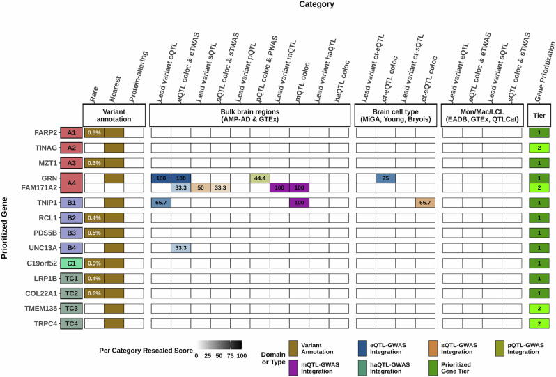
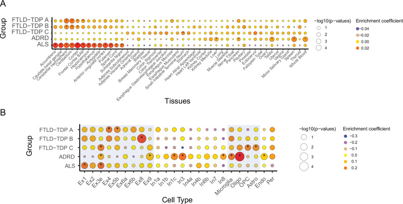
Frontotemporal lobar degeneration with neuronal inclusions of the TAR DNA-binding protein 43 (FTLD-TDP) is a fatal neurodegenerative disorder with only a limited number of risk loci identified. We report our comprehensive genome-wide association study as part of the International FTLD-TDP Whole-Genome Sequencing Consortium, including 985 patients and 3,153 controls compiled from 26 institutions/brain banks in North America, Europe and Australia, and meta-analysis with the Dementia-seq cohort. We confirm UNC13A as the strongest overall FTLD-TDP risk factor and identify TNIP1 as a novel FTLD-TDP risk factor. In subgroup analyzes, we further identify genome-wide significant loci specific to each of the three main FTLD-TDP pathological subtypes (A, B and C), as well as enrichment of risk loci in distinct tissues, brain regions, and neuronal subtypes, suggesting distinct disease aetiologies in each of the subtypes. Rare variant analysis confirmed TBK1 and identified C3AR1, SMG8, VIPR1, RBPJL, L3MBTL1 and ANO9, as novel subtype-specific FTLD-TDP risk genes, further highlighting the role of innate and adaptive immunity and notch signaling pathway in FTLD-TDP, with potential diagnostic and novel therapeutic implications.


Conflict of interest statement

Competing interests: S.J.H. serves on the scientific advisory board (SAB) of Proximity Therapeutics, Psy Therapeutics, Frequency Therapeutics, Souvien Therapeutics, Sensorium Therapeutics, 4 M Therapeutics, Ilios Therapeutics, Entheos Labs, Alzheimer’s Drug Discovery Foundation, and the Kissick Family Foundation FTD Grant Program, none of whom were involved in the present study. S.J.H. has also received speaking or consulting fees from Amgen, AstraZeneca, Biogen, Merck, Regenacy Pharmaceuticals, Syros Pharmaceuticals, Juvenescence Life, as well as sponsored research or gift funding from AstraZeneca, JW Pharmaceuticals, Lexicon Pharmaceuticals, Vesigen Therapeutics, Compass Pathways, Atai Life Sciences, and Stealth Biotherapeutics. The funders had no role in the design or content of this article. ZKW serves as PI or Co-PI on Biohaven Pharmaceuticals, Inc. (BHV4157-206), Vigil Neuroscience, Inc. (VGL101-01.002, VGL101-01.201, PET tracer development protocol, Csf1r biomarker and repository project, and ultra-high field MRI in the diagnosis and management of CSF1R-related adult-onset leukoencephalopathy with axonal spheroids and pigmented glia), and ONO-2808-03 projects/grants. He serves as Co-PI of the Mayo Clinic APDA Center for Advanced Research and as an external advisory board member for the Vigil Neuroscience, Inc., and as a consultant for Eli Lilly & Company and for NovoGlia, Inc. R.R. and I.R.M. receive royalties from progranulin-related patent. The remaining authors declare no competing interests.

Figures

Fig. 1
Fig. 1. Genome-wide association study on common variants.
Association studies were performed using logistic regression with allele dosage as the predictor assuming log-additive allele effects adjusted for relevant covariates. Raw two-sided P are reported as −log10(P). A Manhattan plot of the FTLD-TDP All patients versus controls association study (inflation, λ = 1.05). B Manhattan plot of the FTLD-TDP A patients versus controls association study (λ = 1.07). C Manhattan plot of the FTLD-TDP B patients versus controls association study (λ = 1.06). D Manhattan plot of the FTLD-TDP C patients versus controls association study (λ = 1.05). The red-dotted line represents the genome-wide significance level (p = 5 × 10−8).
Fig. 2
Fig. 2. Gene prioritization results for FTLD-TDP subgroups.
A visual summary of weighted evidence category scores for the prioritized genes within genome-wide significant loci in related FTLD subtype-specific GWAS summary statistics. Using the gene prioritization strategy in these selected loci, we prioritized a total of 13 genes in 12 genome-wide significant loci at two different confidence levels (10 tier 1 and 3 tier 2 prioritized genes). The leftmost squares, which are colored in red for FTLD-TDP A, in blue for FTLD-TDP B, in lighter green for FTLD-TDP C, and in darker green for FTLD-TDP C* specific analyzes, indicate the locus index numbers for the genome-wide loci. The types of evidence for each category are colored according to the six different domains to which they belonged. Weighted scores for each evidence category are rescaled to a 0–100 scale based on the maximum score a candidate gene can obtain from a category (see Supplementary Data 3). The darker colors represent higher scores in categories, while tier 1 prioritized genes are displayed in dark green, and tier 2 prioritized genes are displayed in light green. Only tier 1 and tier 2 genes are shown for each locus, whereas all candidate genes considered and scored can be found in Supplementary Data 4. MAFs (based on gnomAD v4 non-Finnish European samples) and CADD (v1.7) PHRED scores for rare and/or protein-altering rare variants are labeled in white within the respective squares. eQTL expression QTL, sQTL splicing QTL, mQTL methylation QTL, pQTL protein-expression QTL, haQTL histone acetylation QTL, coloc colocalization, eTWAS expression transcriptome-wide association study, sTWAS splicing transcriptome-wide association study, PWAS proteome-wide association study, Mon Mac monocytes and macrophages, LCL lymphoblastoid cell line, QTLCat the eQTL catalog.
Fig. 3
Fig. 3. Top 5 Gene Ontology terms enriched in FTLD-TDP subgroups.
Hierarchical GO analysis of biological process terms considering genes in genetic loci prioritized for FTLD-TDP All, FTLD-TDP A, FTLD-TDP B, and FTD-TDP C. Raw two-sided p values are represented.
Fig. 4
Fig. 4. Enrichment of brain regions and cell types in FTLD subgroups.
A Enrichment of genes in multiple tissues, including 13 brain regions, and based on GTEX data in FTLD subgroups, ADRD, and ALS. Color represents the enrichment coefficient, and size indicates two-sided −log10 (FDR-adjusted Ps) of enrichment obtained by the linear regression model in the MAGMA gene property analysis. B Central nervous system cell type enrichment analyzes in FTLD subgroups, ADRD, and ALS. Color represents the enrichment coefficient, and size indicates two-sided −log10 (FDR-adjusted Ps) of enrichment obtained by the linear regression model in the MAGMA gene property analysis. Excitatory neurons and glial cells are highlighted in blue. Excitatory and inhibitory neurons from the PsychENCODE dataset were labeled based on their transcriptional profile from 1 to 8. Asterisks denote brain regions or cell types enriched with FDR P < 0.05. Cx cortex, Ex* Excitatory neuron, In* inhibitory neurons, Oligo oligodendrocytes, OPCs oligodendrocyte progenitor cells, Astro astrocytes, Endo endothelial cells, Per pericytes.
Fig. 5
Fig. 5. Locus zoom plots for UNC13A and TNIP1 loci.
A Genetic colocalization between the UNC13A locus in FTLD (meta-analysis) and ALS signal. B Genetic colocalization between the TNIP1 locus in FTLD (meta-analysis) and ALS. C Genetic colocalization between the TNIP1 locus in FTLD (meta-analysis) and ADRD. For A, B, C, chromosome position is located on the x-axis, and −log10 transformed raw two-sided P is represented on the y-axis. Each dot represents a SNV tested in the dataset for its association with disease status. Purple diamonds are the index SNVs reported. Linkage disequilibrium with index SNV is indicated by r2.
Fig. 6
Fig. 6. Rare loss of function and predicted pathogenic variants in proteins associated with FTLD.
Schematic representation of C3AR1, SMG8, VIPR1, L3MBTL1, and RBPL protein structure (source Uniprot) showing a map of nonsense, splicing, frameshift, and missense rare variants with a REVEL score > 0.75 in patients and controls. Variants identified in patients are colored in orange, and variants identified in controls are colored in blue. n = number of carriers. When no number is indicated, the variant was observed in a single individual. Total number of subjects included in the analyzes was FTLD-TDP A (n = 193), FTLD-TDP B (n = 288), FTLD-TDP C (n = 467), and FTLD-TDP C* (n = 199), and controls (n = 3153).
Fig. 7
Fig. 7. Schematic representation of findings from the International FTLD-TDP WGS phase II.
Genome-wide significant single variant loci, exome-wide significant genes, enriched gene ontology pathways, and tissues- and cell-types enriched for genome-wide significant risk loci are shown for each FTLD-TDP pathological subtype in rings moving from the center (genome-wide significant single variant loci in FTLD-TDP All) to the outer rings. Orange background shades correspond to FTLD-TDP A findings, green background shades to FTLD-TDP B findings, and blue background shades to FTLD-TDP C findings. Gene names in green font were exome-wide significant using a gene-based approach with common variants, while gene names in red font were exome-wide significant using a gene-based approach with rare variants. In addition to unique associations, some overlap between FTLD-TDP A and B exists (TBK1, lysosomal function, and inflammatory response), whereas FTLD-TDP C showed a unique and non-overlapping genetic profile. Note that genetic associations with LRP1B, COL22A1, TRPC4, and TMEM135 (identified in the FTLD-TDP C* GWAS focused solely on pathologically confirmed FTLD-TDP C patients) are not shown.

Update of

  • Deciphering Distinct Genetic Risk Factors for FTLD-TDP Pathological Subtypes via Whole-Genome Sequencing.
    Pottier C, Küçükali F, Baker M, Batzler A, Jenkins GD, van Blitterswijk M, Vicente CT, De Coster W, Wynants S, Van de Walle P, Ross OA, Murray ME, Faura J, Haggarty SJ, van Rooij JG, Mol MO, Hsiung GR, Graff C, Öijerstedt L, Neumann M, Asmann Y, McDonnell SK, Baheti S, Josephs KA, Whitwell JL, Bieniek KF, Forsberg L, Heuer H, Lago AL, Geier EG, Yokoyama JS, Oddi AP, Flanagan M, Mao Q, Hodges JR, Kwok JB, Domoto-Reilly K, Synofzik M, Wilke C, Onyike C, Dickerson BC, Evers BM, Dugger BN, Munoz DG, Keith J, Zinman L, Rogaeva E, Suh E, Gefen T, Geula C, Weintraub S, Diehl-Schmid J, Farlow MR, Edbauer D, Woodruff BK, Caselli RJ, Donker Kaat LL, Huey ED, Reiman EM, Mead S, King A, Roeber S, Nana AL, Ertekin-Taner N, Knopman DS, Petersen RC, Petrucelli L, Uitti RJ, Wszolek ZK, Ramos EM, Grinberg LT, Gorno Tempini ML, Rosen HJ, Spina S, Piguet O, Grossman M, Trojanowski JQ, Keene DC, Lee-Way J, Prudlo J, Geschwind DH, Rissman RA, Cruchaga C, Ghetti B, Halliday GM, Beach TG, Serrano GE, Arzberger T, Herms J, Boxer AL, Honig LS, Vonsattel JP, Lopez OL, Kofler J, White CL, Gearing M, Glass J, Rohrer JD, Irwin DJ, Lee EB, Van Deerlin V, Castellani R, Mesulam MM, Tartaglia MC, Finger EC, Troak… See abstract for full author list ➔ Pottier C, et al. medRxiv [Preprint]. 2024 Jun 25:2024.06.24.24309088. doi: 10.1101/2024.06.24.24309088. medRxiv. 2024. Update in: Nat Commun. 2025 Apr 25;16(1):3914. doi: 10.1038/s41467-025-59216-0. PMID: 38978643 Free PMC article. Updated. Preprint.

References

    1. Rascovsky, K. et al. Sensitivity of revised diagnostic criteria for the behavioural variant of frontotemporal dementia. Brain134, 2456–2477 (2011). - PMC - PubMed
    1. Gorno-Tempini, M. L. et al. Classification of primary progressive aphasia and its variants. Neurology76, 1006–1014 (2011). - PMC - PubMed
    1. Lee, E. B. et al. Expansion of the classification of FTLD-TDP: distinct pathology associated with rapidly progressive frontotemporal degeneration. Acta Neuropathol.134, 65–78 (2017). - PMC - PubMed
    1. Mackenzie, I. R. et al. A harmonized classification system for FTLD-TDP pathology. Acta Neuropathol.122, 111–113 (2011). - PMC - PubMed
    1. Chatterjee, M. et al. Plasma extracellular vesicle tau and TDP-43 as diagnostic biomarkers in FTD and ALS. Nat. Med.30, 1771–1783 (2024). - PMC - PubMed

Grants and funding