. 2025 Apr 25;8(1):662.
doi: 10.1038/s42003-025-07811-8.

Soil-mimicking microfluidic devices reveal restricted flagellar motility of Bradyrhizobium diazoefficiens under microconfinement

Affiliations

Soil-mimicking microfluidic devices reveal restricted flagellar motility of Bradyrhizobium diazoefficiens under microconfinement

Moniellen Pires Monteiro et al. Commun Biol. .

Abstract

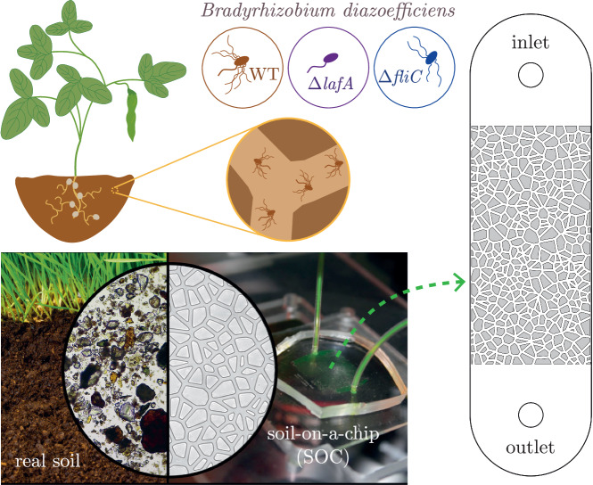
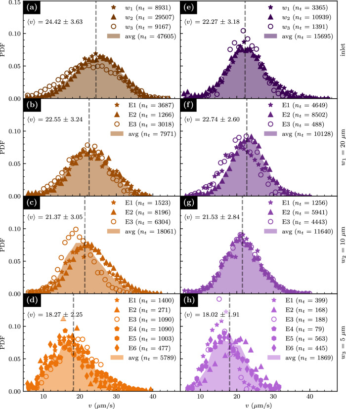
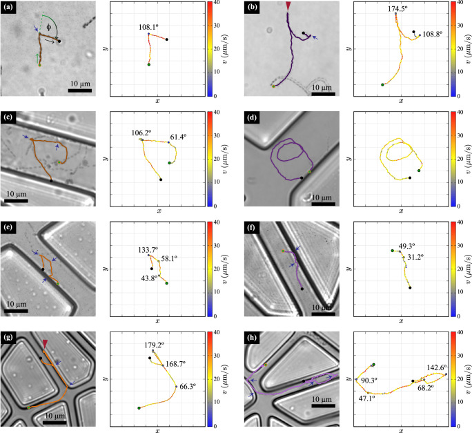
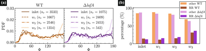
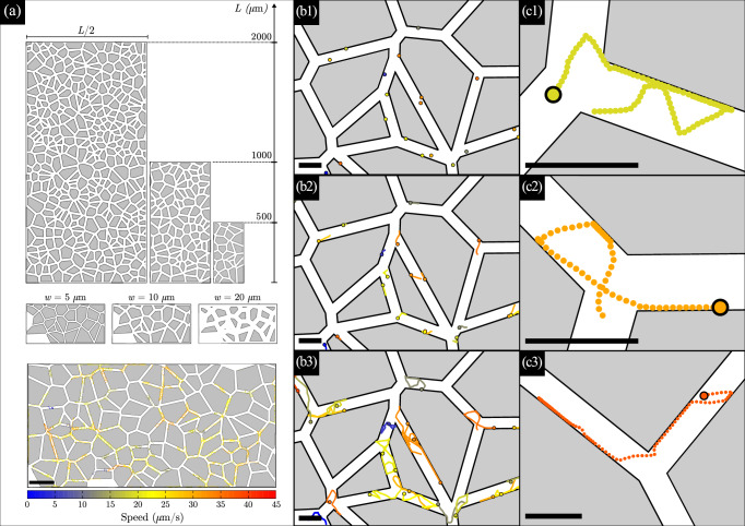
Bradyrhizobium diazoefficiens is a nitrogen-fixing symbiont of soybean, worldwide used as biofertilizer. This soil bacterium possesses two flagellar systems enabling it to swim in water-saturated soils. However, the motility in soil pores, which may be crucial for competitiveness for root nodulation, is difficult to predict. To address this gap, we fabricated microfluidic devices with networks of connected microchannels surrounding grains. In them, we directly visualise bacterial behaviour in transparent geometries mimicking minimalist soils-on-a-chip (SOCs). We measured the population velocities and changes of direction for two strains: the wild-type and a mutant with only a subpolar flagellum. A detailed statistical analysis revealed that both strains exhibited reduced speeds and increased changes of direction of 180°, in channels of decreasing cross sectional area, down to a few microns in width. Interestingly, while the wild-type strain displayed faster swimming in unconfined spaces, this advantage was negated in the SOCs with the narrowest microchannels. We employed the measured motility parameters to propose a realistic model and simulate B. diazoefficiens confined dynamics being able to reproduce their behaviour, which additionally can be extended enabling further predictions for long time and macro scales. This multidisciplinary work, combining design, microfabrication, microbiology and modelling, offers useful methods to study soil bacteria and may be readily extended to other beneficial/harmful soil species.


Conflict of interest statement

Competing interests: The authors declare no competing interests.

Figures

Fig. 1
Fig. 1. Direct observation of soil bacteria into transparent chambers with microchannels of different widths.
In this scheme, flagellated soil bacteria navigate the soil by swimming through flooded pores between soil particles and grains (left semicircle). To visualize its swimming through a complex geometry with different degrees of confinement, transparent chambers with microchannels of different widths were used (right semicircle, showing a microchannel network of 20 μm channel width). Microfluidic devices, as sketched at the right, were fabricated based on Voronoi tessellations (see Materials and Methods), with channel widths of increasing confinement (20 μm, 10 μm and 5 μm). Three different strains of Bradyrhizobium diazoefficiens were inoculated in the devices, a wild type (WT) with two flagellar systems and two mutants, one lacking the lateral flagellar system (ΔlafA) and the other lacking the subpolar one (ΔfliC) depicted as light-brown, violet, and blue bacteria, respectively. Credit for the real soil image (left semicircle): Katelyn Solbakk, Mikroliv.
Fig. 2
Fig. 2. Speed distributions of bacteria in microfluidic devices.
Speed probability density function (PDF) of WT (ad) and ΔlafA (eh) B. diazoefficiens strains are shown for measurements into the inoculation site at the inlet (a) and (e), or a porous geometry with channels of width w1 = 20 μm (b) and (f), w2 = 10 μm (c) and (g), and w3 = 5 μm (d) and (h). Symbols represent experimental distributions from 3 to 6 biological replicas (Ei, with i = 1, …, 6), with a number of observed tracks nt indicated for each case in the legends. Solid light-coloured backgrounds represent the average of all experiments (avg). Mean speed values  ± error (in units of μm/s) are given in all panels and indicated with a dashed vertical line.
Fig. 3
Fig. 3. Average speed and speed variation ratio in microfluidic devices with different degrees of confinement.
Speed PDF for WT and ΔlafA mutant in the inoculation chamber (a) and in microfluidic devices with different degrees of confinement: w1 = 20 μm (b), w2 = 10 μm (c), and w3 = 5 μm (d). e Bacteria average speed in the microfluidic devices as a function of the channel width. f SVR for each strain as a function of the channel width (see Eq. (1)). Vertical bars in both cases indicate the data corresponding errors.
Fig. 4
Fig. 4. Swimming behaviour of B. diazoefficiens in unconfined space and in microchannels.
Examples of trajectories followed by WT (left) and ΔlafA (right) at the unconfined inlets (a, b) and in the microchannels with increasing confinement [w1 = 20 μm (c) and (d); w2 = 10 μm (e) and (f); and w3 = 5 μm (g) and (h)]. For each case, the trajectory is shown at the left and at the right, the bacterium speed is presented along their path in colour scale, with the angle values for each detected change of direction, Φ. Several snapshots are overlaid to show the trajectory of the bacteria for the time span of the tracks, and only one bacterium track is shown. The green and black dots mark, respectively, the start and end of the track. The blue arrows and red arrowheads mark changes of direction (CHD) and run and reverses (RR), respectively. In (a), the reorientation event is marked with the definition of the reorientation angle, Φ. Videos of the trajectories are available as Supplementary Movies 2–9.
Fig. 5
Fig. 5. Changes of Directions of B. diazoefficiens measured in the microfluidic devices.
a Reorientation angle PDF of WT (left) and ΔlafA (right) in the inoculation chambers and in the different confined networks with channels width w1 = 20 μm, w2 = 10 μm, and w3 = 5 μm. b Classification of CHDs in the inlets and in the different microchannels, according to their angle: RR for Φ > 160°, and “other” otherwise.
Fig. 6
Fig. 6. Simulated B. diazoefficiens swimming behaviour in microdevices mimicking a porous media with 50 μm mean size grains.
a Designed arrays composed of islands and channels, with total area L/2 × L. In the second row, an array with L = 500 μm and three channel widths is shown: w = 5, 10, 20 μm (left to right respectively). In the lower panel, a few bacteria trajectories are drawn inside a complete array with L = 1000 μm, w = 10 μm. Scale bar: 100 μm. The colour of the tracks corresponds to the speed of each simulated bacterium (proportional to their motor force of self-propulsion), according to the speed colour scale at the bottom. The same colour scale is used in all panels. b1 Initial positions, at t = 0. Dots representing bacteria are enlarged for visualization. b2 Starting bacteria trajectories until t = 0.5 s. The black circles represent the final positions. b3 Simulated tracks of duration 3.5 s. Most of the bacteria have performed several changes of direction into the middle of the channels or due to interactions. Note the swimming along walls, with some straight paths of more than 30 μm. c1c3 show three examples of simulated tracks of duration 5.5 s, 5 s, and 3.75 s, respectively. Scale bars in (b) and (c) panels correspond to 20 μm.
Fig. 7
Fig. 7. Microfluidics devices for Studying B. diazoefficiens Confined Swimming behaviour: Experimental vs. Theoretical approaches.
The central circle highlights the reached goals of this work: nice agreements between real-time microbial tracking in microfluidic devices and predictive theoretical models and calculations, utilizing high-performance computing.

References

    1. Garrido-Sanz, D. et al. Phylogenomic analyses of Bradyrhizobium reveal uneven distribution of the lateral and subpolar flagellar systems, which extends to Rhizobiales. Microorganisms7, 50 (2019). - PMC - PubMed
    1. Catroux, G., Hartmann, A. & Revellin, C. Trends in rhizobial inoculant production and use. Plant Soil230, 21–30 (2001).
    1. Basile, L. A. & Lepek, V. C. Legume–rhizobium dance: an agricultural tool that could be improved? Microb. Biotechnol.14, 1897–1917 (2021). - PMC - PubMed
    1. Aroney, S. T. N., Poole, P. S. & Sánchez-Cañizares, C. Rhizobial chemotaxis and motility systems at work in the soil. Front. Plant Sci.12, 725338 (2021). - PMC - PubMed
    1. Kanbe, M., Yagasaki, J., Zehner, S., Göttfert, M. & Aizawa, S.-I. Characterization of two sets of subpolar flagella in Bradyrhizobium japonicum. J. Bacteriol. 189, 1083–1089 (2007). - PMC - PubMed

Supplementary concepts

LinkOut - more resources