. 2025 Apr 14;15(8):1129.
doi: 10.3390/ani15081129.

Morphological, Histological and Morphometrical Aspects of Auditory Ossicles in Pig Fetuses (Sus scrofa domestica)

Affiliations

Morphological, Histological and Morphometrical Aspects of Auditory Ossicles in Pig Fetuses (Sus scrofa domestica)

Cristian Olimpiu Martonos et al. Animals (Basel). .

Abstract

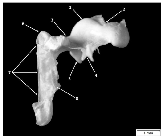
The detailed study of the morphology and morphometrics of the auditory ossicles in swine became a topic for investigation due to their resemblance to the human ear. The methods used in this study cover the typical macroscopical investigation of gross morphology: a detailed metrical and histological assessment through H&E standard protocol on auditory ossicles originating from eight pig fetuses originating from four distinctive sows. The ossicular assembly in the malleus, incus and stapes present in 66-day fetuses shows all the features generally described in swine. The malleus comprises two uneven laminae of mineralized hyaline cartilage and a medullary cavity. The areas of the head and neck show a high degree of vascularization. The incus has two similar cortical fascicles separated by a compartmentalized medullary cavity, with the highest degree of mineralization found at the distal part of the long process. Stapes show an early degree of mineralization at the level of the crura, lacking medullary cavities. The ossicular chain shows typical morphological elements, similar to adults, and from a dimensional perspective, our investigations point to an uneven degree of development of the ossicles, according to gestational age: the malleus and stapes reach almost 80% and the incus about 50-60% of their adult sizes.

Keywords: Sus scrofa; fetus; gross anatomy; histology; incus; lever ratio; malleus; morphometry; ossicula auditoria; stapes.


Conflict of interest statement

The authors declare no conflicts of interest.

Figures

Figure 1
Figure 1
Incus and malleus joined in the incudo-malleal joint: 1. head of the malleus, 2. body of the incus, 3. incudo-malleal joint, 4. bony lame, 5. anterior process of the malleus, 6. muscular process of the malleus, 7. lateral process of the malleus, 8. neck of the malleus, 9. handle of the malleus, 10. the short process of the incus, 11. the long process of the incus and 12. lenticular process.
Figure 2
Figure 2
Malleus—anatomic features. 1. head of the malleus, 2. articular surface, 3. neck of the malleus, 4. anterior process, 5. muscular process, 6. lateral process, 7. handle of the malleus and 8. tympanic membrane.
Figure 3
Figure 3
Histological features of the malleus in 66-day-old pig fetuses. (A) malleal head area/H&E staining; (B) malleal neck area/H&E staining; (C) lateral tubercle/H&E staining; (D) malleal handle bottom extremity/H&E staining; (E) malleus, macroscopical features; (F) malleal handle/H&E staining; (G) malleal handle, proximal end/H&E staining; ct—cartilaginous trabeculae; ecf—external cortical fascicles; fct—fibrous connective tissue; icf—internal cortical fascicles; mc—marrow cavity; mt—mineralized trabeculae; tm—tympanic membrane; and black arrows—blood vessels.
Figure 4
Figure 4
Incus—anatomic features. 1. Body of the incus, 2. articular surface of the incus, 3. short process of the incus, 4. long process of the incus, and 5. lenticular process.
Figure 5
Figure 5
Histological features of the incus in 66-day-old pig fetuses. (A) Incudal articular surface/H&E staining; (B) incus, macroscopical features; (C) incudal articular surface/H&E staining; (D) long process/H&E staining; (E) short process/H&E staining; (F) incudal body/H&E staining; ct—cartilaginous trabeculae; dcf—dorsal cortical fascicles; fct—fibrous conjunctive tissue; mc—marrow cavity; mhc—mineralized hyaline cartilage; mt—mineralized trabeculae; vcf—ventral cortical fascicles; and red asterisk—malleo-incal joint capsule.
Figure 6
Figure 6
Stapes—anatomical features. 1. Head of the stapes, 2. articular surface, 3. insertion surface for the stapedial muscle, 4. stapedial muscle, 5. posterior crus of the stapes, 6. anterior crus of the stapes, 7. intercusal foramen, and 8. stapedial footplate of the stapes.
Figure 7
Figure 7
Histological features of the stapes in 66-day-old pig fetuses. (A) Posterior crus/H&E staining; (B) head of the stapes/H&E staining; (C) anterior crus/H&E staining; (D) caudal segment of the footplate/H&E staining; (E) stapes, macroscopical features; (F) footplate/H&E staining; (G) rostral segment of the footplate/H&E staining; mpc—mineralized posterior crus; mac—mineralized anterior crus; af—acoustical fascicle of the footplate; if—intercrural foramen; isc—incudostapedial joint capsule; hc—hyaline cartilage; mc—marrow cavity; mhc—mineralized hyaline cartilage; vf—vestibular compact fascicle of the footplate; and red asterisk—annular ligament.
Figure 8
Figure 8
Graphical representation of the functional length of the malleus and functional length of the incus for the reference species.

Similar articles

References

    1. Simaei N., Soltanalinejad F., Najafi G., Shalizar Jalali A. Anatomical and Morphometrical Study of Middle Ear Ossicles in 2 to 3-Month-Old Makouei Sheep Fetuses. Vet. Res. Forum. 2017;8:237–241. - PMC - PubMed
    1. Erdogan S., Kilinc M. Gross Anatomy and Arterial Vascularization of the Tympanic Cavity and Osseous Labyrinth in Mid-Gestational Bovine Fetuses. Anat. Rec. 2010;293:2083–2093. doi: 10.1002/ar.21269. - DOI - PubMed
    1. Parodi A.-L., Barone R., Simoes P. Anatomie Comparée Des Mammifères Domestiques, Paris, Vigot Édit., 2010, 7, Neurologie 2, 836 p. Bull. Acad. Natl. Med. 2011;195:1148–1150. doi: 10.1016/s0001-4079(19)32028-x. - DOI
    1. Robert M.P., Carstens A., de Beer F.C., Hoffman J.W., Steenkamp G. Micro-Anatomy of the Ear of the Southern White Rhinoceros (Ceratotherium simum simum) Anat. Histol. Embryol. 2021;50:316–323. doi: 10.1111/ahe.12632. - DOI - PubMed
    1. Barone R., Bortolami R. Anatomie Comparee Des Mammiferes Domestiques. Tome Sixieme. Neurologie I. Systeme Nerveux Central. Vigot Frerres; Paris, France: 2004.

LinkOut - more resources