. 2025 Apr 8;14(4):387.
doi: 10.3390/biology14040387.

Integration of Microarray and Single-Cell RNA-Seq Data and Machine Learning Allows the Identification of Key Histone Modification Gene Changes in Spermatogonial Stem Cells

Affiliations

Integration of Microarray and Single-Cell RNA-Seq Data and Machine Learning Allows the Identification of Key Histone Modification Gene Changes in Spermatogonial Stem Cells

Ali Shakeri Abroudi et al. Biology (Basel). .

Abstract

Histone modifications play a critical role in regulating gene expression and maintaining the functionality of spermatogonial stem cells (SSCs), which are essential for male fertility and spermatogenesis. In this study, we integrated microarray and single-cell RNA-sequencing (scRNA-seq) data to identify key histone modification gene changes associated with SSC function and aging. Through differential expression analysis, we identified 2509 differentially expressed genes (DEGs) in SSCs compared to fibroblasts. Among these, genes involved in histone modification, such as KDM5B, SCML2, SIN3A, and ASXL3, were highlighted for their significant roles in chromatin remodeling and gene regulation. Protein-protein interaction (PPI) networks and gene ontology (GO) enrichment analysis revealed critical biological processes such as chromatin organization, histone demethylation, and chromosome structure maintenance. Weighted gene co-expression network analysis (WGCNA) further revealed three key modules of co-expressed genes related to spermatogonial aging. Additionally, ligand-receptor interaction scoring based on tumor microenvironment analysis suggested potential signaling pathways that could influence the stemness and differentiation of SSCs. Our findings provide new insights into the molecular mechanisms underlying SSC aging, highlighting histone modification genes as potential therapeutic targets for preserving male fertility and improving SSC-culturing techniques. This study advances our understanding of histone modification in SSC biology and will serve as a valuable resource for future investigations into male fertility preservation.

Keywords: bioinformatics; gene ontology; germ cell; microarray; spermatogonia stem cell.


Conflict of interest statement

It is declared by the remaining authors that there are no commercial or financial relationships that might conflict with this research.

Figures

Figure 1
Figure 1
In-vitro-cultivated human spermatogonia after matrix and CD49f selection. During culturing, it was observed that the spermatogonia had the expected shape. Connected spermatogonia existed in all the cell cultures, either singly, in pairs, in chains, or in colonies. The cells were cultured for an extended duration using inactivated CF1 feeder cells. (A1) hSSC testicular cell expansion in the culture, (A2) SSEA4 expression in hSSCs, (A3) DAPI, (A4) merge, (B1) hSSC testicular cell expansion in the culture, (B2) VASA expression in hSSCs, (B3) DAPI, and (B4) merging. Scale bar: 25 μm.
Figure 2
Figure 2
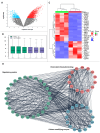
Analysis of histone-modifying enzyme gene expression by microarray: (A) volcano plot of differentially expressed genes based on microarray analysis, (B) correlation plot of SSCs and fibroblasts, (C) heatmap of histone-modifying enzyme DEGs, and (D) PPI of histone-modifying enzymes.
Figure 3
Figure 3
Results of performing gene ontology (GO) enrichment analysis on the genes inside the module. The colors correspond to the corrected p-values (BH), while the sizes of the dots correspond to the number of genes. This picture refers to (A) the biological processes, (B) molecular functions, (C) cellular components and signaling pathways, and signaling pathway analysis, (D) Signaling pathway analysis.
Figure 4
Figure 4
Signaling pathways. (A) Using the KEGG database, we investigated the key genes’ signaling pathways to find out how they contribute to signaling pathways. (B) When looking at the MCC scores of the genes, the red and yellow nodes reflect genes with high and low values, respectively.
Figure 5
Figure 5
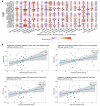
WGCNA of the GEO datasets. Clustering the DEGs from the GEO datasets yielded a gene dendrogram (A). Different colors are used to indicate a total of three modules: MEblue, MEturquoise, and MEgrey. The consensus MEs and phenotypes in the GEO datasets are explained in (B) correlation of WGCNA. (C) The gene dendrogram of the TCGA dataset, produced by clustering the DEGs. Differently colored markers are used to identify four separate modules: MEblue, MEbrown, MEgrey, and MEturquoise. (D) Phenotypic correlations from the TCGA dataset with respect to the consensus MEs. Abbreviations: differentially expressed gene (DEG), Gene Expression Omnibus (GEO), The Cancer Genome Atlas (TCGA), and module eigengene (ME).
Figure 6
Figure 6
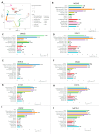
Single-cell RNA-sequencing investigation of human adult spermatogonia demonstrated histone-modifying enzyme genes similar to those seen in humans: (A) UMAP plot depiction of germ cells from merged single-cell RNA-sequencing data and (B) NCOR3, (C) JARID2, (D) STAG1, (E) SCML2, (F) SIN3A, (G) H1FNT, (H) CHD1L, (I) ATAD2, and (J) NAP1L4 gene expression in germ cells.
Figure 7
Figure 7
Analysis of cellular communication: (A) communication between individual cells that is influenced by genes that modify histones (Ligand–Receptor interactions 1–10 and their niche organization) and (B) communication between cells in a network that is influenced by the co-expression of genes that modify histones.

Similar articles

References

    1. Aballa L., Chraa M., Louhab N., Kissani N. Extensive anaplastic multi-centric ependymoma in a young adult: Case report and literature review. Egypt. J. Neurol. Psychiatry Neurosurg. 2023;59:67. doi: 10.1186/s41983-023-00663-1. - DOI
    1. Voigt A., Dardari R., Lara N., He T., Steele H., Dufour A., Orwig K., Dobrinski I. Multiomics approach to profiling Sertoli cell maturation during development of the spermatogonial stem cell niche. Mol. Hum. Reprod. 2023;29:gaad004. - PMC - PubMed
    1. Voigt A.L., Thiageswaran S., de Lima e Martins Lara N., Dobrinski I. Metabolic requirements for spermatogonial stem cell establishment and maintenance in vivo and in vitro. Int. J. Mol. Sci. 2021;22:1998. doi: 10.3390/ijms22041998. - DOI - PMC - PubMed
    1. Liu W., Du L., Li J., He Y., Tang M. Microenvironment of spermatogonial stem cells: A key factor in the regulation of spermatogenesis. Stem Cell Res. Ther. 2024;15:294. - PMC - PubMed
    1. Hui L., Ziyue Z., Chao L., Bin Y., Aoyu L., Haijing W. Epigenetic regulations in autoimmunity and cancer: From basic science to translational medicine. Eur. J. Immunol. 2023;53:2048980. doi: 10.1002/eji.202048980. - DOI - PubMed

LinkOut - more resources