. 2025 Mar 30;16(4):404.
doi: 10.3390/genes16040404.

The Interconnection Between UbcH10, p53, and EGFR in Lung Cancer Cells and Their Involvement in Treatment Response

Affiliations

The Interconnection Between UbcH10, p53, and EGFR in Lung Cancer Cells and Their Involvement in Treatment Response

Cristina Quintavalle et al. Genes (Basel). .

Abstract

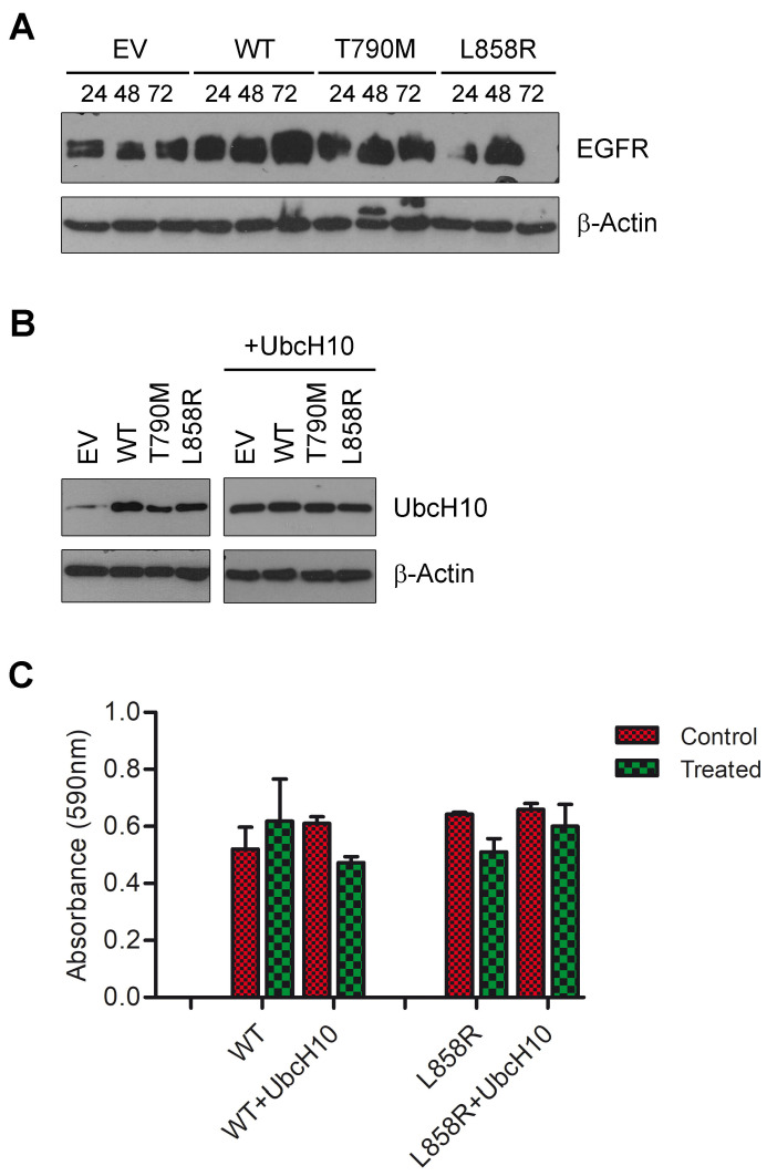
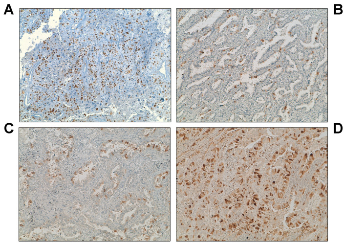
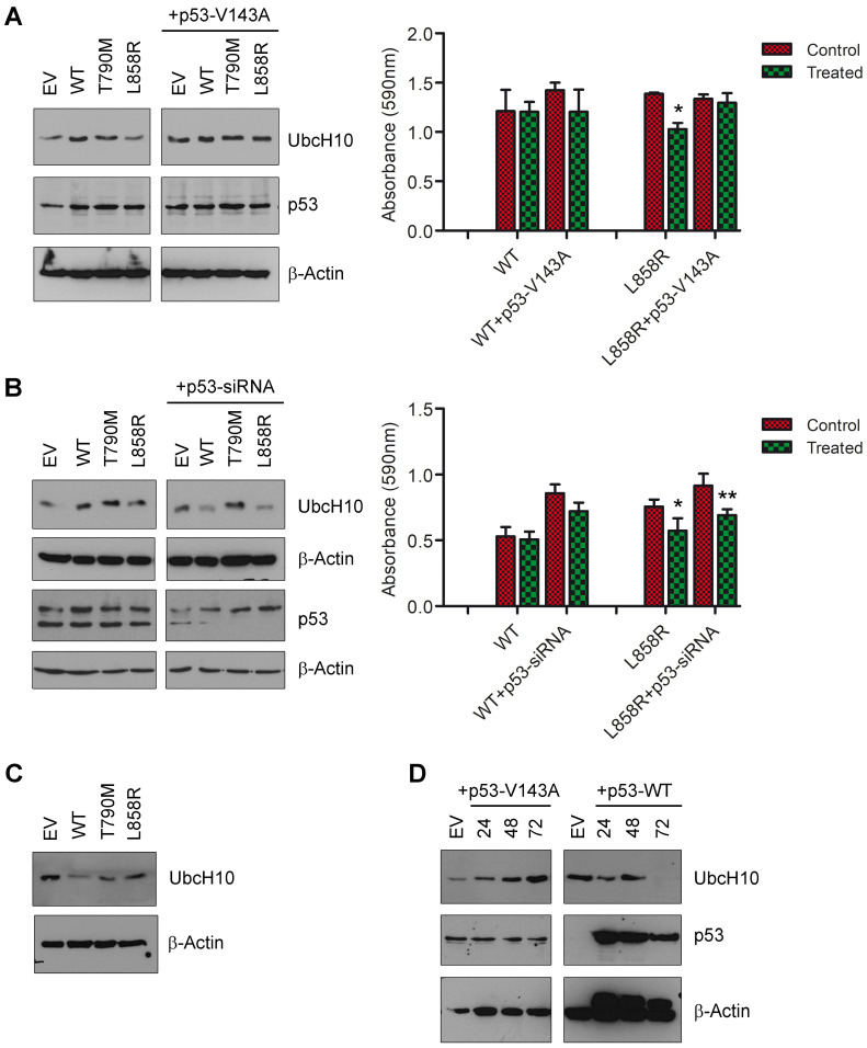
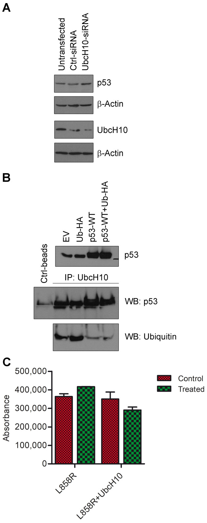
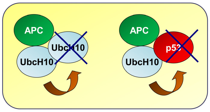
Background/Objectives: The UbcH10 protein plays an important role in a variety of human malignancies, including thyroid, breast, ovarian, and colorectal carcinomas. It has been previously reported that UbcH10 is overexpressed in non-small cell lung cancer (NSCLC) compared to normal lungs and that its expression is directly and inversely correlated with the mutational status of p53 and EGFR, respectively. Methods: We transfected lung cancer cells with wild-type and mutant forms of EGFR, modulated the expression of UbcH10 and p53, and treated these cells with tyrosine kinase inhibitor (TKI) erlotinib. Using Western blotting, we evaluated the expression of UbcH10 induced by EGFR and p53. Finally, we employed immunohistochemistry to assess the levels of UbcH10 expression in a subset of NSCLC patients receiving TKI therapy. Results: We reported a possible modulation of UbcH10 expression by the overexpression of wild-type and mutant EGFR in H460 lung cancer cells, potentially through p53. The enforced expression of UbcH10 in cells transfected with mutant EGFR suggested a potential increase in resistance to erlotinib treatment. Finally, immunohistochemical analysis of samples from NSCLC patients with mutant EGFR indicated a possible connection between UbcH10 expression levels and progression-free survival. Conclusions: In NSCLC, UbcH10 may play a role in the regulation of TKI response via a molecular pathway potentially involving p53 and EGFR. However, further research is needed to fully understand this mechanism.

Keywords: EGFR; UbcH10; adenocarcinoma; non-small cell lung cancer; p53; resistance.


Conflict of interest statement

U.M., Consulting or advisory role (unrelated to the current work): Boehringer Ingelheim, MSD, Roche, Amgen, Lilly, Thermo Fisher Scientific, Diaceutics, Merck, Glaxo Smith Kline, Astra Zeneca; Speakers’ Bureau (unrelated to the current work): Boehringer Ingelheim, Roche, AstraZeneca, MSD, Merck, Amgen, Thermo Fisher Scientific, Diaceutics, Lilly, Glaxo Smith Kline. F.P., Relationship unrelated to the current work (advisory fees, honoraria, grants, travel accommodation and expenses, and non-financial support) with Menarini and Roche. All other authors declare no conflicts of interest. The funder had no role in the design of the study; in the collection, analyses, or interpretation of data; in the writing of the manuscript; or in the decision to publish the results.

Figures

Figure 1
Figure 1
UbcH10 modulates the drug sensitivity of EGFR-L858R H460 cells to erlotinib treatment. (A) The empty vector (EV) and the three EGFR constructs (WT, T790M, and L858R) were transfected into H460 cells, and the expression of the EGFR protein was checked at three different time points (24, 48, and 72 h after transfection). β-Actin was assessed as a normalization control. Representative images. (B) Levels of UbcH10 protein were evaluated using Western blotting after transfection of EGFR-WT, EGFR-T790M, and EGFR-L858R in H460 cells (left blot). The enforced expression of UbcH10 following co-transfection of EGFR-WT, EGFR-T790M, EGFR-L858R, and UbcH10 in H460 cells (right blot) was also evaluated. The left and right blots are from two different Western blots. β-Actin was evaluated as a normalization control. Representative images. EV, empty vector. (C) H460 cells were treated with 0.1 μM erlotinib after transfection of EGFR-WT and EGFR-L858R, and their viability was assessed 24 h after treatment (left). H460 cells were also treated with 0.1 μM erlotinib after co-transfection of EGFR-WT, EGFR-L858R, and UbcH10, and their viability was assessed 24 h after treatment (right). Data are in triplicate and are shown as mean ± sd. Two-way ANOVA: treatment, p = 0.005.
Figure 2
Figure 2
Higher levels of UbcH10 are associated with short progression-free survival in NSCLC patients. At the time of diagnosis, UbcH10 immunohistochemical expression and EGFR mutational status were assessed. Objective response was evaluated according to RECIST criteria with the first evaluation performed 8 weeks after the start of the treatment. The response rate was linked to UbcH10 levels at diagnosis. (A) Exon 19 and 21 wild-type patient (W1) characterized by high UbcH10 expression levels. (B) EGFR mutant patient (M5) showing low UbcH10 expression levels and a complete response to treatment. (C) EGFR mutant patient (M9) showing intermediate UbcH10 expression levels and stable disease. (D) EGFR mutant patient (M3) showing moderate to high UbcH10 expression levels and partial response.
Figure 3
Figure 3
The expression of UbcH10 in lung cancer cells is under the control of p53. (A) Left panel. Levels of UbcH10 and p53 protein were evaluated after transfection of H460 cells with EGFR-WT, EGFR-T790M, and EGFR-L858R. UbcH10 protein expression was also evaluated after co-transfection of H460 cells with EGFR different forms and a vector expressing p53-V143A. β-Actin was evaluated to normalize the amount of protein loaded. Representative images. EV, empty vector. Right panel. Viability of H460 cells co-transfected with EGFR-WT, EGFR-L858R, and p53-V143A was evaluated 24 h after treatment with 0.1 μM erlotinib. Data are in triplicate and are shown as mean ± sd. Two-way ANOVA: EGFR-L858R treated vs. EGFR-L858R, * p < 0.05. (B) Left panel. Levels of UbcH10 and p53 (two different Western blots) were evaluated after the co-transfection of EGFR-WT, EGFR-T790M, and EGFR-L858R with siRNA suppressing the p53 expression. UbcH10 levels were evaluated 48 h after silencing p53 expression. β-Actin was evaluated to normalize the amount of protein loaded. Representative images. EV, empty vector. Right panel. Viability of H460 cells co-transfected with EGFR-WT, EGFR-L858R, and p53-siRNA was evaluated 24 h after treatment with 0.1 μM erlotinib. Data are in triplicate and are shown as mean ± sd. Two-way ANOVA: treatment, p = 0.0021; +p53-siRNA, p < 0.0001; EGFR-L858R treated vs. EGFR-L858R, * p < 0.05; EGFR-L858R+p53-siRNA treated vs. EGFR-L858R+p53-siRNA, ** p < 0.01. (C) Levels of UbcH10 protein were evaluated after transfection of Calu-1 cells with EGFR-WT, EGFR-T790M, and EGFR-L858R. β-Actin was evaluated as a normalization control. EV, empty vector. (D) UbcH10 protein expression was evaluated at 24, 48, and 72 h after transfecting p53-V143A and p53-WT in H460 (left blot) and Calu-1 (right blot) cells, respectively. β-Actin was assessed as a control for normalization. EV, empty vector.
Figure 4
Figure 4
UbcH10 and p53 are part of a self-regulatory mechanism. (A) P53 and UbcH10 expression levels (two different Western blots) were evaluated 48 h after transfection of A549 cells with siRNA able to reduce the expression of UbcH10. β-Actin was evaluated as a normalization control. (B) HEK 293 cells were transfected with empty vector, ubiquitin-HA, p53-WT, and p53-WT+ubiquitin-HA, and p53 protein expression levels were evaluated. Protein lysates were immunoprecipitated with anti-UbcH10 antibodies, and levels of UbcH10/ubiquitin complexes were evaluated using Western blotting with an anti-ubiquitin antibody. EV, empty vector. (C) H460 cells were co-transfected with EGFR-L858R and UbcH10. Caspase activity was evaluated using caspase assay 24 h after treatment with 0.1 μM erlotinib. Data are in duplicate and are shown as mean ± sd. Two-way ANOVA: interaction, p = 0.0303; +UbcH10, p = 0.0201.
Figure 5
Figure 5
EGFR-p53-UbcH10 loop in the modulation of NSCLC cells to treatment. Schematic model likely showing UbcH10 overexpression in the presence of p53. In the absence of p53 target molecules, UbcH10 self-degrades, preventing accumulation. When p53 is present, UbcH10 targets it for degradation via the ubiquitin–proteasome system, resulting in UbcH10 accumulation and the activation of its oncogenic potential. APC, anaphase-promoting complex.

Similar articles

References

    1. Bray F., Laversanne M., Sung H., Ferlay J., Siegel R.L., Soerjomataram I., Jemal A. Global Cancer Statistics 2022: GLOBOCAN Estimates of Incidence and Mortality Worldwide for 36 Cancers in 185 Countries. CA Cancer J. Clin. 2024;74:229–263. doi: 10.3322/caac.21834. - DOI - PubMed
    1. Barr T., Ma S., Li Z., Yu J. Recent Advances and Remaining Challenges in Lung Cancer Therapy. Chin. Med. J. 2024;137:533–546. doi: 10.1097/CM9.0000000000002991. - DOI - PMC - PubMed
    1. Reck M., Remon J., Hellmann M.D. First-Line Immunotherapy for Non–Small-Cell Lung Cancer. J. Clin. Oncol. 2022;40:586–597. - PubMed
    1. Mok T.S., Wu Y.-L., Ahn M.-J., Garassino M.C., Kim H.R., Ramalingam S.S., Shepherd F.A., He Y., Akamatsu H., Theelen W.S.M.E., et al. Osimertinib or Platinum–Pemetrexed in EGFR T790M–Positive Lung Cancer. N. Engl. J. Med. 2017;376:629–640. doi: 10.1056/NEJMoa1612674. - DOI - PMC - PubMed
    1. Ramalingam S.S., Vansteenkiste J., Planchard D., Cho B.C., Gray J.E., Ohe Y., Zhou C., Reungwetwattana T., Cheng Y., Chewaskulyong B., et al. Overall Survival with Osimertinib in Untreated, EGFR -Mutated Advanced NSCLC. N. Engl. J. Med. 2020;382:41–50. doi: 10.1056/nejmoa1913662. - DOI - PubMed

MeSH terms