

Anatomical Variations in the Formation of the Sural Nerve: A Pilot Study in a Sample of Lithuanian Cadavers
- PMID: 40282962
- PMCID: PMC12029134
- DOI: 10.3390/medicina61040671
Anatomical Variations in the Formation of the Sural Nerve: A Pilot Study in a Sample of Lithuanian Cadavers
Abstract
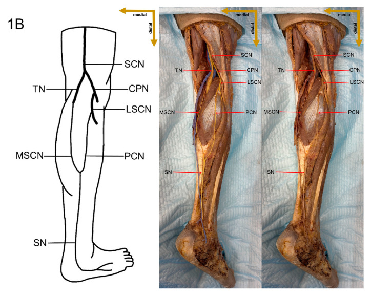
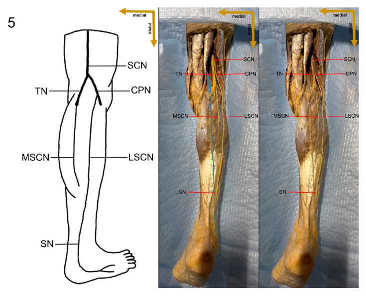
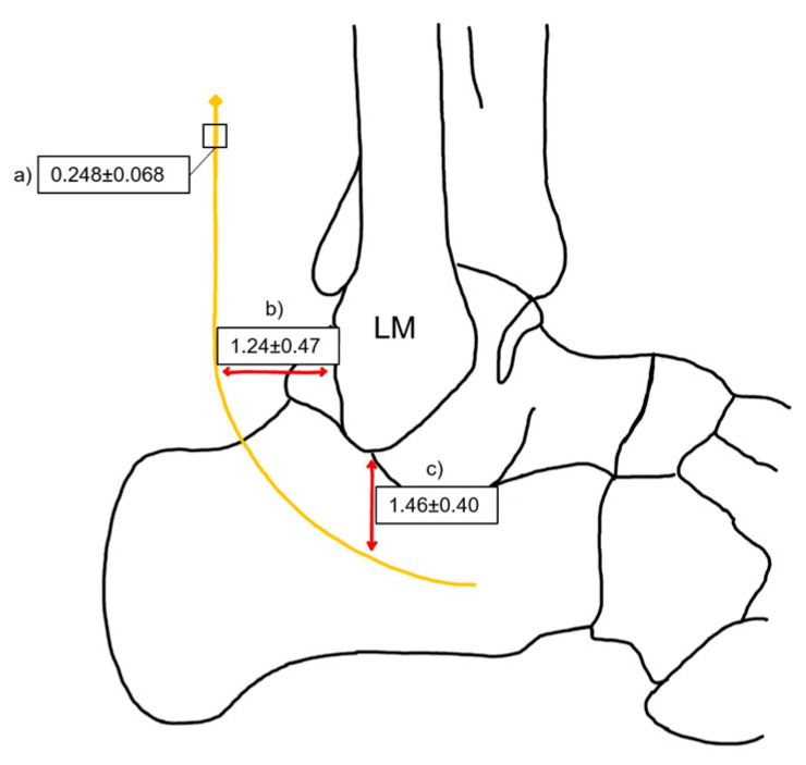
Background and Objectives: The sural nerve (SN) is a pure sensory nerve that supplies the lateral aspect of the ankle and foot. Its anatomical variability has been extensively documented, with multiple classifications describing its different formation patterns. The SN is commonly used for nerve grafting and is a critical structure in lower-limb surgeries. Due to its superficial course, it is vulnerable to iatrogenic injuries, particularly in procedures involving the Achilles tendon. The presence of anatomical variations in SN formation and trajectory has significant implications for surgical planning, diagnostics, and nerve conduction studies. Understanding these formation variations is essential to minimize surgical complications and optimize clinical outcomes. Materials and Methods: A pilot cross-sectional cadaveric study was conducted on nine formalin-fixed adult cadavers at the Department of Anatomy, Histology, and Anthropology, Vilnius University Faculty of Medicine, Lithuania. Standard dissection techniques were employed to examine the formation and trajectory of the SN. Morphometric parameters, including nerve diameter and length, were measured using an RS PTO Digital Caliper with 0.01 mm precision. Variations in SN formation were classified according to the system proposed by P.K. Ramakrishnan et al. Statistical analyses were performed using SPSS 26.0 and RStudio, with a significance threshold set at p ≤ 0.05. Results: The most prevalent SN formation variation observed in the Lithuanian cadaveric sample was Type 3, which was found in 8 out of 18 limbs (44.4%), while Type 6 was not identified. Additionally, a symmetric formation was observed bilaterally in 5 out of the 9 cadavers (55.6%). The SN was significantly thicker in two-contributor formations (3.17 mm) compared to single-contributor formations (1.93 mm, p = 0.001). The SN was also significantly longer in two-contributor formations (25.80 cm) than in single-contributor formations (18.96 cm, p = 0.016). No significant differences in SN morphology were found between left and right lower limbs. Conclusions: This study highlights the substantial anatomical variability of the SN in the Lithuanian population. The findings suggest a correlation between SN diameter and formation type, which may have clinical implications for nerve grafting and surgical planning. The predominance of Type 3 formation and the observed symmetry rate provide valuable anatomical insights for lower limb surgeries. Further large-scale studies are necessary to establish population-specific SN variations and their relevance in clinical practice.
Keywords: anatomical variations; cadaveric study; sural nerve.
Conflict of interest statement
The authors declare no conflicts of interest.
Figures


References
-
- Moore K.L., Dalley A.F. Clinically Oriented Anatomy. 4th ed. Lippincott Williams & Wilkins; Philadelphia, PE, USA: 1999. pp. 572, 601.
MeSH terms
LinkOut - more resources
Full Text Sources
