Review
. 2025 Apr 10;61(4):696.
doi: 10.3390/medicina61040696.

Imaging Diagnosis of Major Kidney and Urinary Tract Disorders in Children

Affiliations
Review

Imaging Diagnosis of Major Kidney and Urinary Tract Disorders in Children

Ahmad Aldughiem. Medicina (Kaunas). .

Abstract

Background and Objectives: Diagnostic imaging is essential for evaluating urinary tract disorders, offering critical insights into renal pathology. This review examines the strengths, limitations, and clinical applications of various imaging modalities, with a focus on pediatric populations. Materials and Methods: A narrative review was conducted, synthesizing current literature on ultrasound (US), computed tomography (CT), magnetic resonance imaging (MRI), nuclear medicine, and voiding cystourethrography (VCUG). Relevant studies were selected based on diagnostic accuracy, clinical utility, and safety considerations. Results: US is the preferred first-line imaging due to its safety, accessibility, and cost-effectiveness. CT excels in detecting renal calculi, trauma, and malignancies but is limited by radiation exposure. MRI offers superior soft tissue contrast without radiation but is costly and often requires sedation. Nuclear medicine evaluates renal function and scarring, while VCUG remains the gold standard for diagnosing vesicoureteral reflux and posterior urethral valves. Conclusions: Imaging modalities are vital for diagnosing and managing urinary tract disorders, with selection based on clinical needs, patient age, and safety. Ultrasound is the primary choice for its non-invasiveness and cost-effectiveness, while CT, MRI, nuclear medicine, and VCUG provide essential structural and functional insights. A balanced approach ensures accuracy while minimizing patient risk, especially in pediatrics.

Keywords: computed tomography (CT); magnetic resonance imaging (MRI); nuclear medicine; ultrasound (US); voiding cystourethrography (VCUG).


Conflict of interest statement

The authors declare no conflict of interest.

Figures

Figure 14
Figure 14
A CT scan reveals a horseshoe kidney, with fusion of the lower poles indicated by green arrows.
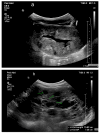
Figure 1
Figure 1
(a) Normal ultrasound of a 1-month-old infant’s kidney, showing prominent hypoechoic medullary pyramids. (b) Normal renal ultrasound of a 4-year-old child.
Figure 2
Figure 2
Renal ultrasound (US) showing hydronephrosis in a 5-day-old male infant with ureteropelvic junction obstruction.
Figure 3
Figure 3
CT of a normal 8-year-old boy: (A) Coronal image, (B) Sagittal image, (C) Axial image.
Figure 4
Figure 4
Voiding cystourethrogram. (a) Normal voiding image of a 2-year-old female. (b) Normal voiding image of a 2-year-old male.
Figure 5
Figure 5
IVP shows right crossed renal ectopia (indicated by arrows).
Figure 6
Figure 6
CT shows right sided pyelonephritis (indicated by the arrow) in a 4-year-old female.
Figure 7
Figure 7
A CT scan of a 15-year-old female with a history of recurrent pyelonephritis shows a scarred and atrophic right kidney (indicated by green arrow).
Figure 8
Figure 8
US reveals a small echogenic right kidney with a renal cyst (highlighted by green arrows) in a 2-year-old female.
Figure 9
Figure 9
(a) US shows right MCDK in a 23-month-old male. (b) Renal scan shows no uptake in the right kidney.
Figure 10
Figure 10
Right ureteropelvic junction obstruction in a neonate. (a) US shows severe dilatation of the right renal pelvis. (b) Nuclear medicine shows retention of radiotracer in the right kidney. (c) Mild drainage occurs in the right kidney after Lasix, with a T ½ exceeding 30 min.
Figure 11
Figure 11
CT scan showing left ureteropelvic junction obstruction.
Figure 12
Figure 12
Voiding cystourethrogram shows dilatation of the posterior urethra (arrow) in a 4-week-old boy with PUV.
Figure 13
Figure 13
VCUG shows Grade 4–5 VUR into the lower pole of the duplex system. VUR is superimposed on residual contrast in the upper pole.
Figure 15
Figure 15
VCUG reveals a ureterocele on the right side of the bladder, identified as a filling defect marked by the green arrow.
Figure 16
Figure 16
VCUG shows: (a) right VUR grade II in a 4-year-old female, (b) left VUR grade III in a 4-year-old female, and (c) left duplicated ureters with left VUR grade VI and right VUR grade III in an 8-week-old female.
Figure 16
Figure 16
VCUG shows: (a) right VUR grade II in a 4-year-old female, (b) left VUR grade III in a 4-year-old female, and (c) left duplicated ureters with left VUR grade VI and right VUR grade III in an 8-week-old female.
Figure 17
Figure 17
Voiding cystourethrogram showing VUR to a transplanted kidney in an 18-year-old female.
Figure 18
Figure 18
(a) ARPKD in a 5-day-old male identified by enlarged, echogenic kidneys with poor corticomedullary differentiation, without visible renal cysts. (b) A 6-year-old female with ARPKD, presenting with significantly enlarged kidneys and macrocysts (highlighted by green arrows).
Figure 19
Figure 19
Autosomal dominant polycystic kidney disease in a 16-year-old female.
Figure 20
Figure 20
An abdominal MRI of an 18-year-old female with tuberous sclerosis reveals a large angiomyolipoma in the left kidney (marked by green arrows) and a smaller angiomyolipoma in the right kidney (marked by a red arrow).
Figure 21
Figure 21
Abdominal MRI of a 16-year-old female with tuberous sclerosis, showing bilateral renal cysts (indicated by green arrows).
Figure 22
Figure 22
Nephrolithiasis in a 16-year-old female (indicated by number 1). (a) CT reveals a left intrarenal stone. (b) CT highlights a stone at the right ureterovesical junction (indicated by the green arrow).
Figure 23
Figure 23
Largely infarcted right kidney in an 18-year-old female involved in a motor vehicle collision.
Figure 24
Figure 24
Angiography demonstrating focal stenosis (indicated by the arrows) in the segmental artery supplying the midportion of the right kidney in a 12-year-old male.
Figure 25
Figure 25
(a) Thrombosis in the renal vein (green arrows) in a 7-day-old male. (b) Thrombosis in the inferior vena cava (IVC) (green arrows) in a 7-day-old male. (c) Doppler ultrasound (US) shows minimal flow around the thrombus in the right renal vein (green arrows). (d) Doppler US shows no flow around the thrombus in the IVC (green arrows).
Figure 26
Figure 26
CT with angiography shows the compression of the left renal vein between the aorta and the proximal superior mesenteric artery as indicated by green arrows (Nutcracker Syndrome).
Figure 27
Figure 27
MRI shows bilateral nephroblastomatosis with bilateral nephroblastoma in a 2-year-old female.

Similar articles

  • Pediatric urologic imaging.
    Palmer LS. Palmer LS. Urol Clin North Am. 2006 Aug;33(3):409-23. doi: 10.1016/j.ucl.2006.03.009. Urol Clin North Am. 2006. PMID: 16829274 Review.
  • Pediatric urologic advanced imaging: techniques and applications.
    Renjen P, Bellah R, Hellinger JC, Darge K. Renjen P, et al. Urol Clin North Am. 2010 May;37(2):307-18. doi: 10.1016/j.ucl.2010.03.007. Urol Clin North Am. 2010. PMID: 20569808 Review.
  • Morphologic Urologic Imaging.
    Mojtahed A, Anderson MA, Gee MS. Mojtahed A, et al. Urol Clin North Am. 2025 Feb;52(1):1-12. doi: 10.1016/j.ucl.2024.07.003. Epub 2024 Aug 8. Urol Clin North Am. 2025. PMID: 39537296 Review.
  • Imaging of the Pediatric Urinary System.
    Chung EM, Soderlund KA, Fagen KE. Chung EM, et al. Radiol Clin North Am. 2017 Mar;55(2):337-357. doi: 10.1016/j.rcl.2016.10.010. Epub 2016 Dec 24. Radiol Clin North Am. 2017. PMID: 28126219 Review.
  • State-of-the-Art Renal Imaging in Children.
    Viteri B, Calle-Toro JS, Furth S, Darge K, Hartung EA, Otero H. Viteri B, et al. Pediatrics. 2020 Feb;145(2):e20190829. doi: 10.1542/peds.2019-0829. Epub 2020 Jan 8. Pediatrics. 2020. PMID: 31915193 Free PMC article.

References

    1. Fried J.G., Morgan M.A. Renal Imaging: Core Curriculum 2019. Am. J. Kidney Dis. 2019;73:552–565. doi: 10.1053/j.ajkd.2018.12.029. - DOI - PubMed
    1. Viteri B., Calle-Toro J.S., Furth S., Darge K., Hartung E.A., Otero H. State-of-the-Art Renal Imaging in Children. Pediatrics. 2020;145:e20190829. doi: 10.1542/peds.2019-0829. - DOI - PMC - PubMed
    1. Maliborski A., Zegadło A., Placzyńska M., Sopińska M., Lichosik M., Jobs K. The role of modern diagnostic imaging in diagnosing and differentiating kidney diseases in children. Dev. Period Med. 2018;22:81–87. doi: 10.34763/devperiodmed.20182201.8187. - DOI - PMC - PubMed
    1. Emma F., Goldstein S.L., Bagga A., Bates C.M., Shroff R., editors. Pediatric Nephrology. 8th ed. Springer; Cham, Switzerland: 2022. Diagnostic imaging in pediatric nephrology; pp. 173–211.
    1. Kher K., Schnaper H.W., Greenbaum L.A. Clinical Pediatric Nephrology. 3rd ed. CRC Press; Boca Raton, FL, USA: 2017. Diagnostic imaging of the urinary tract; pp. 87–103.

MeSH terms

LinkOut - more resources