

AMPK Knockout Impairs the Formation of Three-Dimensional Spheroids
- PMID: 40283080
- PMCID: PMC12028351
- DOI: 10.3390/life15040525
AMPK Knockout Impairs the Formation of Three-Dimensional Spheroids
Abstract
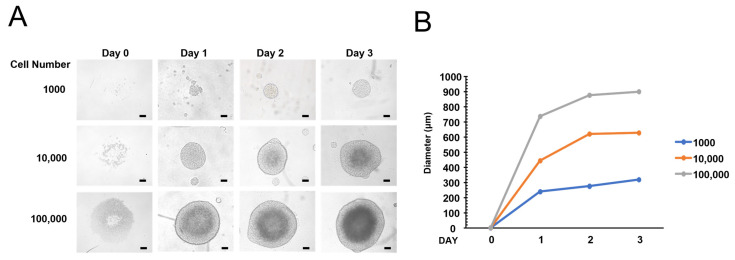
AMP-activated protein kinase (AMPK) is an important regulator of cellular energy homeostasis, and AMPK contributes to cell growth, apoptosis, and autophagy. Although most cell studies have been performed using two-dimensional (2D) cell culture, recent studies have demonstrated that the three-dimensional (3D) spheroid technique is helpful in various cell research fields, such as tumor biology, due to its resemblance to the 3D tissue structure. However, the role of AMPK in 3D spheroid formation has not been characterized clearly. This study used the AMPK knockout cell line to examine the role of AMPK in 3D spheroid formation and is the first report describing the generation of 3D spheroids using AMPK knockout cells. While control cells produced round spheroids with a similar length-to-width ratio, AMPK knockout produced an oval shape with a more significant length-to-width ratio. We demonstrate that AMPK knockout spheroids contain significantly more prominent lysosomes in each cell, indicating that autophagic flux is impaired in 3D spheroids. Finally, flow cytometry analysis showed that AMPK knockout spheroids contain more apoptotic cells than control cells. These results indicate that AMPK is required for efficient 3D spheroid formation.
Keywords: 3D culture; AMPK; knockout; lysosome; spheroid.
Conflict of interest statement
The authors have no conflicts of interest to declare.
Figures


Similar articles
-
Activated CAMKKβ-AMPK signaling promotes autophagy in a spheroid model of ovarian tumour metastasis.J Ovarian Res. 2020 May 11;13(1):58. doi: 10.1186/s13048-020-00660-5. J Ovarian Res. 2020. PMID: 32393385 Free PMC article.
-
Real-time viability and apoptosis kinetic detection method of 3D multicellular tumor spheroids using the Celigo Image Cytometer.Cytometry A. 2017 Sep;91(9):883-892. doi: 10.1002/cyto.a.23143. Epub 2017 Jun 15. Cytometry A. 2017. PMID: 28618188
-
Three-dimensional spheroids of mesenchymal stem/stromal cells promote osteogenesis by activating stemness and Wnt/β-catenin.Biochem Biophys Res Commun. 2020 Mar 5;523(2):458-464. doi: 10.1016/j.bbrc.2019.12.066. Epub 2019 Dec 24. Biochem Biophys Res Commun. 2020. PMID: 31882121
-
Exploring Mitochondrial Energy Metabolism of Single 3D Microtissue Spheroids Using Extracellular Flux Analysis.J Vis Exp. 2022 Feb 3;(180). doi: 10.3791/63346. J Vis Exp. 2022. PMID: 35188130
-
A Facile, Transfection-Free Approach to siRNA Delivery in In Vitro 3D Spheroid Models.Curr Protoc. 2024 Sep;4(9):e1121. doi: 10.1002/cpz1.1121. Curr Protoc. 2024. PMID: 39225471
References
Grants and funding
LinkOut - more resources
Full Text Sources
