Review
. 2025 Apr 2;16(4):426.
doi: 10.3390/mi16040426.

Advances in Microfluidic Single-Cell RNA Sequencing and Spatial Transcriptomics

Affiliations
Review

Advances in Microfluidic Single-Cell RNA Sequencing and Spatial Transcriptomics

Yueqiu Sun et al. Micromachines (Basel). .

Abstract

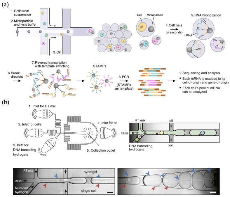
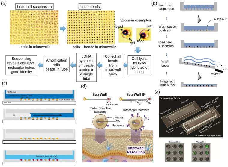
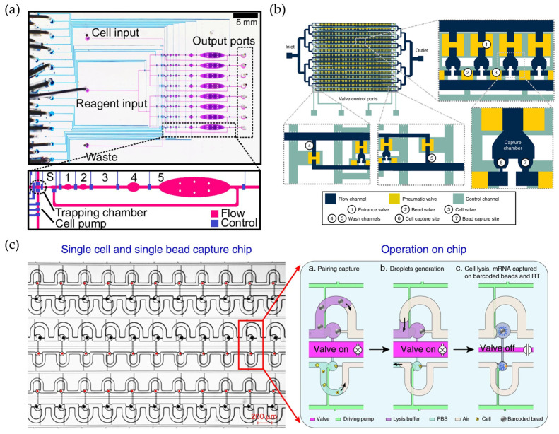
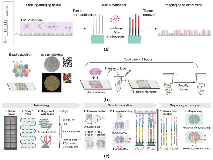
The development of micro- and nano-fabrication technologies has greatly advanced single-cell and spatial omics technologies. With the advantages of integration and compartmentalization, microfluidic chips are capable of generating high-throughput parallel reaction systems for single-cell screening and analysis. As omics technologies improve, microfluidic chips can now integrate promising transcriptomics technologies, providing new insights from molecular characterization for tissue gene expression profiles and further revealing the static and even dynamic processes of tissues in homeostasis and disease. Here, we survey the current landscape of microfluidic methods in the field of single-cell and spatial multi-omics, as well as assessing their relative advantages and limitations. We highlight how microfluidics has been adapted and improved to provide new insights into multi-omics over the past decade. Last, we emphasize the contributions of microfluidic-based omics methods in development, neuroscience, and disease mechanisms, as well as further revealing some perspectives for technological advances in translational and clinical medicine.

Keywords: microfluidics; single cell; single-cell RNA sequencing (scRNA-seq); spatial transcriptome.


Conflict of interest statement

The authors declare no conflict of interest.

Figures

Figure 1
Figure 1
(a) Schematic diagram of the workflow of the Drop-seq technique. Reproduced with permission from Ref. [23]. Single cells are encapsulated in tiny droplets along with uniquely barcoded primer beads, enabling large-scale, highly parallel analysis. (b) Microfluidic device used in inDrop technology. Reproduced with permission from Ref. [20]. Cells are captured and barcoded in nanoliter droplets with high capture efficiency. Arrows indicate cells (red), hydrogels (blue), and flow direction (black).
Figure 2
Figure 2
(a) Schematic and imaging data for CytoSeq. Reproduced with permission from Ref. [39]. The platform enables routine, digitized gene expression profiling of thousands of single-cell genes. (b) Experimental procedure for Microwell-Seq. Reproduced with permission from Ref. [32]. A high-throughput, low-cost platform capable of constructing mouse cellular maps at the single-cell level. (c) A schematic of the basic workflow for single-cell RNA printing. Reproduced with permission from Ref. [36]. Microwells in this platform enable low-cost printing of RNA on glass or capturing of RNA on beads. (d) Comparison between Seq-Well and Seq-Well S3. Reproduced with permission from Ref. [41]. Based on Seq-Well, Seq-Well S3 improves transcript capture and sensitivity. (e) scFTD-seq platform and fluorescence imaging data. Reproduced with permission from Ref. [42]. Portable and easy-to-use devices, as well as simplified and modular workflows facilitate clinical use.
Figure 3
Figure 3
(a) Micrograph of the microfluidic device. Reproduced with permission from Ref. [22]. Injection of single-cell suspensions and reagents, and recovery of double-stranded cDNA from the output for single-cell whole transcriptome analysis. Lines indicate the control channels (blue), and the flow channels (purple). (b) Schematic diagram of Hydro-Seq technology. Reproduced with permission from Ref. [43]. The platform is capable of cleaning contaminants such as erythrocytes and could be used to isolate rare cells. (c) Design of Paired-seq chip. Reproduced with permission from Ref. [44]. The device is capable of efficiently isolating and removing cell-free mRNAs.
Figure 4
Figure 4
(a) Illustration of the DNA-barcoded antibodies used in CITE-seq and analysis of mixtures of mouse and human cells. Reproduced with permission from Ref. [64]. CITE-seq enables simultaneous detection of single cell transcriptomes and protein marks. (b) Schematic of ASAP-seq workflow. Reproduced with permission from Ref. [66]. ASAP-seq is capable of demonstrating chromatin accessibility on a genome-wide scale. (c) Schematic of NEAT-seq workflow. Reproduced with permission from Ref. [74]. NEAT-seq is a droplet-based microfluidics platform that can simultaneously analyze intracellular proteins, chromatin accessibility, as well as transcripts through intranuclear staining.
Figure 5
Figure 5
(a) Spatially localized cDNA synthesis. Reproduced with permission from Ref. [87]. ST was the first use of surface capture and commercialized as the Visium platform. (b) Slide-seq workflow. Reproduced with permission from Ref. [88]. Slide-Seq method deposited 10 μm magnetic beads on the plane of the slide to capture tissue mRNA. (c) Schematic diagram of HDST. Reproduced with permission from Ref. [89]. HDST method modified 2 μm magnetic beads in the microwells of the slide to capture tissue mRNA.
Figure 6
Figure 6
(a) Microfluidic device used in DBiT-seq. Reproduced with permission from Ref. [91]. DBiT-seq is capable of analyzing the proteome, transcriptome, and transcriptome of the same tissue section while maintaining a high resolution. (b) Schematic diagram and validation of Well-ST-Seq technology. Reproduced with permission from Ref. [25]. Well-ST-Seq avoids channel blockage problems associated with tissue deformation.
Figure 7
Figure 7
(a) Location, period, study design, and number of cells sequenced information. Reproduced with permission from Ref. [100]. (b) Overview of study design for intestinal development atlas. Reproduced with permission from Ref. [105]. (c) A schematic displaying the key cell types, stages, and markers involved in human hematopoietic stem and progenitor cell (HSPC) specification, emergence, and maturation. Reproduced with permission from Ref. [84]. (d) Schematic of DDC-induced injury and repair. Reproduced with permission from Ref. [109]. Spatiotemporal analyses of cholestatic injury and repair processes in mice reveal the potential of the spatiotemporal transcriptome for tissue injury and regeneration studies.
Figure 8
Figure 8
(a) Schematic for alignment of scATAC-seq and scRNA-seq data. Reproduced with permission from Ref. [128]. scATAC-seq has simultaneously tagged and fragmented DNA sequences, such as 10x Multiome, in open chromatin regions and further paved the way for other technologies that profile both the epigenome and transcriptome in single nuclei. (b) Schematic for alignment of snRNA-seq and Stereo-seq. Reproduced with permission from Ref. [129]. Multimodal intersection analysis (MIA) across the two datasets has revealed the spatial distribution of the cell populations and subpopulations. (c) Slide-DNA and slide-RNA integration. Reproduced with permission from Ref. [123]. Slide-DNA-seq has captured spatially resolved DNA sequences from intact tissue sections. The integration of slide-DNA-seq and spatial transcriptomics has uncovered distinct sets of genes that are associated with clone-specific genetics.

Similar articles

Cited by

References

    1. Neumann P.E., Neumann E.E. General Histological Woes: Definition and Classification of Tissues. Clin. Anat. 2021;34:794–801. doi: 10.1002/ca.23741. - DOI - PubMed
    1. Davis M.M., Tato C.M., Furman D. Systems Immunology: Just Getting Started. Nat. Immunol. 2017;18:725–732. doi: 10.1038/ni.3768. - DOI - PMC - PubMed
    1. Lee J., Hyeon D.Y., Hwang D. Single-Cell Multiomics: Technologies and Data Analysis Methods. Exp. Mol. Med. 2020;52:1428–1442. doi: 10.1038/s12276-020-0420-2. - DOI - PMC - PubMed
    1. Travaglini K.J., Nabhan A.N., Penland L., Sinha R., Gillich A., Sit R.V., Chang S., Conley S.D., Mori Y., Seita J., et al. A Molecular Cell Atlas of the Human Lung from Single-Cell RNA Sequencing. Nature. 2020;587:619–625. doi: 10.1038/s41586-020-2922-4. - DOI - PMC - PubMed
    1. Wang Y., Navin N.E. Advances and Applications of Single-Cell Sequencing Technologies. Mol. Cell. 2015;58:598–609. doi: 10.1016/j.molcel.2015.05.005. - DOI - PMC - PubMed

LinkOut - more resources