Review
. 2025 Apr 14;16(4):462.
doi: 10.3390/mi16040462.

Recent Advances in Microfluidics-Based Monitoring of Waterborne Pathogens: From Isolation to Detection

Affiliations
Review

Recent Advances in Microfluidics-Based Monitoring of Waterborne Pathogens: From Isolation to Detection

Guohao Xu et al. Micromachines (Basel). .

Abstract

Waterborne pathogens seriously threaten human life and can cause diarrhea, gastrointestinal disorders, and more serious systemic infections. These pathogens are usually caused by contaminated water sources that contain disease-causing microorganisms, such as bacteria, viruses, and parasites, which cause infection and disease when they enter the human body through drinking water or other means. Due to the wide range of transmission routes and the high potential risk of waterborne pathogens, there is an urgent need for an ultrasensitive, rapid, and specific pathogenic microorganism monitoring platform to meet the critical monitoring needs of some water bodies' collection points daily monitoring needs. Microfluidics-based pathogen surveillance methods are an important stage towards automated detection through real-time and multi-targeted monitoring, thus enabling a comprehensive assessment of the risk of exposure to waterborne pathogens and even emerging microbial contaminants, and thus better protection of public health. Therefore, this paper reviews the latest research results on the isolation and detection of waterborne pathogens based on microfluidic methods. First, we introduce the traditional methods for isolation and detection of pathogens. Then, we compare some existing microfluidic pathogen isolation and detection methods and finally look forward to some future research directions and applications of microfluidic technology in waterborne pathogens monitoring.

Keywords: detection; isolation; lab-on-a-chip (LOC); microfluidic chip; nucleic acid analysis; sample processing; waterborne pathogens.


Conflict of interest statement

The authors declare no conflicts of interest.

Figures

Figure 2
Figure 2
Conventional separation and detection methods (A) (i) Schematic illustrating a direct hydrothermal growth method for fabrication of TNM. (ii) Typical SEM image of the porous titanium membrane substrate. (iii) Typical SEM image of an as-synthesized TNM. (iv) Typical SEM image of an annealed TNM [13]. Copyright 2009, Elsevier. (B) Immunomagnetic separation (IMS) method for isolating and concentrating target bacteria from samples [31]. Copyright 2020, MDPI.
Figure 3
Figure 3
Static mechanisms (A) Overview of the multiplexed cross-flow filtration micromode [48]. Copyright 2018, Springer Nature. (B) An integrated membrane valve microfluidic platform for ultrafiltration control [49]. Copyright 2022, Springer Nature. (C) Schematic diagram of a microfluidic detection chip based on finger-driven mixing and core-track membrane filtration for rapid and sensitive detection of Salmonella [50]. Copyright 2023, Elsevier. (D) Bio-inspired anti-fouling membrane filtration device enabled by 3D printing-on-membrane [51]. Copyright 2022, Springer Nature.
Figure 5
Figure 5
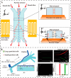
Electrical separation. (A) A schematic of the experimental setup used for concentration of E. coli by ICP experiments [63]. Copyright 2020, Wiley. (B) A discrete dielectrophoresis device for the separation of malaria-infected cells [64]. Copyright 2022, Wiley.
Figure 6
Figure 6
Fluidic separation. (A) Schematic illustration of the microfluidic device and separation mechanism using a SSAW field [69]. Copyright 2013, American Chemical Society (B) Separation of bacterivorous jakobid flagellate by sharp velocity gradient-induced soft inertial forces [70]. Copyright 2018, Royal Society of Chemistry.
Figure 10
Figure 10
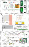
Paper-based microfluidics for pathogen detection. (A) The paper-based separation device detects E. coli, and two visible color bands display the detection results in [33]. Copyright 2019, American Chemical Society. (B) Smartphone detection of Salmonella and cross-reaction with E. coli resistance and detection results from, the angle of incident light to paper (angle a) and the angle of light scatter from paper (angle b). [99]. Copyright 2013, Royal Society of Chemistry. (C) E. coli was detected using a μPAD and a smartphone reader [100]. Copyright 2023, American Chemical Society.
Figure 1
Figure 1
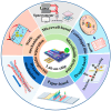
Microfluidics technology realizes efficient separation and highly sensitive detection of pathogens through integrated chip design (e.g., centrifugation, droplet encapsulation, paper-based detection, and other modules) and can be used in diverse application scenarios ranging from clinical diagnosis to environmental monitoring.
Figure 4
Figure 4
Optical separation. Salmonella bacteria bind to MBs conjugated with antiSalmonella antibody, thereby forming the Salmonella–bead complex, which is subsequently captured using infrared (IR) laser-based OT [57]. Copyright 2020, MDPI.
Figure 7
Figure 7
Microwell-based detection of waterborne pathogens. (A) Schematic drawing of the KPC-2 E. coli detection system [78]. Copyright 2020, Elsevier. (B) Microfluidic device for detecting E. coli using Raman scattering technology [79]. Copyright 2020, Elsevier. (C) Real-time monitoring and surface domain-based fluorescence analysis of single E. coli EK-19 detection in a microporous array [80]. Copyright 2022, Elsevier.
Figure 8
Figure 8
Centrifugal microfluidic detection methods. (A) A schematic diagram of a centrifugal chip for bacteria detection and detection results from [82]. Copyright 2017, Springer Nature. (B) Centrifugal microfluidic chip performs rotating fluorescence scanning detection by installing a fluorescence detector on the channel [83]. Copyright 2024, MDPI.
Figure 9
Figure 9
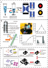
Droplet-based microfluidic detection chip. (A) An open-surface droplet microfluidic device for monitoring microcystin-LR in reservoirs and detection principle [86]. Copyright 2020, American Chemical Society. (B) Microdroplet-based PCR structure [88]. Copyright 2008, American Chemical Society. (C) LC film sensing device, diagram of the ordered state of 5cb droplets at the live and dead E. coli water suspension LC interface [89]. Copyright 2018, Elsevier. (D) CRISPR/Cas13a-based droplet microfluidics for multiplexed bacterial detection [90]. Copyright 2024, Elsevier.

Similar articles

References

    1. Wang M., Bodirsky B.L., Rijneveld R., Beier F., Bak M.P., Batool M., Droppers B., Popp A., Van Vliet M.T.H., Strokal M. A Triple Increase in Global River Basins with Water Scarcity Due to Future Pollution. Nat. Commun. 2024;15:880. doi: 10.1038/s41467-024-44947-3. - DOI - PMC - PubMed
    1. Khan J.R., Hossain M.B., Chakraborty P.A., Mistry S.K. Household Drinking Water E. coli Contamination and Its Associated Risk with Childhood Diarrhea in Bangladesh. Environ. Sci. Pollut. Res. 2022;29:32180–32189. doi: 10.1007/s11356-021-18460-9. - DOI - PubMed
    1. Al-Halhouli A., Doofesh Z., Albagdady A., Dietzel A. High-Efficiency Small Sample Microparticle Fractionation on a Femtosecond Laser-Machined Microfluidic Disc. Micromachines. 2020;11:151. doi: 10.3390/mi11020151. - DOI - PMC - PubMed
    1. Zhang Y., Hu X., Wang Q. Review of Microchip Analytical Methods for the Determination of Pathogenic Escherichia coli. Talanta. 2021;232:122410. doi: 10.1016/j.talanta.2021.122410. - DOI - PubMed
    1. Shukhratovich Abdullaev S., H Althomali R., Raza Khan A., Sanaan Jabbar H., Abosoda M., Ihsan A., Aggarwal S., Mustafa Y.F., Hammoud Khlewee I., Jabbar A.M. Integrating of Analytical Techniques with Enzyme-Mimicking Nanomaterials for the Fabrication of Microfluidic Systems for Biomedical Analysis. Talanta. 2024;273:125896. doi: 10.1016/j.talanta.2024.125896. - DOI - PubMed

LinkOut - more resources