Review
. 2025 Apr 13;14(8):2657.
doi: 10.3390/jcm14082657.

Proenkephalin 119-159 in Heart Failure: From Pathophysiology to Clinical Implications

Affiliations
Review

Proenkephalin 119-159 in Heart Failure: From Pathophysiology to Clinical Implications

Dionysis Matsiras et al. J Clin Med. .

Abstract

Heart failure (HF) is a challenging clinical syndrome with high morbidity and mortality rates. Along the spectrum of cardiovascular diseases, HF constitutes an ever-expanding area of research aiming at combating the associated mortality and improving the prognosis of patients with HF. Although natriuretic peptides have an established role among biomarkers in HF diagnosis and prognosis, several novel biomarkers reflecting the complex pathophysiology of HF are under investigation for their ability to predict adverse clinical outcomes in HF. Proenkephalin 119-159 (PENK119-159) is a non-functional peptide belonging to the enkephalin family of the endogenous opioid system and is considered a surrogate biomarker of the biologically active enkephalin peptides. PENK119-159 has demonstrated promising results in predicting short- and long-term mortality, readmission rates, and worsening renal function in patients with HF. Indeed, in the setting of HF, the levels of both active enkephalins and their surrogate PENK119-159 are elevated and are associated with a dismal prognosis. However, the biological effects of PENK119-159 remain largely unknown. Thus, it is crucial to gain a deeper insight into both the physiology of the enkephalin peptide family and the enkephalin-mediated cardiovascular regulation. In order to elucidate the complex pathophysiological mechanisms that lead to the upregulation of enkephalins in patients with HF, as well as the potential clinical implications of elevated enkephalins and PENK119-159 levels in this patient population, the present review will describe the physiology and distribution of the endogenous opioid peptides and their corresponding opioid receptors, with a particular focus on the action of enkephalins. The effects of the enkephalin peptides will be analyzed in both healthy subjects and patients with HF, especially with regard to their role in the regulation of cardiovascular and renal function. The review will also discuss the findings of recent studies that have explored the prognostic value of PENK119-159 in patients with HF.

Keywords: enkephalins; heart failure; opioid peptide receptors; pathophysiology; proenkephalin 119–159; worsening renal function.


Conflict of interest statement

All authors declare no conflicts of interest related to the work under consideration.

Figures

Figure 1
Figure 1
Human proenkephalin peptide processing. The 243-amino-acid-long proenkephalin is cleaved at different sites to yield the intermediate-sized peptides Synenkephalin (1–70), peptide F (83–116), PENK 119–159, peptide E (186–210), and peptide B (218–243), as well as the small-sized biologically active peptides Met5-enkephalin (76–80) and Octapeptide (162–169). Further cleavage of intermediate peptides generates a larger number of active peptides, namely two copies of Met5-enkephalin (83–87) and (112–116) from peptide F; one Met5-enkephalin (186–190) from sequential processing of peptide E into metorphamide (MO); one Leu5-enkephalin (206–210) directly from peptide E; and finally, one heptapeptide (237–243) from peptide B. Collectively, one human proenkephalin molecule gives rise to active enkephalin peptides at a ratio of 4 Met5-enkephalin: 1 Leu5-enkephalin: 1 heptapeptide: 1 octapeptide [3,10,11,12,13,14].
Figure 2
Figure 2
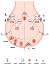
Modulation of presynaptic signaling in cholinergic nerve terminals by enkephalins. Enkephalins modulate presynaptic signaling in cholinergic nerve terminals innervating the sinoatrial node by acting on two functionally different subtypes of δ (delta)-opioid receptors (ORs), namely δ1-ORs and δ2-ORs. These two subtypes of δ-ORs are activated by enkephalins in a concentration-dependent manner, thereby resulting in bimodal effects on acetylcholine (Ach) release [27,29]. Low doses of enkephalins activate the δ1-OR, a stimulatory G protein-coupled receptor (Gs), which augments Ach release and induces vagotonic effects. On the contrary, higher levels of enkephalins activate the δ2-OR, an inhibitory G protein-coupled receptor (Gi), which inhibits Ach release and promotes vagolytic effects [27,28]. δ1-OR activation by enkephalins initially leads to the dissociation of the Gαs subunit from the Gβ/γ heterodimer. In turn, the dissociated Gαs subunit increases the activity of adenylyl cyclase and the production of cyclic adenosine monophosphate (cAMP), while the Gβ/γ subunit promotes calcium (Ca) entry into the nerve terminal (green arrow), thereby enhancing Ach release [38,39]. Concerning δ2-OR activation, the mechanism of action likely involves several pathways. Upon δ2-OR activation, the Gαi subunit reduces adenylyl cyclase activity, while the Gβ/γ subunit inhibits voltage-gated Ca channels (red arrow), through which Ca enters the nerve terminals to activate calcium sensor proteins and trigger the initiation of docking and fusion processes. The net result of decreased Ca influx is attenuated cholinergic transmission due to decreased Ach release in the synaptic cleft [28,31,32]. Moreover, the dissociated Gβ/γ subunit modulates the function of the soluble N-ethylmaleimide-sensitive factor attachment protein receptor (SNARE) complex, which is a protein apparatus embedded on both vesicular and plasma membranes that is actively involved in the docking and fusion of synaptic vesicles and subsequent neurotransmitter exocytosis [32]. The Gβ/γ subunit appears to interact with the SNARE complex mainly through competing with synaptotagmin (a Ca sensor protein that promotes the fusion of the vesicles with the plasma membrane) for binding sites on the SNARE complex, thus resulting in inhibition of Ach exocytosis [31,32,33]. In parallel, it is also possible that δ2-OR activation interferes with the function of synapsin, which has been implicated in the processes of tethering, docking, mobilization, and fusion of the synaptic vesicles. Under resting states, synaptic vesicles are reversibly tethered to the meshwork of the actin cytoskeleton by synapsin, which thereby acts by linking adjacent synaptic vesicles with each other and keeping them clustered and anchored in a distal reserve pool away from the plasma membrane. Apart from its tethering action, synapsin is also involved in the docking, post-docking, and fusion events. Upon phosphorylation of synapsin, synaptic vesicles are mobilized from the reserve pool, dissociate from the cytoskeleton and from each other, and move to the readily releasable pool, close to the synaptic cleft, where they are now free to fuse with the plasma membrane and release their content through exocytosis [34]. Accordingly, following δ2-OR activation, it could be postulated that the δ2-OR-coupled Gi protein inhibits synapsin phosphorylation and the downstream events, eventually resulting in reduced Ach release from the presynaptic cholinergic nerve terminal. Finally, δ2-OR signaling seems to modulate vesicular acetylcholine transporter (VAChT) turnover by inhibiting VAChT, which is a vesicular membrane-bound transporter that facilitates entry and storage of Ach into the synaptic vesicles. Due to VAchT inhibition, Ach cannot move into the synaptic vesicles and thus cannot be secreted in the synaptic cleft [29,35].
Figure 3
Figure 3
Modulation of opioid receptor-mediated postsynaptic signaling by enkephalins. On the postsynaptic membrane, the activation of the opioid receptor (OR) by enkephalins generates a sympatholytic effect via a cross-talk process between the OR and the beta 1 (b1)-adrenergic receptor (AR) [40]. Normally, upon norepinephrine binding to the b1-AR, a conformational change in the G protein-coupled receptor occurs, which leads to the dissociation of the Gαs subunit from the Gβ/γ subunit. This results in the sequential activation (green arrow) of adenylyl cyclase (AC) and protein kinase A (PKA), which in turn triggers the opening of both L-type calcium channels (LTCC) on the T-tubules and the ryanodine receptors (RyR) on the membrane of the sarcoplasmic reticulum (SR) [30,43]. Accordingly, both the amplification of calcium (Ca) influx through LTCC and the augmented release of Ca from the SR through RyR result in the increase in intracellular Ca levels and the subsequent stimulation of the excitation–contraction coupling [43]. During adrenergic stimulation, enkephalins are co-released with norepinephrine in the synaptic cleft and bind to the OR. Subsequent OR activation leads to the dissociation of the inhibitory Gαi subunit from the trimeric G protein. Thereafter, the Gαi subunit inhibits (red arrow) adenylyl cyclase and results in the reduction of all downstream messengers, thereby blunting the positive inotropic effects mediated by the b1-AR signaling [30,40,41]. It should additionally be noted that, apart from the postsynaptic membrane, ORs are also present in the T-tubule (in proximity with LTCC), as well as in the SR (colocalized with isoform 2 RyR) [5,6]. In parallel, OR activation appears to further inhibit excitation–contraction coupling via a non-b1-AR cross-talk manner, whereby enkephalins stimulate sarcolemmal δ-ORs of the Gq family [44]. In this case, dissociation of the Gaq subunit leads to the activation of phospholipase C (PLC) with subsequent synthesis of diacylglycerol (DAG) and inositol 1,4,5-trisphosphate (IP3). In turn, IP3 activates the IP3 receptor on the SR membrane and promotes Ca release from the SR [30,44]. Although initially, an increase in cytosolic Ca favors excitation–contraction coupling, it appears that enkephalins eventually deplete SR Ca stores and lead to a reduction in cytosolic Ca transient [44]. Additionally, DAG normally activates protein kinase C (PKC), which stimulates sodium–hydrogen (Na+/H+) antiporter to move Na+ inside the cell in exchange for H+. However, it seems that another alternative Gi OR-mediated pathway exists, which leads to the inhibition of PKC and the subsequent inhibition of Na+/H+ exchanger, thereby resulting in the reduction of intracellular pH and consequently in the decreased responsiveness of myosin filaments to Ca [45,46].
Figure 4
Figure 4
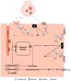
The interplay between neurohormonal and enkephalinergic overactivity in heart failure. Signals relating to the decreased cardiac output (CO) and the subsequent cardiac volume overload are correspondingly sensed by the aortic and carotid bodies on the one hand and by the cardiac sensory nerves on the other and are then relayed via the IX and X cranial nerves (C.N.IX and X), as well as via the thoracic spinal sensory tracts, respectively, to the cardiovascular (CV) center in the medulla oblongata. After all inputs are integrated and processed in the brainstem, the appropriate response arises, which is then conveyed through the efferent pathways to a variety of organs in order to maintain a certain degree of homeostasis [23]. In the setting of heart failure (HF), the net result is the adrenergic stimulation of the pre- and post-ganglionic sympathetic fibers, which innervate the cardiac, vascular, renal, and adrenal medullary tissues [47]. Simultaneously, an enkephalinergic response is mounted, which is characterized by an overproduction of enkephalins, coupled with overexpression of opioid receptors (OR), in an attempt to combat the underlying sympathetic hyperactivity. The upregulation of the enkephalinergic system blunts the b-adrenergic-mediated increase in cardiac contractility [40,41] in an effort to decrease cardiac workload and counteract the effects of increased sympathetic tone while it also stimulates renal diuresis [49] in order to achieve volume offloading. As HF progresses and renal deterioration ensues, plasma levels of PENK119–159 increase as a result of both enkephalin overproduction and decreased glomerular filtration. Although the counterregulatory mechanisms of the activated enkephalinergic system are initially intended to protect the cardiovascular system from the HF-driven sympathetic surge, they eventually become maladaptive, leading to further decline in cardiac function and renal deterioration. Abbreviations: C.N. = cranial nerve; CO = cardiac output; CV = cardiovascular; HF = heart failure; OR = opioid receptors; PENK = proenkephalin; SNS = sympathetic nervous system; WHF = worsening heart failure; WRF = worsening renal function.

Similar articles

References

    1. Chaudhry S.-P., Stewart G.C. Advanced Heart Failure. Heart Fail. Clin. 2016;12:323–333. doi: 10.1016/j.hfc.2016.03.001. - DOI - PubMed
    1. Siranart N., Laohasurayotin K., Phanthong T., Sowalertrat W., Ariyachaipanich A., Chokesuwattanaskul R. Proenkephalin as a Novel Prognostic Marker in Heart Failure Patients: A Systematic Review and Meta-Analysis. Int. J. Mol. Sci. 2023;24:4887. doi: 10.3390/ijms24054887. - DOI - PMC - PubMed
    1. Ernst A., Köhrle J., Bergmann A. Proenkephalin A 119–159, a Stable Proenkephalin A Precursor Fragment Identified in Human Circulation. Peptides. 2006;27:1835–1840. doi: 10.1016/j.peptides.2006.03.008. - DOI - PubMed
    1. Schulz C.-A., Christensson A., Ericson U., Almgren P., Hindy G., Nilsson P.M., Struck J., Bergmann A., Melander O., Orho-Melander M. High Level of Fasting Plasma Proenkephalin-A Predicts Deterioration of Kidney Function and Incidence of CKD. J. Am. Soc. Nephrol. 2017;28:291–303. doi: 10.1681/ASN.2015101177. - DOI - PMC - PubMed
    1. Treskatsch S., Feldheiser A., Shaqura M., Dehe L., Habazettl H., Röpke T.K., Shakibaei M., Schäfer M., Spies C.D., Mousa S.A. Cellular Localization and Adaptive Changes of the Cardiac Delta Opioid Receptor System in an Experimental Model of Heart Failure in Rats. Heart Vessels. 2016;31:241–250. doi: 10.1007/s00380-014-0620-6. - DOI - PubMed

LinkOut - more resources