

Bridging the Gap Between hiPSC-CMs Cardiotoxicity Assessment and Clinical LVEF Decline Risk: A Case Study of 21 Tyrosine Kinase Inhibitors
- PMID: 40283889
- PMCID: PMC12030206
- DOI: 10.3390/ph18040450
Bridging the Gap Between hiPSC-CMs Cardiotoxicity Assessment and Clinical LVEF Decline Risk: A Case Study of 21 Tyrosine Kinase Inhibitors
Abstract
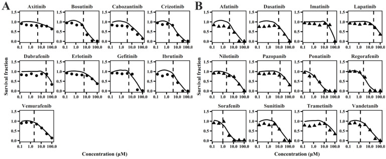
Objectives: There is growing concern over tyrosine kinase inhibitor (TKI)-induced cardiotoxicity, particularly regarding left ventricular dysfunction and heart failure in clinical treatment. These adverse effects often lead to treatment discontinuation, severely impacting patient outcomes. Therefore, there is an urgent need for more precise risk assessment methods. This study aimed to assess the cardiotoxicity of TKIs, refine in vitro to in vivo extrapolation (IVIVE) methodologies to improve predictive accuracy, and identify critical in vitro parameters for assessment. Methods: By leveraging high-throughput cardiotoxicity screening with human induced pluripotent stem cell-derived cardiomyocytes (hiPSC-CMs), a mechanism-based toxicodynamic (TD) model for TKIs was constructed. A QSP-PK-TD model was developed by integrating pharmacokinetic (PK) and quantitative systems pharmacology (QSP) models. This model incorporates critical drug exposure factors, such as plasma protein binding, tissue-plasma partitioning, and drug distribution heterogeneity to enhance extrapolation accuracy. Results: The QSP-PK-TD model validated the reliability of IVIVE and identified the area under the curve of drug effects on mitochondrial membrane potential (AEMMP) and cardiomyocyte contractility (AEAAC) as key in vitro parameters for assessing TKI-induced cardiotoxicity. Incorporating critical drug exposure factors obviously improved qualitative and quantitative extrapolation accuracy. Conclusions: This study established a framework for predicting in vivo cardiotoxicity from in vitro parameters, enabling efficient translation of preclinical data into clinical risk assessment. These findings provide valuable insights for drug development and regulatory decision-making, offering a powerful tool for evaluating TKI-induced cardiotoxicity.
Keywords: QSP-PK-PD model; TKIs; cardiotoxicity; hiPSC-CMs; in vitro to in vivo extrapolation (IVIVE).
Conflict of interest statement
The authors declare no conflicts of interest.
Figures


References
-
- Santoni M., Guerra F., Conti A., Lucarelli A., Rinaldi S., Belvederesi L., Capucci A., Berardi R. Incidence and risk of cardiotoxicity in cancer patients treated with targeted therapies. Cancer Treat. Rev. 2017;59:123–131. - PubMed
-
- Kenigsberg B., Wellstein A., Barac A. Left Ventricular Dysfunction in Cancer Treatment: Is it Relevant? JACC Heart Fail. 2018;6:87–95. - PubMed
-
- Banke A., Fosbøl E.L., Ewertz M., Videbæk L., Dahl J.S., Poulsen M.K., Cold S., Jensen M.-B., Gislason G.H., Schou M., et al. Long-Term Risk of Heart Failure in Breast Cancer Patients After Adjuvant Chemotherapy With or Without Trastuzumab. JACC Heart Fail. 2019;7:217–224. - PubMed
-
- Kemp C.D., Conte J.V. The pathophysiology of heart failure. Cardiovasc. Pathol. 2012;21:365–371. - PubMed
Grants and funding
LinkOut - more resources
Full Text Sources
Miscellaneous
