

Is Copper-61 the New Gallium-68? Automation and Preclinical Proof-of-Concept of 61Cu-Based Radiopharmaceuticals for Prostate Cancer Imaging
- PMID: 40283906
- PMCID: PMC12030277
- DOI: 10.3390/ph18040469
Is Copper-61 the New Gallium-68? Automation and Preclinical Proof-of-Concept of 61Cu-Based Radiopharmaceuticals for Prostate Cancer Imaging
Abstract
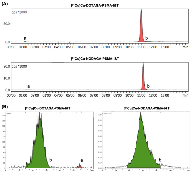
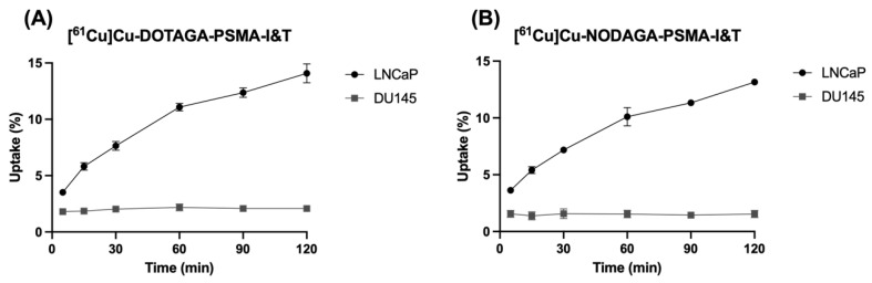
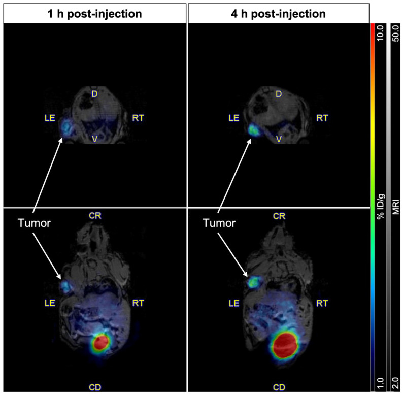
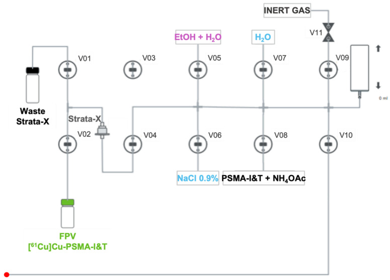
Background: While gallium-68 has traditionally dominated PET imaging in oncology, copper radionuclides have sparked interest for their potential applications in nuclear medicine and theranostics. Considering the advantageous physical decay properties of copper-61 compared to those of gallium-68, we describe a fully automated GMP-compliant synthesis process for 61Cu-based radiopharmaceuticals and demonstrate their in vivo application for targeting the overexpressed PSMA by PET/MR imaging. Methods: Copper-61 was obtained through the irradiation of natural zinc liquid targets in a biomedical cyclotron. [61Cu]Cu-DOTAGA-PSMA-I&T and [61Cu]Cu-NODAGA-PSMA-I&T were produced without manual intervention in two Synthera® Extension modules. Radiochemical purity was analyzed by radio-HPLC and iTLC. Cellular uptake was evaluated in LNCaP and DU145 cells. In vivo PET/MRI was performed in control mice to evaluate the biodistribution of both radiopharmaceuticals, and in tumor-bearing mice to assess the targeting ability towards PSMA. Results: The fully automated process developed proved to be effective for the synthesis of 61Cu-based radiopharmaceuticals, with appropriate molar activities. The final products exhibited high radiochemical purity (>98%) and remained stable for up to 6 h after the EOS. A time-dependent increase in cellular uptake was observed in LNCaP cells, but not in DU145 cells. As opposed to [61Cu]Cu-NODAGA-PSMA-I&T, [61Cu]Cu-DOTAGA-PSMA-I&T exhibited poor kinetic stability in vivo. Subsequent PET/MR imaging with [61Cu]Cu-NODAGA-PSMA-I&T showed tumor uptake lasting up to 4 h post-injection, predominant renal clearance, and no detectable accumulation in non-targeted organs. Conclusions: These results demonstrate the feasibility of the implemented process, which yields adequate amounts of high-quality radiopharmaceuticals and can be adapted to any standard production facility. This streamlined approach enhances reproducibility and scalability, bringing copper-61 closer to widespread clinical use, to the detriment of the conventionally accepted gallium-68.
Keywords: automation; copper-61; positron emission tomography; prostate cancer; prostate specific membrane antigen; radiopharmaceutical.
Conflict of interest statement
The authors declare no conflicts of interest.
Figures


References
-
- Prostate SEER Relative Survival Rates by Time Since Diagnosis, 2000–2020. [(accessed on 22 October 2024)]; Available online: https://seer.cancer.gov/statistics-network/explorer/application.html?sit....
-
- Mouraviev V., Madden J.F., Broadwater G., Mayes J.M., Burchette J.L., Schneider F., Smith J., Tsivian M., Wong T., Polascik T.J. Use of 111In-Capromab Pendetide Immunoscintigraphy to Image Localized Prostate Cancer Foci Within the Prostate Gland. J. Urol. 2009;182:938–948. doi: 10.1016/j.juro.2009.05.047. - DOI - PubMed
-
- Sodee D.B., Ellis R.J., Samuels M.A., Spirnak J.P., Poole W.F., Riester C., Martanovic D.M., Stonecipher R., Bellon E.M. Prostate cancer and prostate bed SPECT imaging with ProstaScint®: Semiquantitative correlation with prostatic biopsy results. Prostate. 1998;37:140–148. doi: 10.1002/(SICI)1097-0045(19981101)37:3<140::AID-PROS3>3.0.CO;2-Q. - DOI - PubMed
-
- Ellis R.J., Kaminsky D.A., Zhou E.H., Fu P., Chen W.-D., Brelin A., Faulhaber P.F., Bodner D. Ten-Year Outcomes: The Clinical Utility of Single Photon Emission Computed Tomography/Computed Tomography Capromab Pendetide (Prostascint) in a Cohort Diagnosed with Localized Prostate Cancer. Int. J. Radiat. Oncol. Biol. Phys. 2011;81:29–34. doi: 10.1016/j.ijrobp.2010.05.053. - DOI - PubMed
Grants and funding
LinkOut - more resources
Full Text Sources
Research Materials
Miscellaneous
