

Fibroblast Activation Protein Inhibitor (FAPI)-Based Theranostics
- PMID: 40283957
- PMCID: PMC12030087
- DOI: 10.3390/ph18040522
Fibroblast Activation Protein Inhibitor (FAPI)-Based Theranostics
Abstract
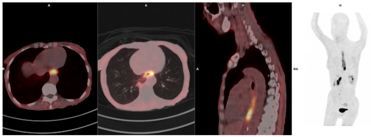
Fibroblast activation protein (FAP) is a serine protease selectively expressed in cancer-associated fibroblasts (CAFs), fibrotic tissues, and areas of active tissue remodeling, making it an attractive target for diagnostic imaging across a spectrum of disease. FAP inhibitors (FAPIs) labeled with PET tracers have rapidly advanced as a novel imaging modality with broad clinical applications that offers several advantages, including rapid tumor accumulation, low background uptake, and high tumor-to-background ratios. In oncology, FAPI PET has demonstrated excellent performance in visualizing a wide range of malignancies, including those with low glycolytic activity, such as pancreatic cancer, cholangiocarcinoma, and certain sarcomas. Its high sensitivity and specificity for the stromal component enables improved tumor delineation, staging, and response assessment. Additionally, the potential to guide theranostic approaches, where the same tracer can be labeled with therapeutic radionuclides, positions FAPI as a key player in precision oncology. Beyond oncology, FAPI PET has shown promise in imaging conditions characterized by fibrotic and inflammatory processes. In the cardiovascular field, FAPI PET imaging is being investigated for its ability to detect myocardial fibrosis and active cardiac remodeling, crucial in conditions like heart failure, post-myocardial infarction remodeling, and hypertrophic cardiomyopathy. This review highlights the expanding clinical applications of FAPI-based PET imaging across oncology, inflammation, and cardiovascular disease. While the current data are promising, further large-scale studies and multicenter trials are essential to validate these findings and establish standardized protocols. The versatility and broad applicability of FAPI PET underscore its potential as a transformative tool in precision medicine.
Keywords: FAPI; PET; theranostics.
Conflict of interest statement
The authors declare no conflict of interest.
Figures


Similar articles
-
Design and Development of 99mTc-Labeled FAPI Tracers for SPECT Imaging and 188Re Therapy.J Nucl Med. 2020 Oct;61(10):1507-1513. doi: 10.2967/jnumed.119.239731. Epub 2020 Mar 13. J Nucl Med. 2020. PMID: 32169911 Free PMC article.
-
Cardiac fibroblast activation detected by Ga-68 FAPI PET imaging as a potential novel biomarker of cardiac injury/remodeling.J Nucl Cardiol. 2021 Jun;28(3):812-821. doi: 10.1007/s12350-020-02307-w. Epub 2020 Sep 25. J Nucl Cardiol. 2021. PMID: 32975729 Free PMC article.
-
Theranostics Targeting Fibroblast Activation Protein in the Tumor Stroma: 64Cu- and 225Ac-Labeled FAPI-04 in Pancreatic Cancer Xenograft Mouse Models.J Nucl Med. 2020 Apr;61(4):563-569. doi: 10.2967/jnumed.119.233122. Epub 2019 Oct 4. J Nucl Med. 2020. PMID: 31586001 Free PMC article.
-
Fibroblast Activation Protein Inhibitor (FAPI) PET Imaging in Sarcomas: A New Frontier in Nuclear Medicine.Semin Nucl Med. 2024 May;54(3):340-344. doi: 10.1053/j.semnuclmed.2024.01.001. Epub 2024 Feb 16. Semin Nucl Med. 2024. PMID: 38365545 Review.
-
FAPI PET: Fibroblast Activation Protein Inhibitor Use in Oncologic and Nononcologic Disease.Radiology. 2023 Feb;306(2):e220749. doi: 10.1148/radiol.220749. Epub 2023 Jan 3. Radiology. 2023. PMID: 36594838 Review.
References
-
- Busek P., Mateu R., Zubal M., Kotackova L., Sedo A. Targeting fibroblast activation protein in cancer—Prospects and caveats. Front. Biosci. (Landmark Ed.) 2018;23:1933–1968. - PubMed
Publication types
LinkOut - more resources
Full Text Sources
Miscellaneous
