. 2025 Apr 8;18(4):544.
doi: 10.3390/ph18040544.

Neuroprotective Effects of Peanut Skin Extract Against Oxidative Injury in HT-22 Neuronal Cells

Affiliations

Neuroprotective Effects of Peanut Skin Extract Against Oxidative Injury in HT-22 Neuronal Cells

Jinlan Huang et al. Pharmaceuticals (Basel). .

Abstract

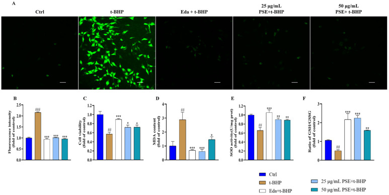
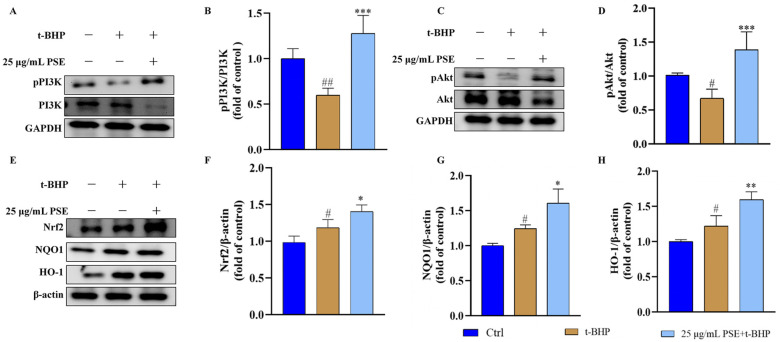
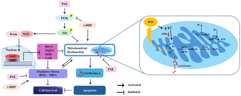
Background: Oxidative stress is a key therapeutic target in neurological disorders. As processing wastes from the peanut industry, peanut skins are great sources of antioxidants and possess potential in neuroprotection. Methods: We prepared a peanut skin extract (PSE) and investigated its protective effects against tert-butyl hydroperoxide (t-BHP)-induced oxidative injury in HT-22 neuronal cells. Results: PSE was rich in phenolic compounds (123.90 ± 0.46 mg GAE/g), comprising flavonoids (75.97 ± 0.23 mg RE/g) and proanthocyanidins (53.34 ± 1.58 mg PE/g), and displayed potent radical scavenging activities in chemical-based assays. In HT-22 cells, PSE pretreatment restored oxidative balance and endogenous antioxidant defense disrupted by t-BHP, as evidenced by significant reductions in ROS generation and lipid peroxidation levels, along with enhanced endogenous antioxidants. Specifically, 25 μg/mL PSE pretreatment reduced ROS levels by 53.03%, decreased MDA content by 78.82%, enhanced superoxide dismutase (SOD) activity by 12.42%, and improved the ratio of glutathione (GSH) to oxidized glutathione (GSSG) by 80.34% compared to the t-BHP group. Furthermore, PSE rescued mitochondrial membrane potential collapse, inhibited cytochrome c (Cyt.c) release, and prevented subsequent apoptotic death. Notably, the neuroprotective efficacy of PSE was comparable to that of edaravone, an approved neuroprotective drug. Mechanistic investigations combining network pharmacology and experimental validation revealed that the PI3K/Akt/Nrf2 signaling pathway played a pivotal role in mediating the neuroprotective effects of PSE. Compared to t-BHP-treated cells, 25 µg/mL PSE pretreatment significantly upregulated PI3K/Akt phosphorylation, the expression of Nrf2, and its downstream antioxidant proteins heme oxygenase-1 (HO-1) and NAD(P)H dehydrogenase quinone 1 (NQO1). Conclusions: Collectively, these findings demonstrate the potential of PSE as a natural protective agent against oxidative-related neurological disorders.

Keywords: Nrf2; PI3K/Akt; antioxidant; neuroprotection; peanut skins.


Conflict of interest statement

The authors declare that they have no competing financial interests or personal relationships that could influence the work reported in this paper.

Figures

Figure 1
Figure 1
Antioxidant activity of PSE in HT-22 cells. (A) Intracellular ROS were stained using DCFH-DA and visualized under a fluorescence microscope (scale bar: 50 μm). (B) Quantitative analysis of the mean fluorescence density. (C) A CCK-8 assay was used to assess the effect of PSE on t-BHP-induced cytotoxicity in HT-22 cells. (DF) Levels of MDA, SOD, and the ratio of GSH/GSSG in HT-22 cells were measured with the corresponding assay kits. All results were expressed as the means ± SD of at least three independent experiments. ## p < 0.01, ### p < 0.001 versus control; * p < 0.05, ** p < 0.01, *** p < 0.001 versus t-BHP group.
Figure 2
Figure 2
PSE inhibited mitochondrial dysfunction and apoptosis in t-BHP-damaged HT-22 cells. JC-1 staining was used to evaluate the ΔΨm of the HT-22 cells. (A,B) Images of JC-1-stained cells were captured using a fluorescence microscope (scale bar: 100 μm). The red signal represents normal ΔΨm, while the green signal represents decreased ΔΨm, and quantitative analysis of the ratio of red to green fluorescence intensity. (C,D) Apoptosis of HT-22 cells was examined using TUNEL staining. Images were captured using a confocal laser scanning microscope (CLSM) (scale bar: 100 μm) and quantitatively analyzed using Image J software (×64). (E,F) Western blotting was used to analyze the expression level of Cyt.c with glyceraldehyde-3-phosphate dehydrogenase (GAPDH) as the internal control. All results were expressed as means ± SD of at least three independent experiments. ## p < 0.01, ### p < 0.001 versus control; * p < 0.05, ** p < 0.01, *** p < 0.001 versus t-BHP group.
Figure 3
Figure 3
Network pharmacological analysis of the potential neuroprotective targets of PSE. (A) Venn diagram of the overlapping targets between the targets of peanut skin (PS) phenolic compounds and neuroprotection-related targets. (B) PPI network of 195 overlapping targets. The larger the red circle and font size, the greater the degree of connection within the PPI network. (C) The top 15 targets with the highest degree of connection in the PPI network.
Figure 4
Figure 4
GO and KEGG enrichment analysis. (A) GO enrichment analysis was conducted, revealing the top 30 GO functional items based on their p-values. In the histogram, the ordinate represents the GO term, while the abscissa indicates the number of genes associated with each term. The length of each bar signifies the number of targets within each GO term. (B) KEGG enrichment analysis was performed. In the bubble chart, the ordinate represents the KEGG term, while the abscissa signifies the proportion of targets associated with each term. The size of each circle indicates the number of enriched targets within the specific KEGG term. The color of the circles represents the p-value, with a redder color indicating a higher degree of enrichment and a correspondingly smaller p-value.
Figure 5
Figure 5
PSE activated the PI3K/Akt/Nrf2 pathway in HT-22 cells. (A,C,E) Western blot was used to detect the expression levels of PI3K, pPI3K, Akt, pAkt, Nrf2, NQO1, and HO-1 in total proteins from HT-22 cells, with GAPDH and β-actin as the internal control separately. (B,D) Quantitative analysis of the ratio of pPI3K/PI3K and pAkt/Akt in different groups. (FH) Quantitative analysis of the expression levels of Nrf2, NQO1, and HO-1 in different groups. All results were expressed as means ± SD of at least three independent experiments. # p < 0.05, ## p < 0.01 versus control; * p < 0.05, ** p < 0.01, *** p < 0.001 versus t-BHP group.
Figure 6
Figure 6
Neuroprotective mechanisms of PSE against t-BHP-induced oxidative stress. PSE quenched the free radicals generated in response to t-BHP treatment. Meanwhile, PSE enhanced the phosphorylation of PI3K and Akt, which activated Nrf2, leading to an upregulation of the antioxidant system. This process improved cellular resistance to oxidative stress. Notably, this pathway specifically enhances the expression of mitochondrial-specific antioxidant enzymes that help eliminate ROS. Additionally, phosphorylated Akt can directly influence mitochondrial activity and prevent mitochondrial dysfunction.

Similar articles

References

    1. Liu T., Li X., Zhou X., Chen W., Wen A., Liu M., Ding Y. PI3K/AKT signaling and neuroprotection in ischemic stroke: Molecular mechanisms and therapeutic perspectives. Neural Regen. Res. 2025;20:2758–2775. doi: 10.4103/nrr.Nrr-d-24-00568. - DOI - PMC - PubMed
    1. Verkhratsky A., Butt A., Li B., Illes P., Zorec R., Semyanov A., Tang Y., Sofroniew M.V. Astrocytes in human central nervous system diseases: A frontier for new therapies. Signal Transduct. Target. Ther. 2023;8:396. - PMC - PubMed
    1. Cao D.F., Zhou X.Y., Guo Q., Xiang M.Y., Bao M.H., He B.S., Mao X.Y. Unveiling the role of histone deacetylases in neurological diseases: Focus on epilepsy. Biomark. Res. 2024;12:142. - PMC - PubMed
    1. Hu E., Li Z., Li T., Yang X., Ding R., Jiang H., Su H., Cheng M., Yu Z., Li H., et al. A novel microbial and hepatic biotransformation-integrated network pharmacology strategy explores the therapeutic mechanisms of bioactive herbal products in neurological diseases: The effects of Astragaloside IV on intracerebral hemorrhage as an example. Chin. Med. 2023;18:40. - PMC - PubMed
    1. Chamorro Á., Lo E.H., Renú A., van Leyen K., Lyden P.D. The future of neuroprotection in stroke. J. Neurol. Neurosurg. Psychiatry. 2021;92:129–135. doi: 10.1136/jnnp-2020-324283. - DOI - PubMed

LinkOut - more resources