. 2025 Apr 14;18(4):569.
doi: 10.3390/ph18040569.

Tauroursodeoxycholic Acid Protects Retinal Ganglion Cells and Reduces Inflammation in Mice Following Optic Nerve Crush

Affiliations

Tauroursodeoxycholic Acid Protects Retinal Ganglion Cells and Reduces Inflammation in Mice Following Optic Nerve Crush

Nan Zhang et al. Pharmaceuticals (Basel). .

Abstract

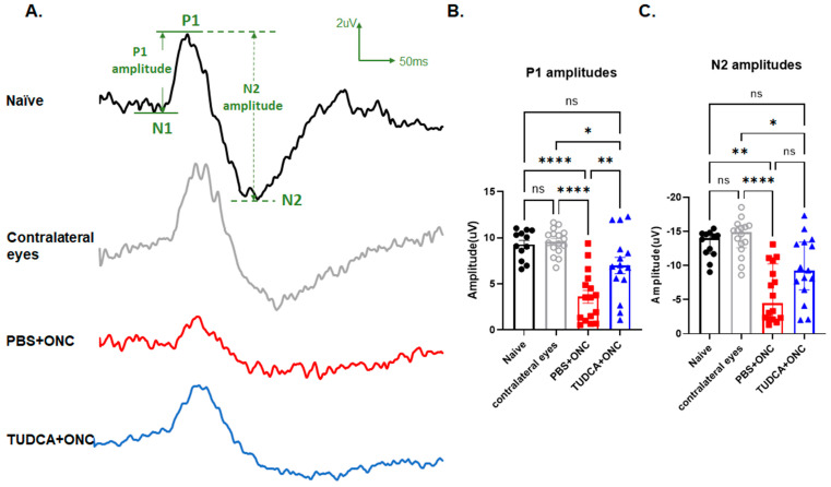
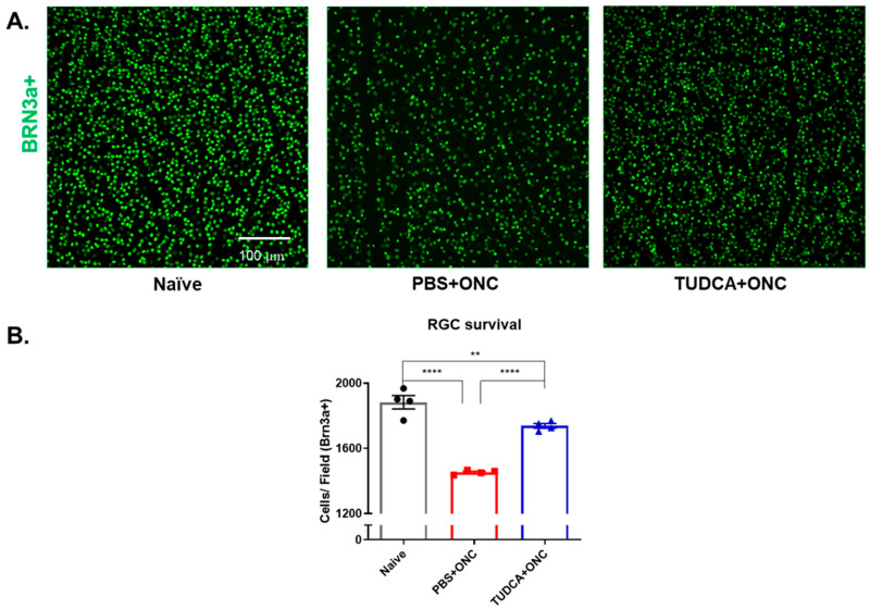
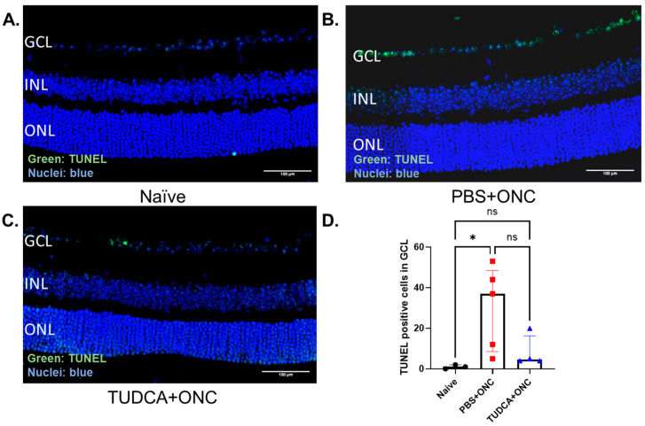
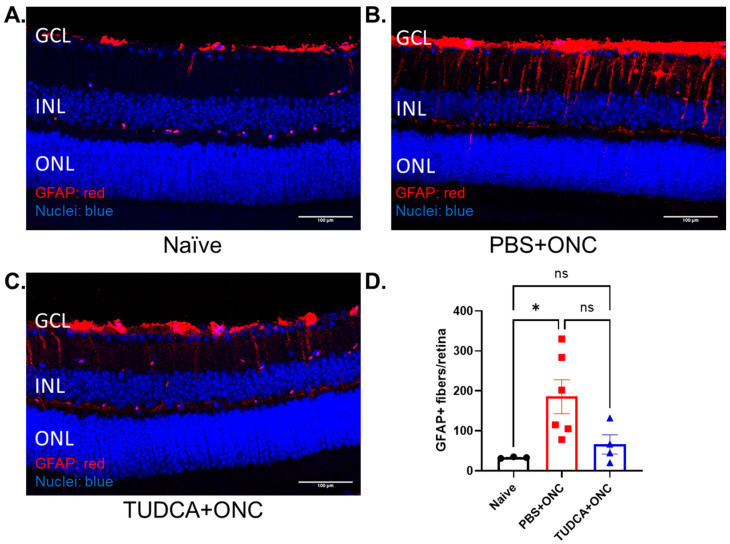
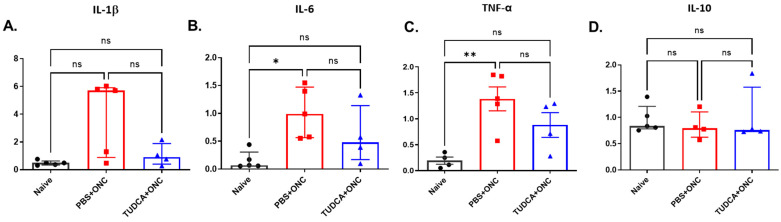
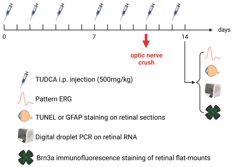
Purpose: The aim of this study was to investigate the protective effects of systemically administered tauroursodeoxycholic acid (TUDCA) in an optic nerve crush (ONC) mouse model of retinal ganglion cell (RGC) death. Methods: C57BL/6J mice were injected intraperitoneally (i.p.) three times per week with TUDCA (500 mg/kg) for two weeks, after which unilateral ONC was performed. A control cohort was identically treated with a drug vehicle (phosphate buffered saline; PBS). A separate cohort did not undergo any injections or surgeries (this was termed the "Naïve" group). Pattern electroretinography (PERG) was recorded 3 days after ONC. Retinas were harvested for whole-mount immunofluorescence staining with an antibody against RGC marker Brn3a and imaged by fluorescent confocal microscopy. Apoptotic cells in the ganglion cell layer (GCL) were detected by Terminal Deoxynucleotidyl Transferase-Mediated dUTP Nick End Labeling (TUNEL) performed on fixed retina sections. Glial fibrillary acidic protein (GFAP) immunostaining on fixed retina sections was conducted to detect the activation of Müller cells. Total RNA was extracted from retinas and expression of interleukin (IL)-1β, IL-6, tumor necrosis factor (TNF)-α, and IL-10 was determined by digital droplet PCR (ddPCR). Results: TUDCA treatment preserved visual function as assessed by PERG. P1 and N2 amplitudes from the PBS-treated ONC group were significantly diminished compared to those of the Naïve group (p < 0.001). TUDCA treatment prevented this diminution. The amplitudes of P1 and N2 in the TUDCA-treated ONC group were statistically indistinguishable from those of the Naïve group and were higher than the PBS-treated ONC group (TUDCA+ONC vs. PBS+ONC, P1: 6.99 ± 0.89 µV vs. 3.60 ± 0.69 µV, p < 0.01; N2: -9.30 (IQR: -13.43--6.44) µV vs. -4.47 (IQR: -10.26--2.17) µV). TUDCA treatment preserved RGCs. The ONC-vehicle-only group had 25% fewer RGCs (Brn3a-positive cells) than Naïve eyes (p < 0.0001). TUDCA treatment nearly completely prevented this loss, preserving all but 7.7% of the RGCs, and the number of RGCs in the TUDCA-treated ONC group was significantly higher than in the PBS-treated ONC group (TUDCA+ONC vs. PBS+ONC, 1738.00 ± 14.43 cells per field vs. 1454.00 ± 6.55 cells per field, p < 0.0001). The number of TUNEL-positive cells in the GCL (Naïve vs. PBS+ONC group: 1.00 (IQR: 0.00-2.00) % vs. 37.00 (IQR: 8.50-48.50) %, p < 0.05) and GFAP-positive fibers transversing retina sections (Naïve vs. PBS+ONC group: 33.00 ± 1.15 vs. 185.70 ± 42.37 fibers/retina, p < 0.05), and the expression of IL-6, TNF-α were significantly greater in the PBS-treated ONC group compared to that of the Naïve group (Naïve vs. PBS+ONC group, IL-6: 0.07 (IQR: 0.06-0.31) vs. 0.99 (IQR: 0.56-1.47), p < 0.05, TNF-α: 0.19 ± 0.069 vs. 1.39 ± 0.23; p < 0.01), an increase not observed with TUDCA treatment. Conclusions: Systemic TUDCA treatment significantly preserved RGC function and survival in the mouse ONC model of RGC damage. TUDCA treatment prevented RGC apoptosis, Müller glial cell activation, and retinal expression of several inflammatory cytokines. These data suggest that TUDCA is a promising therapeutic candidate for preserving RGC numbers and function.

Keywords: bile acid; inflammation; mice; mouse; mus musculus; optic nerve crush; retinal ganglion cell; tauroursodeoxycholic acid.


Conflict of interest statement

The authors declare no conflicts of interest.

Figures

Figure 1
Figure 1
TUDCA treatment preserved RGC function 3 days after optic nerve crush. (A) Representative PERG waveforms from each group. (B) Quantification of P1 amplitudes 3 days after ONC. P1 amplitudes from PBS+ONC group were significantly diminished compared to those of the Naïve or Contralateral eye groups. TUDCA treatment preserved the P1 amplitudes significantly (p < 0.01). (C) Quantification of N2 amplitudes 3 days after ONC. N2 amplitudes from PBS-treated ONC group were significantly diminished compared to those of the Naïve group or Contralateral eye groups. TUDCA treatment preserved the N2 amplitudes significantly (p < 0.05). The results are represented as mean ± SEM (B), median ± IQR (C). A one-way ANOVA with Tukey’s multiple comparisons test (B) and Kruskal–Wallis test with Dunn‘s multiple comparison test (C) was conducted between the mean cells/field in all combinations. ns: no statistical difference, * p < 0.05, ** p < 0.01, **** p < 0.0001. n = 12 mice in Naïve group; n = 16 mice in PBS+ONC group and for the Contralateral eyes of that group; n = 15 mice in TUDCA + ONC group. PERG: pattern electroretinogram; RGC: retinal ganglion cell; PBS: phosphate buffered solution; ONC: optic nerve crush; TUDCA: tauroursodeoxycholic acid.
Figure 2
Figure 2
TUDCA treatment protects against ONC-induced RGC loss. (A) Representative images of retina flatmounts stained against Brn3a from different groups. (B) Quantification of Brn3a-positive cells, assumed to be RGCs, 3 days after ONC. The number of Brn3a-positive cells was significantly less in the PBS-treated ONC group 3 days after ONC compared to Naïve group (25% decrease). TUDCA treatment partially but significantly prevented this loss. The number of Brn3a-positive cells in TUDCA-treated ONC group was slightly lower than Naïve group (1883 ± 41.0 vs. 1738 ± 14.4 cells/field). The results are represented as mean ± SEM. A one-way ANOVA with Tukey’s multiple comparisons test was conducted between the mean cells/field in all combinations. ** p < 0.01, **** p < 0.0001. n = 4 mice per group. Scale bar = 100 microns. RGC: retinal ganglion cell; PBS: phosphate buffered solution; ONC: optic nerve crush; TUDCA: tauroursodeoxycholic acid.
Figure 3
Figure 3
TUDCA treatment prevented ONC-induced RGC apoptosis. (AC) Representative morphologic images of TUNEL staining from the region of 250–750 μm from the optic nerve. (D) Quantification of percentage of TUNEL positive RGC in GCL. RGC apoptosis significantly increased 3 days after ONC (p < 0.05). TUDCA treatment prevented the increase in TUNEL-positive cells of the GCL observed in retinas from eyes that had undergone ONC. TUNEL-positive cells in GCL were counted by masked observers across entire sections, which include optic nerve head. The results are represented as median ± IQR. Kruskal–Wallis test with Dunn‘s multiple comparison test was conducted between the mean percent in all combinations. ns: no statistical difference, * p < 0.05. n = 3 mice in Naïve group; n = 5 mice in PBS group; n = 4 mice in TUDCA group. Scale bar = 100 microns. GCL: ganglion cell layer; INL: inner nuclear layer; ONL: outer nuclear layer; TUNEL: terminal deoxynucleotidyl transferase dUTP nick end labeling; PBS: phosphate buffered solution; ONC: optic nerve crush; TUDCA: tauroursodeoxycholic acid.
Figure 4
Figure 4
TUDCA treatment suppresses ONC-induced Müller cell activation. (AC) Representative fluorescent images of GFAP staining from the region of 250–750 μm from the optic nerve. (D) Quantification of GFAP-positive fibers per retina. GFAP-positive fibers significantly increased 3 days after ONC. TUDCA treatment prevented the increase in number of GFAP-positive fibers in mouse retina caused by ONC damage. No significant difference was been found between Naïve and TUDCA treated groups. GFAP-positive fibers fully penetrating the INL were counted across entire sections by masked observers. The results are represented as mean ± SEM. A one-way ANOVA with Tukey’s multiple comparisons test was conducted between the mean cells/field in all combinations. ns: no statistical difference, * p < 0.05, ns: no significant difference. n = 3 mice in Naïve group; n = 6 mice in PBS group; n = 4 mice in TUDCA group. Scale bar = 100 microns. GCL: ganglion cell layer; INL: inner nuclear layer; ONL: outer nuclear layer; GFAP: glial fibrillary acidic protein; PBS: phosphate buffered solution; ONC: optic nerve crush; TUDCA: tauroursodeoxycholic acid. Control cohort “PBS+ONC” was shared across the experiment reported in Figure 4 in [55].
Figure 5
Figure 5
TUDCA treatment prevented ONC-induced overexpression of pro-inflammatory factors. The mRNA levels of IL-1β (A), IL-6 (B), TNF-α (C), IL-10 (D) were examined 3 days after ONC. Y-axis in all channels represent the mRNA ratio to HPRT multiplied by one thousand. The results are represented as mean ± SEM (C), median ± IQR (A,B,D). Kruskal–Wallis test with Dunn‘s multiple comparison test was conducted between the mean ratio to HPRT in all combinations (A,B,D). One-way ANOVA with Tukey’s tests was conducted between the mean ratio to HPRT in all combinations (C). ns: no statistical difference, * p < 0.05, ** p < 0.01, ns: no significant difference, n = 5 mice in Naïve group/PBS+ONC group; n = 4 mice in TUDCA+ONC group. IL-1β: interleukin 1β; IL-6: interleukin 6; IL-10: interleukin 10; TNF-α: tumor necrosis factor-α; PBS: phosphate buffered solution; ONC: optic nerve crush; TUDCA: tauroursodeoxycholic acid.
Figure 6
Figure 6
Experimental timeline. Details in text of Section 4.1.

Similar articles

References

    1. Tham Y.C., Li X., Wong T.Y., Quigley H.A., Aung T., Cheng C.Y. Global prevalence of glaucoma and projections of glaucoma burden through 2040: A systematic review and meta-analysis. Ophthalmology. 2014;121:2081–2090. doi: 10.1016/j.ophtha.2014.05.013. - DOI - PubMed
    1. Gupta D., Chen P.P. Glaucoma. Am. Fam. Physician. 2016;93:668–674. - PubMed
    1. Almasieh M., Levin L.A. Neuroprotection in Glaucoma: Animal Models and Clinical Trials. Annu. Rev. Vis. Sci. 2017;3:91–120. doi: 10.1146/annurev-vision-102016-061422. - DOI - PubMed
    1. McMonnies C.W. Glaucoma history and risk factors. J. Optom. 2017;10:71–78. doi: 10.1016/j.optom.2016.02.003. - DOI - PMC - PubMed
    1. Quigley H.A. Glaucoma. Lancet. 2011;377:1367–1377. doi: 10.1016/S0140-6736(10)61423-7. - DOI - PubMed

LinkOut - more resources