. 2025 Apr 15;18(4):574.
doi: 10.3390/ph18040574.

Lipid-Based Nanoformulations of [6]-Gingerol for the Chemoprevention of Benzo[a] Pyrene-Induced Lung Carcinogenesis: Preclinical Evidence

Affiliations

Lipid-Based Nanoformulations of [6]-Gingerol for the Chemoprevention of Benzo[a] Pyrene-Induced Lung Carcinogenesis: Preclinical Evidence

Faris Alrumaihi et al. Pharmaceuticals (Basel). .

Abstract

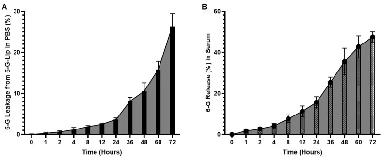
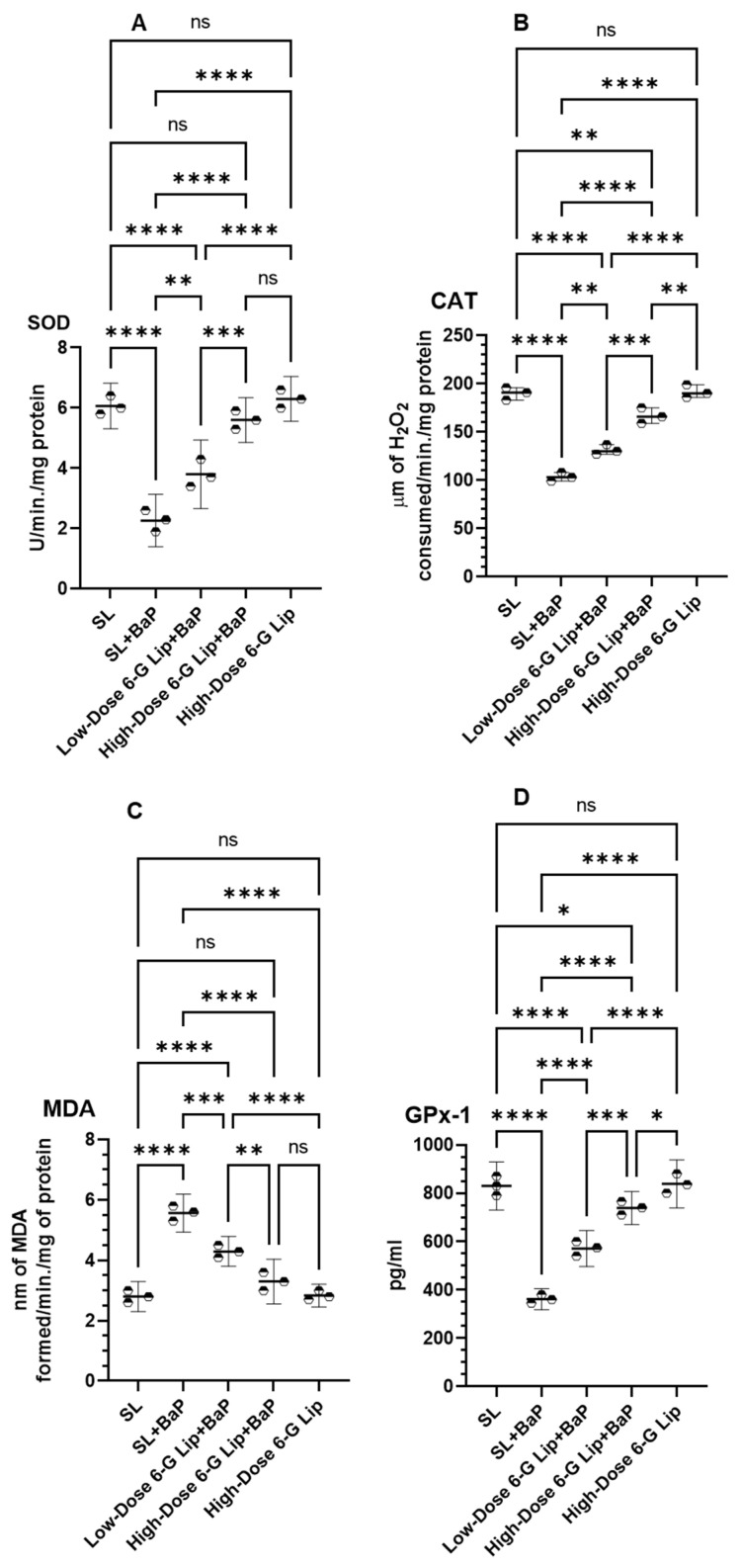
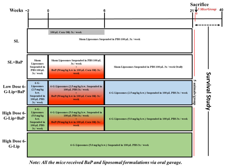
Background/Objectives: [6]-Gingerol ([6]-G), a bioactive compound derived from Zingiber officinale (ginger), exhibits strong anticancer potential but is hindered by poor aqueous solubility and low bioavailability. This study aimed to develop and evaluate PEGylated liposomal [6]-G (6-G-Lip) to enhance its stability, bioavailability, and chemopreventive efficacy in benzo[a]pyrene (BaP)-induced lung carcinogenesis. Methods: 6-G-Lip was synthesized using a modified thin-film hydration technique and characterized for size, polydispersity index (PDI), zeta potential, encapsulation efficiency (EE%), and release kinetics. The chemopreventive effects were assessed in BaP-induced lung cancer in Swiss albino mice, with prophylactic 6-G-Lip administration from two weeks before BaP exposure through 21 weeks. Cancer biomarkers, antioxidant enzyme activity, reactive oxygen species (ROS) generation, induction of apoptosis, and histopathological alterations were analyzed. Results: 6-G-Lip exhibited a particle size of 129.7 nm, a polydispersity index (PDI) of 0.16, a zeta potential of -18.2 mV, and an encapsulation efficiency (EE%) of 91%, ensuring stability and effective drug loading. The formulation exhibited a controlled release profile, with 26.5% and 47.5% of [6]-G released in PBS and serum, respectively, at 72 h. 6-G-Lip significantly lowered cancer biomarkers, restored antioxidant defenses (SOD: 5.60 U/min/mg protein; CAT: 166.66 μm H2O2/min/mg protein), reduced lipid peroxidation (MDA: 3.3 nm/min/mg protein), and induced apoptosis (42.2%), highlighting its chemopreventive efficacy. Conclusions: This study is the first to prepare, characterize, and evaluate PEGylated [6]-G-Lip for the chemoprevention of lung cancer. It modulates oxidative stress, restores biochemical homeostasis, and selectively induces apoptosis. These findings support 6-G-Lip as a promising nanotherapeutic strategy for cancer prevention.

Keywords: animal model; cancer therapy; drug delivery system; drug formulation; lung cancer; nanocarrier.


Conflict of interest statement

The author declares no conflicts of interest.

Figures

Figure 1
Figure 1
Characterization of DSPC/Chol/mPEG-DSPE and DSPC/Chol/mPEG-DSPE/6-G Liposomes. (A) Particle size distribution, (B) polydispersity index (PDI), (C) zeta (ζ) potential, and (D) encapsulation efficiency (EE%). Data are presented as mean values, with error bars representing the 95% confidence intervals (CIs) from three independent experiments.
Figure 1
Figure 1
Characterization of DSPC/Chol/mPEG-DSPE and DSPC/Chol/mPEG-DSPE/6-G Liposomes. (A) Particle size distribution, (B) polydispersity index (PDI), (C) zeta (ζ) potential, and (D) encapsulation efficiency (EE%). Data are presented as mean values, with error bars representing the 95% confidence intervals (CIs) from three independent experiments.
Figure 2
Figure 2
In vitro stability and release kinetics of [6]-Gingerol liposomes (6-G-Lip). (A) Stability of 6-G-Lip in phosphate-buffered saline (PBS) at 37 °C over 72 h, demonstrating structural integrity and minimal aggregation. (B) The cumulative release profile of [6]-Gingerol from PEGylated liposomes in serum under physiological conditions indicates controlled and sustained drug release. Data represent the mean ± 95% confidence intervals (CIs) of three independent experiments.
Figure 3
Figure 3
Effect of 6-G-Lip on BaP-induced changes in body weight and survival rates. (A) Average body weight (ABW) variations across experimental groups over the study period. Data are expressed as mean ± 95% confidence intervals (CIs), with a sample size of fifteen mice per group (n = 15). Statistical significance for ABW differences was determined using one-way ANOVA, followed by Tukey’s post hoc test. ‘ns’ indicates no statistically significant difference between groups. Asterisks denote statistical significance: ** p < 0.01 and *** p < 0.001. (B) Kaplan–Meier survival analysis depicting survival probabilities monitored for up to 40 weeks, with ten mice per group (n = 10). The survival differences were analyzed using the log-rank (Mantel–Cox) test.
Figure 4
Figure 4
Effects of 6-G-Lip on serum cancer biomarkers in BaP-induced lung carcinogenesis. This figure illustrates the impact of 6-G-Lip on serum levels of key cancer-associated enzymes in the BaP-induced lung cancer model. (A) Adenosine deaminase (ADA), (B) Gamma-glutamyl transferase (GGT), (C) 5′-Nucleotidase (CD73), and (D) Lactate dehydrogenase (LDH) activity levels across experimental groups. BaP exposure (SL + BaP) resulted in a significant increase in these biomarkers compared to the vehicle control (SL), indicating metabolic alterations associated with tumor progression. Treatment with 6-G-Lip resulted in a dose-dependent decrease in these enzyme levels, with high-dose 6-G-Lip + BaP restoring values closer to those of the control group, suggesting its protective role in counteracting BaP-induced metabolic dysregulation. The high-dose 6-G-Lip-only group exhibited no significant differences from the SL control, confirming the formulation’s safety under physiological conditions. These findings highlight the potential of 6-G-Lip in mitigating tumor-associated metabolic disturbances and reinforce its role in chemoprevention. Data are presented as mean ± 95% confidence intervals (CIs) from three independent experiments. Statistical significance is indicated as (*) p < 0.05, (**) p < 0.01, (***) p < 0.001, and (****) p < 0.0001; ‘ns’ denotes no statistically significant differences between groups.
Figure 5
Figure 5
Protective effects of 6-G-Lip on antioxidant enzyme activities in BaP-induced lung carcinogenesis. This figure illustrates the impact of 6-G-Lip on oxidative stress regulation in lung tissues of BaP-induced small cell lung carcinoma (SCLC) mice. (A) Superoxide dismutase (SOD); (B) catalase (CAT); (C) malondialdehyde (MDA), a key marker of lipid peroxidation; and (D) glutathione peroxidase 1 (GPx1) across experimental groups. BaP exposure (SL + BaP) resulted in a marked depletion of SOD, CAT, and GPx1 activities, accompanied by a significant increase in MDA levels, reflecting severe oxidative stress and lipid peroxidation. Treatment with 6-G-Lip restored antioxidant enzyme activity in a dose-dependent manner, with high-dose 6-G-Lip + BaP achieving values comparable to the SL control group, indicating effective mitigation of BaP-induced oxidative damage. Notably, the high-dose 6-G-Lip-only group exhibited no significant deviations from the SL control, confirming its safety under physiological conditions. These findings underscore the potential of 6-G-Lip in counteracting oxidative stress-driven carcinogenesis. Data are presented as the mean ± 95% confidence intervals (CIs) from three independent experiments. Statistical significance is denoted as (*) p < 0.05, (**) p < 0.01, (***) p < 0.001, and (****) p < 0.0001; ‘ns’ indicates no statistically significant differences between groups.
Figure 6
Figure 6
Effect of 6-G-Lip on intracellular reactive oxygen species (ROS) levels in lung cells. This figure illustrates the impact of 6-G-Lip on intracellular ROS levels in a BaP-induced SCLC model, as measured using the 2′,7′-dichlorofluorescein diacetate (DCFDA) assay via flow cytometry. The mean fluorescence intensity (MFI) represents ROS production across experimental groups. Exposure to BaP (SL + BaP) did not lead to a substantial increase in ROS compared to the vehicle control (SL), suggesting an adaptive cellular response that may facilitate tumor progression. In contrast, administration of 6-G-Lip resulted in a significant, dose-dependent elevation in ROS levels, with both low- and high-dose 6-G-Lip + BaP groups exhibiting markedly increased ROS accumulation relative to SL + BaP. This surge in ROS suggests that 6-G-Lip enhances oxidative stress in transformed cells, potentially driving apoptosis through ROS-mediated cytotoxicity. Notably, the high-dose 6-G-Lip-only group exhibited ROS levels comparable to those of the control, indicating that 6-G-Lip does not induce oxidative stress under non-malignant conditions. Data are presented as mean ± 95% confidence intervals (CIs) from three independent experiments. Statistical significance is denoted as (*) p < 0.05, (***) p < 0.001, (****) p < 0.0001, while ‘ns’ indicates no statistically significant difference between groups.
Figure 7
Figure 7
6-G-Lip-induced apoptosis in lung cells, evaluated by annexin V-FITC/PI flow cytometry. This figure illustrates the pro-apoptotic effects of 6-G-Lip in lung cells from a BaP-induced SCLC model, assessed using Annexin V-FITC and propidium iodide (PI) staining via flow cytometry. The analysis differentiates between viable, early apoptotic, late apoptotic, and necrotic cell populations across experimental groups. The SL + BaP group exhibited minimal apoptosis, indicating an inadequate activation of intrinsic cell death pathways in response to BaP-induced carcinogenesis. In contrast, 6-G-Lip treatment significantly increased apoptosis in a dose-dependent manner, with the low-dose 6-G-Lip + BaP group showing 36% apoptotic cells and the high-dose 6-G-Lip + BaP group reaching 42.2%, demonstrating a strong activation of programmed cell death mechanisms. The high-dose 6-G-Lip-only group exhibited apoptosis levels comparable to the vehicle control (SL), confirming that 6-G-Lip does not induce cytotoxicity under normal physiological conditions. Data are presented as mean ± 95% confidence intervals (CIs) from three independent experiments. Statistical significance is indicated as (**) p < 0.01, (****) p < 0.0001, while ‘ns’ denotes no statistically significant difference between groups.
Figure 8
Figure 8
Histopathological assessment of lung tissues in BaP-induced SCLC and the effects of 6-G-Lip. Hematoxylin and eosin (H&E)-stained lung sections from experimental groups. SL + BaP group exhibits tumor cell proliferation, hemorrhagic regions (blue arrows), dilated bronchioles (red stars), and carcinoma invasion (green arrows). The lower panel highlights mitotic figures, hyperchromatic nuclei (green stars), and luminal narrowing (yellow stars). Low-dose 6-G-Lip + BaP group shows mild to moderate parenchymal alterations, hemorrhage (blue stars), emphysema (black arrow), and absence of carcinoma invasion (purple arrow). The high-dose 6-G-Lip + BaP group exhibits preserved lung architecture, characterized by mild chronic bronchitis (yellow star) and leukocytic infiltration with alveolar wall thickening (red star). SL and high-dose 6-G-Lip groups maintain normal bronchioles and alveolar structures. Upper panel: 100× magnification, bar = 100 µm; lower panel: 400× magnification, bar = 50 µm.
Figure 9
Figure 9
Experimental design overview. This figure illustrates the experimental design assessing the chemopreventive efficacy of [6]-gingerol-loaded PEGylated liposomes (6-G-Lip) in a benzo[a]pyrene (BaP)-induced small cell lung carcinoma (SCLC) model. Seventy-five female Swiss albino mice were randomly assigned to five groups (n = 15 per group) and received all treatments via oral gavage to ensure precise and consistent dosing. The vehicle control group (SL) received empty liposomes in phosphate-buffered saline (PBS) from week−2 to week 21. The SL + BaP group was administered BaP (50 mg/kg in corn oil) three times a week for four weeks to induce SCLC, and sham liposomes were administered. Two treatment groups received 6-G-Lip in a dose-dependent manner: low-dose 6-G-Lip + BaP (2.5 mg/kg) and high-dose 6-G-Lip + BaP (5.0 mg/kg), both administered from week 2 to week 21 alongside BaP. An additional high-dose 6-G-Lip group received 6-G-Lip (5.0 mg/kg) alone to evaluate its safety under normal physiological conditions. At week 22, five mice in each group were sacrificed for biochemical and histopathological assessments. The remaining ten mice in each group were monitored for survival analysis until week 40.

Similar articles

References

    1. Bray F., Laversanne M., Sung H., Ferlay J., Siegel R.L., Soerjomataram I., Jemal A.l. Global cancer statistics 2022: GLOBOCAN estimates of incidence and mortality worldwide for 36 cancers in 185 countries. CA Cancer J. Clin. 2024;74:229–263. doi: 10.3322/caac.21834. - DOI - PubMed
    1. Thai A.A., Solomon B.J., Sequist L.V., Gainor J.F., Heist R.S. Lung cancer. Lancet. 2021;398:535–554. doi: 10.1016/S0140-6736(21)00312-3. - DOI - PubMed
    1. Carroll R., Bortolini M., Calleja A., Munro R., Kong S., Daumont M.J., Penrod J.R., Lakhdari K., Lacoin L., Cheung W.Y. Trends in treatment patterns and survival outcomes in advanced non-small cell lung cancer: A Canadian population-based real-world analysis. BMC Cancer. 2022;22:255. doi: 10.1186/s12885-022-09342-5. - DOI - PMC - PubMed
    1. Basse C., Carton M., Milder M., Geiss R., Du Rusquec P., Daniel C., Massiani M., Liwartowski A., Girard N. Real-World Survival Impact of New Treatment Strategies for Lung Cancer: A 2000–2020 French Cohort. Cancers. 2024;16:2768. doi: 10.3390/cancers16152768. - DOI - PMC - PubMed
    1. Jazieh A., AlGhamdi M., AlGhanem S., AlGarni M., AlKattan K., AlRujaib M., AlMaimi M., Babelli O.M., AlShehri S., AlQahtani R., et al. Saudi lung cancer prevention and screening guidelines. Ann. Thorac. Med. 2018;13:198–204. doi: 10.4103/atm.ATM_147_18. - DOI - PMC - PubMed

LinkOut - more resources