Review
. 2025 Apr 19;14(8):1247.
doi: 10.3390/plants14081247.

Endophytes in Cannabis sativa: Identifying and Characterizing Microbes with Beneficial and Detrimental Effects on Plant Health

Affiliations
Review

Endophytes in Cannabis sativa: Identifying and Characterizing Microbes with Beneficial and Detrimental Effects on Plant Health

Liam Buirs et al. Plants (Basel). .

Abstract

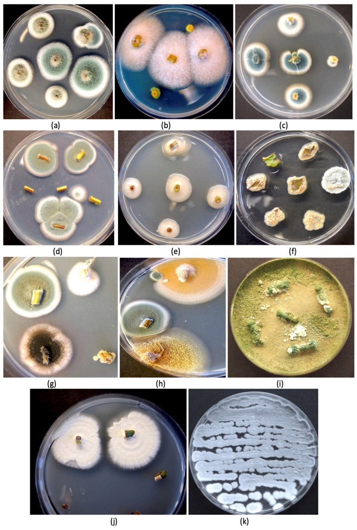
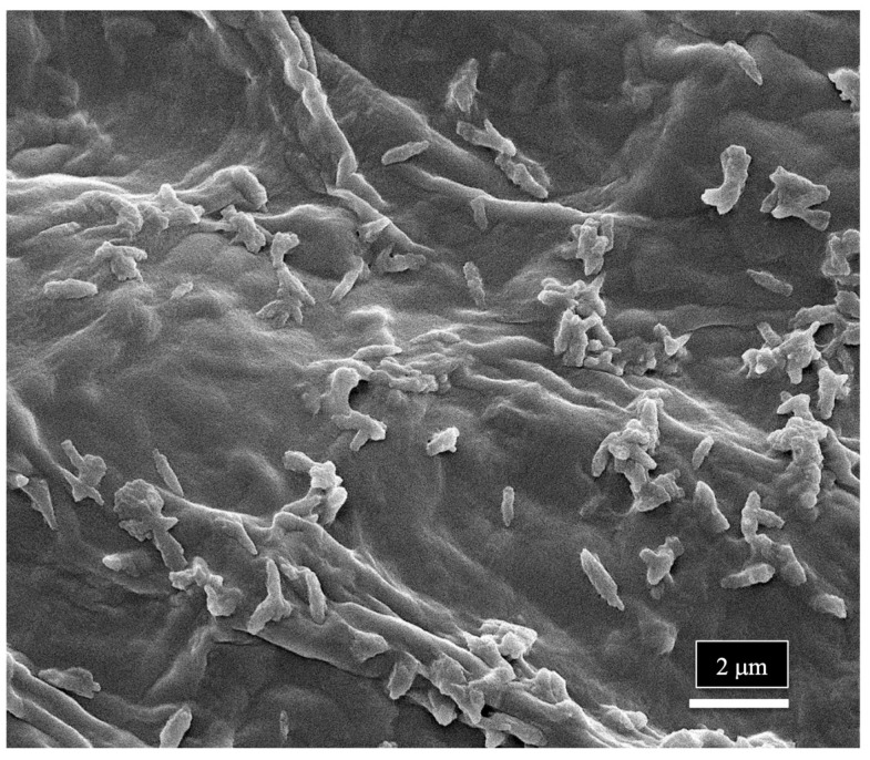
The roles of endophytes in Cannabis sativa (cannabis, hemp) remain poorly explored. While in vitro studies suggest that there can be several benefits, such as plant growth promotion and protection against pathogens, more in planta studies are needed. This review summarizes the bacterial and fungal endophytes previously reported in tissues of C. sativa and discusses the factors influencing their presence, as well as their potential beneficial and detrimental effects. Using genome sequencing and culture-based approaches, we describe the microbial diversity in hydroponically cultivated cannabis plants at several developmental stages. These include mother plants, cuttings, vegetative and flowering plants, and tissue-cultured plantlets. Microbes that were present include fungal, yeast, and bacterial endophytes found in roots, stems, leaves, inflorescences, and seeds. These may have originated from the growing substrate or be transmitted through vegetative propagation. Notable endophytes included Rhizophagus irregularis (a mycorrhizal fungus), Penicillium chrysogenum (an antibiotic producer), and various endophytic yeast species not previously described in C. sativa. Endophytes representing potential plant pathogens, such as Fusarium oxysporum, are also present within cannabis tissues, which can negatively impact plant health. Using scanning electron microscopy, we observed that fungal propagules are present within pith parenchyma cells and xylem vessel elements in stem tissues, illustrating for the first time the in situ localization and distribution of endophytes in cannabis vascular tissues. The mechanism of spread through xylem vessels likely contributes to the spread of endophytes within cannabis and hemp plants. Further research is required to validate the roles of endophytes in cannabis and hemp plants grown under commercial production conditions.

Keywords: biological control; microbial diversity; plant microbiome; plant pathogens; soil microbes.


Conflict of interest statement

Author Liam Buirs is employed by the company Pure Sunfarms Corp. The remaining author (Zamir K. Punja) declares that the research was conducted in the absence of any commercial or financial relationships that could be construed as a potential conflict of interest. The authors declare that this study received funding from Pure Sunfarms Corp. The funder was not involved in the study design, collection, analysis, interpretation of data, the writing of this article or the decision to submit it for publication.

Figures

Figure 1
Figure 1
The different stages of growth of greenhouse-cultivated cannabis plants from which tissue samples were obtained for microbiome and microscopic analyses. (a) Mother (stock) plants approximately 3 months of age. (b) Vegetative cuttings taken from mother plants used for propagation. (c) A vegetative plant approximately 4 weeks following the rooting of cuttings. (d) Flowering plants approximately 4 weeks into flowering.
Figure 2
Figure 2
Genera of endophytic fungi identified in cannabis tissues at different stages of development and in the growing substrate as determined by whole genome sequencing. Numbers represent the percentage of the total reads of the specific genus relative to the total fungal reads in the tissue sample. The “+” and the blue arrows indicate a value of up to 1% of the total reads. Values over 1% are highlighted in yellow or green.
Figure 3
Figure 3
Recovery of a range of fungi from the stem, leaf, and petiole tissues of cannabis mother plants. All tissues were surface-sterilized before plating onto potato dextrose agar containing 140 mg/L of streptomycin sulfate to inhibit bacterial growth. (a) Two different colony types of Penicillium spp. (b) Fusarium oxysporum colonies. (c) Colonies of Penicillium chrysogenum. (d) Colonies of Penicillium sp. (e) Unidentified bacterial colonies. (f) Yeast colonies from leaf segments. (g,h) A range of colonies of different fungi, including Penicillium and Aspergillus (black and yellow colonies). (i) Recovery of Trichoderma harzianum from petiole segments. (j) Colonies of Beauveria bassiana. (k) Streaked culture of P. chrysogenum. All photos are of 9 cm diameter Petri dishes.
Figure 4
Figure 4
Genera of endophytic yeasts present in cannabis tissues and in the growing substrate as determined by whole genome sequencing. Numbers represent the percentage of total reads of the specific genus relative to the total fungal reads in the tissue sample. The “+” and the blue arrows indicate a value of up to 1%. Values over 1% are indicated.
Figure 5
Figure 5
Treatments applied to the cocofibre growing substrate to alter the endophytes in cannabis cuttings. The substrate was autoclaved, received a drench of Luna fungicide, or was amended with formulations of Trichoderma found in Rootshield® and Asperello® after autoclaving. The substrates were then allowed to incubate under ambient laboratory conditions for 7 days before dilution plating was conducted to determine the background levels of mycoflora compared to the untreated control. In (a), the Petri dishes show microbial growth 5 days after dilution plating from each treatment. Note that there was no growth in the autoclaved substrate, while fungicide application reduced all fungal colonies compared to the control. In (b), rooted cuttings were inserted into the growing substrate following the treatments shown in (a) and then allowed to grow for 4 weeks before tissues were collected for microbiome analysis. (c) Comparison of cuttings inserted into the cocofibre substrate at the time the experiment was initiated (left) compared to after 4 weeks of growth (right). Root and stem tissues were collected, surface-sterilized, and prepared for microbiome analysis.
Figure 6
Figure 6
Tissue culture-derived plantlets from a meristem culture (left) and from a nodal culture (right) were assayed to determine the background microbes present. Tissue culture conditions were as described by Punja et al. [108], and leaf tissues were collected and processed for microbiome analysis as described in the methods section.
Figure 7
Figure 7
Effect of treatments applied to the cocofibre growing substrate on the resulting fungal microbiome detected in roots (R), stems (S) and leaves (L) of cannabis cuttings after a 4-week exposure to the treated substrate. Controls (no treatment) represent replicate samples (Rep. 1 and Rep. 2). Numbers represent the percentage of total reads of the specific genus relative to the total fungal reads in the tissue sample. The “+”indicate a value of up to 1% of the total reads. Values over 1% are highlighted in yellow.
Figure 8
Figure 8
(a) Cross-sectional view of the main stem of a 3-month-old cannabis mother plant that shows the central pith (arrow). (b) Cross-sectional views of the side stems on a mother plant showing the central pith regions (arrow). (c) A scanning electron microscopic view of a section through a side stem as shown in (b). The central pith (P) is surrounded by a ring of xylem parenchyma cells that appear white. The largest area of the stem is occupied by the xylem (X) tissue that extends from the region of the pith cells to the outer ring of the phloem and epidermal tissues (arow).
Figure 9
Figure 9
Scanning electron microscopic images of the stem of a cannabis mother plant. (a) The stems were cut in longitudinal sections and were examined for the various tissue types as shown in (b). (c) A closer view of the layers of tissue types from the center of the stem. Moving outwards shows the pith parenchyma cells (P), xylem fibers (X), tracheids (T), and vessel elements (V). (d,e) A close-up view of the xylem fibers (X) and vessel elements (V). (f) A magnified view of the xylem fibers.
Figure 10
Figure 10
Magnified views of the xylem and pith parenchyma cells found in the stem of a cannabis mother plant as seen under the scanning electron microscope. (a) The length-wise arrangement of the tracheids (T) and vessel elements (T) can be seen. (b) A higher magnification view of the tracheids (T) and vessel elements (V). (c,d) A view of the pith parenchyma cells surrounding the pith region.
Figure 11
Figure 11
Magnified images of the vessel elements and tracheids found in the stem of a cannabis mother plant as viewed under the scanning electron microscope. (a) The longitudinal arrangement and size of the vessel elements (V) and tracheids (T) are shown side-by-side. (b) A close-up view of a vessel element cell showing the presence of pit openings (P) along the walls. (c,d) Magnified views of the pit openings that line the walls of the vessel elements. Pit opening apertures range from 3 μm to 10 μm in diameter.
Figure 12
Figure 12
Scanning electron microscopic images of the xylem vessel elements inside the stem of a cannabis mother plant. The surrounding walls of the vessel elements were broken apart to show the presence of fungal spores inside the vessel elements. (a,b) A range of spore types (S) can be seen lining the vessel element. (c,d) Close-up views of the mostly spherical spores that represent different sizes, presumably of different fungal species. Some spores appear to be collapsed. (e,f) Magnified views of different spore types in the xylem vessel elements. Some spores appear to be coated by a biofilm of unknown source.
Figure 13
Figure 13
Scanning electron microscopic images of the spores of various fungi in relation to the size of the xylem vessel element pits. (a) An extremely small spore that can pass through the pit opening, possibly of Penicillium. (b) A moderately sized spore that can pass though the pit opening, possibly the microconidia of Fusarium. (c) A large spore that cannot pass through the pit opening, possibly of Botrytis. (d,e) Very large spores of different fungi that cannot pass through the pit openings, possibly of mycorrhizal fungi. (f) Spores resembling Fusarium macroconidia at the early stages of development.
Figure 14
Figure 14
Spores of Penicillium sp. observed in the xylem tissues of the stem of a cannabis mother plant. (a,b) The spores are produced in distinct chains and (c,d) are echinulate (with spines).
Figure 15
Figure 15
Scanning electron micrograph images of spores and mycelium observed in xylem tissues of the stem of a cannabis mother plant. (a) Spores (S, white arrow) are present inside the pith parenchyma cells. (b) Mycelium (M, white arrows) can be observed growing over the xylem tissue cells.
Figure 16
Figure 16
Crystals presumed to be those of calcium oxalate (white arrows) are present in the xylem vessel elements (a) as well as in the pith parenchyma cells (bd).
Figure 17
Figure 17
Scanning electron microscopic image showing bacterial cells on the surface of the xylem vessels in a stem sample of cannabis from a mother plant.
Figure 18
Figure 18
The morphology and tissue types observed in cuttings of cannabis plants under the scanning electron microscope. (a) Cross sections made at the base of the cuttings of two genotypes show the different stages of development of the central pith tissues. On the left, the pith in the genotype is less developed compared to the genotype on the right. (b) In the genotype from the left side in (a), the pith parenchyma cells can be seen in the center of the stem of the cutting, but the pith opening has not yet developed. (c,d) In the genotype from the right side in (a), the pith has fully developed to form an opening, and the pith parenchyma cells are reduced to a ring around the opening (white arrow in (c)). (e) View of the xylem conducting tissues that comprise tracheids and vessel elements, similar to that seen in the stems of mother plants. The white arrow shows the ring of pith parenchyma cells adjacent to the xylem vessels. (f) Close-up view of the tracheids and vessel elements.
Figure 19
Figure 19
Summary of the distribution of endophytic species of fungi and yeasts during cannabis cultivation under greenhouse conditions. The symbols represent different groups of endophytes that have been found in the various locations based on results presented in this study.

Similar articles

References

    1. Long T., Wagner M., Demske D., Leipe C., Tarasov P.E. Cannabis in Eurasia: Origin of human use and Bronze Age trans-continental connections. Veg. Hist. Archaeobot. 2017;26:245–258. doi: 10.1007/s00334-016-0579-6. - DOI
    1. Small E. Classification of Cannabis sativa L. in relation to agricultural, biotechnological, medical and recreational utilization. In: Chandra S., Lata H., ElSohly M.A., editors. Cannabis sativa L.—Botany and Biotechnology. Springer International Publishing; Cham, Switzerland: 2017. pp. 1–63.
    1. Li H.L. An archaeological and historical account of cannabis in China. Econ. Bot. 1973;28:437–448. doi: 10.1007/BF02862859. - DOI
    1. Fleming M.P., Clarke R.C. Physical evidence for the antiquity of Cannabis sativa L. (Cannabaceae) J. Int. Hemp Assoc. 1998;5:80–92.
    1. Chandra S., Lata H., ElSohly M.A., Walker L.A., Potter D. Cannabis cultivation: Methodological issues for obtaining medical-grade product. Epilepsy Behav. 2017;70:302–312. doi: 10.1016/j.yebeh.2016.11.029. - DOI - PubMed

LinkOut - more resources