. 2025 Apr 18;17(8):1101.
doi: 10.3390/polym17081101.

NIR pH-Responsive PEGylated PLGA Nanoparticles as Effective Phototoxic Agents in Resistant PDAC Cells

Affiliations

NIR pH-Responsive PEGylated PLGA Nanoparticles as Effective Phototoxic Agents in Resistant PDAC Cells

Degnet Melese Dereje et al. Polymers (Basel). .

Abstract

Pancreatic ductal adenocarcinoma (PDAC) is one of the deadliest cancers worldwide due to its resistance to conventional therapies that is attributed to its dense and acidic tumor microenvironment. Chemotherapy based on gemcitabine usually lacks efficacy due to poor drug penetration and the metabolic characteristics of the cells adapted to grow at a more acidic pHe, thus presenting a more aggressive phenotype. In this context, photodynamic therapy (PDT) offers a promising alternative since it generally does not suffer from the same patterns of cross-resistance observed with chemotherapy drugs. In the present work, a novel bromine-substituted heptamethine-cyanine dye (BrCY7) was synthesized, loaded into PEG-PLGA NPs, and tested on the pancreatic ductal adenocarcinoma cell line cultured under physiological (PANC-1 CT) and acidic (PANC-1 pH selected) conditions, which promotes the selection of a more aggressive phenotype. The cytotoxicity of BrCY7-PEG-PLGA is dose-dependent, with an IC50 of 2.15 µM in PANC-1 CT and 2.87 µM in PANC-1 pH selected. Notably, BrCY7-PEG-PLGA demonstrated a phototoxic effect against PANC-1 pH selected cells but not on PANC-1 CT, which makes these findings particularly relevant since PANC-1 pH selected cells are more resistant to gemcitabine as compared with PANC-1 CT cells.

Keywords: PEGylated PLGA nanoparticles; heptamethine-cyanine dyes; photodynamic therapy; resistant PDAC cells.


Conflict of interest statement

The authors declare no conflicts of interest.

Figures

Scheme 1
Scheme 1
Synthesis of BrCY7. (i) 1-iodobutane and acetonitrile, microwave heating, 155 °C for 30 min; (ii) anhydrous potassium acetate and absolute ethanol, microwave heating, 120 °C for 15 min.
Figure 1
Figure 1
Spectroscopic characteristics of BrCY7 in DMSO (Abs: absorption; Em: emission) (A) and comparison of absorbance behavior in DMSO and PBS (B).
Figure 2
Figure 2
Synthetic workflow for the PEG-PLGA and BrCY7-PEG-PLGA nanoparticle preparation via the single-emulsion method.
Figure 3
Figure 3
FE-SEM morphology of (A) BrCY7-PEG-PLGA and (B) empty PEG-PLGA NPs and (C) NTA size distribution profile of empty and Br-CY7-loaded PEG-PLGA NPs.
Figure 4
Figure 4
(A) Spectroscopic characteristics of the free BrCY7 in DMSO and BrCY7-PEG-PLGA in PBS; (B) time-dependent decrease in the DPBF absorption band at 418 nm, monitored under irradiation in the presence of toluidine blue, rose bengal, and BrCY7-loaded PEG-PLGA nanoparticles; (C) evaluation of photodegradation of BrCY7 after the incorporation into PEG-PLGA NPs.
Figure 5
Figure 5
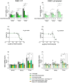
(A,B) In vitro cytotoxicity of BrCY7-PEG-PLGA in PANC-1 CT cells (A) and PANC-1 pH selected cells (B). Cell viability assay performed at 48, 72, and 96 h after incubation with 0, 2, 3, 4, and 5 µM of BrCy7-PEG-PLGA. These concentrations refer to the PS concentration, loaded in, respectively, in 0 µg/mL, 500 µg/mL, 750 µg/mL, 1 mg/mL, and 1.250 mg/mL of PEG-PLGA. Data are normalized on CTR 48 h and are represented as the mean (of at least three independent experiments) ± SEM. Statistical significance versus CTR (RM one-way ANOVA without Geisser–Greenhouse correction with Dunnett’s multiple comparisons post hoc test or Friedman with Dunn’s multiple comparisons post hoc test according to data distribution): *: p-value < 0.05, **: p-value < 0.01, ***: p-value < 0.001. (C,D) IC50 curves of BrCY7-PEG-PLGA in PANC-1 CT cells (C) and PANC-1 pH selected cells (D) 96 h post-treatment. Data are represented as the mean (of at least three independent experiments) ± SEM. (E,F) In vitro photoactivity of 2 µM BrCY7 loaded in 500 µg/mL PEG-PLGA in PANC-1 CT cells (E) and PANC-1 pH selected cells (F). Cell viability was evaluated at 24, 48, and 72 h post-irradiation (LB) for 15 min at 770 nm. Gemcitabine [5 µM] was used as the gold standard for the comparison of the BrCY7-PEG-PLGA [2 µM] effect on both cell lines. Data are normalized on CTR (0 µM) at 24 h and represented as the mean (of at least three independent experiments) ± SEM. Statistical significance (RM one-way ANOVA without Geisser–Greenhouse correction with Dunnett’s multiple comparisons post hoc test or Friedman with Dunn’s multiple comparisons post hoc test according to data distribution): *: p-value < 0.05, **: p-value < 0.01.

Similar articles

References

    1. Mercanti L., Sindaco M., Mazzone M., Di Marcantonio M.C., Piscione M., Muraro R., Mincione G. PDAC, the Influencer Cancer: Cross-Talk with Tumor Microenvironment and Connected Potential Therapy Strategies. Cancers. 2023;15:2923. doi: 10.3390/cancers15112923. - DOI - PMC - PubMed
    1. Webb B.A., Chimenti M., Jacobson M.P., Barber D.L. Dysregulated PH: A Perfect Storm for Cancer Progression. Nat. Rev. Cancer. 2011;11:671–677. doi: 10.1038/nrc3110. - DOI - PubMed
    1. Riemann A., Schneider B., Ihling A., Nowak M., Sauvant C., Thews O., Gekle M. Acidic Environment Leads to ROS-Induced MAPK Signaling in Cancer Cells. PLoS ONE. 2011;6:e22445. doi: 10.1371/journal.pone.0022445. - DOI - PMC - PubMed
    1. Andreucci E., Peppicelli S., Ruzzolini J., Bianchini F., Biagioni A., Papucci L., Magnelli L., Mazzanti B., Stecca B., Calorini L. The Acidic Tumor Microenvironment Drives a Stem-like Phenotype in Melanoma Cells. J. Mol. Med. 2020;98:1431–1446. doi: 10.1007/s00109-020-01959-y. - DOI - PMC - PubMed
    1. Audero M.M., Carvalho T.M.A., Ruffinatti F.A., Loeck T., Yassine M., Chinigò G., Folcher A., Farfariello V., Amadori S., Vaghi C., et al. Acidic Growth Conditions Promote Epithelial-to-Mesenchymal Transition to Select More Aggressive PDAC Cell Phenotypes In Vitro. Cancers. 2023;15:2572. doi: 10.3390/cancers15092572. - DOI - PMC - PubMed

LinkOut - more resources