. 2025 Mar 25;17(4):415.
doi: 10.3390/pharmaceutics17040415.

Anti-Breast Cancer Properties and In Vivo Safety Profile of a Bis-Carbazole Derivative

Affiliations

Anti-Breast Cancer Properties and In Vivo Safety Profile of a Bis-Carbazole Derivative

Jessica Ceramella et al. Pharmaceutics. .

Abstract

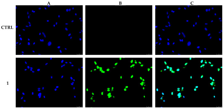
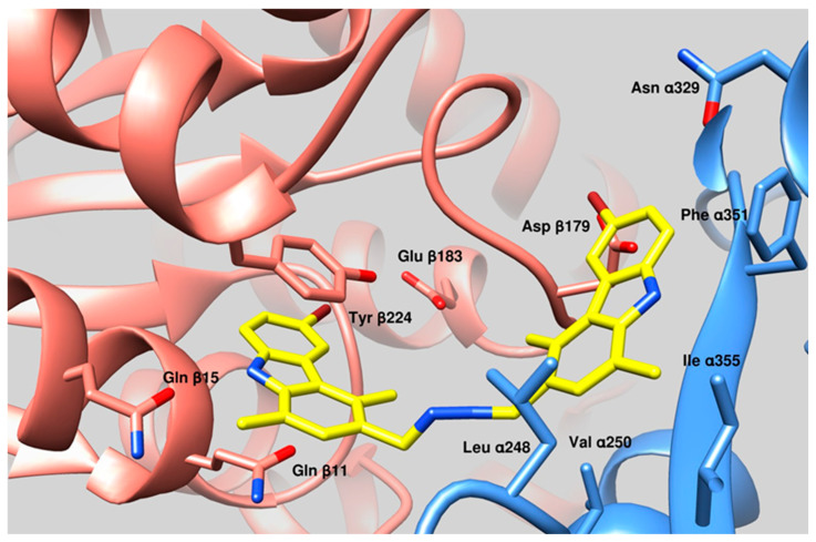
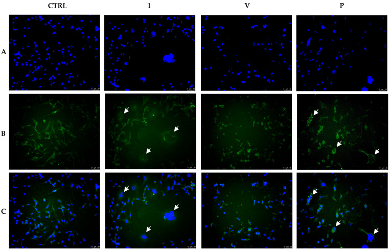
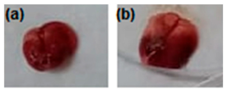
Background: Carbazoles represent one of the most important classes of nitrogen-based tricyclic aromatic heterocycles and are present in natural sources and chemically obtained drugs. Recently, several research groups disclosed their large biological and chemical applications in different fields, leading to an increased interest towards this class of molecules. Some of the obtained derivatives have been successfully employed in the clinical treatment of different tumor types, but the onset of heavy side effects impaired their efficacy and discouraged their use. Pursuing the aim of obtaining carbazoles with less negative features, a lot of chemically modified compounds have been produced and evaluated. Objectives/Methods: In this paper, we describe the in vitro and in vivo evaluation of a bis-carbazole derivative with strong anticancer properties against two breast cancer cell lines. Results: This compound has been found to impact the cell cytoskeleton dynamics, triggering the activation of some key proteins playing a role in the intrinsic and extrinsic apoptotic pathways. Equally important, this derivative has been found to be selective for cancer cells and has shown a safe profile in Balb/c-treated mice. Conclusions: Overall, the disclosed outcomes represent an important landmark for encouraging further studies directed toward the potentiation of this lead to be potentially exploited in both preclinical and clinical applications.

Keywords: anticancer; apoptosis; carbazole derivatives; cytoskeleton; toxicity studies.


Conflict of interest statement

The authors declare no conflicts of interest.

Figures

Figure 1
Figure 1
N,N’-bis-(6-bromo-1,4-dimethyl-9H-carbazol-3-ylmethylene)-hydrazine (1).
Figure 2
Figure 2
MDA-MB-231 cells were exposed to 0.3 µM of compound 1 or vehicle (CTRL) for 24 h. After processing, cells were observed, and images were taken at 20× magnification by means of an inverted fluorescence microscope. The green fluorescence, visible only for compound 1-treated cells, indicates nuclear DNA damage. Panels (A): DAPI (CTRL and 1) excitation/emission wavelength 350 nm/460 nm. Panels (B): CF™488A (CTRL and 1) excitation/emission wavelength 490 nm/515 nm. Panels (C): panels (A) and (B) overlay. Images are representative of three separate experiments.
Figure 3
Figure 3
Determination of caspase-3/7, -8, and -9 activity levels following the treatment of MDA-MB-231 cells with compound 1 (0.3 µM) for 24 h, reported as a percentage versus the DMSO-treated cells, used as a control (CTRL). Data represent the means ± SD of three different experiments. **** p < 0.0001, treated versus CTRL.
Figure 4
Figure 4
MDA-MB-231 cells were treated with 0.3 µM of 1 or vehicle (CTRL) for 24 h, then processed as described in the experimental section and observed at 40× magnification using an inverted fluorescence microscope. Compound 1 treatment induced Bid (green fluorescence, panel (B), 1) translocation into mitochondria (red fluorescence, panel (C), 1), as noticeable in the merge channel (panel (D), 1). Nucleus staining is also shown (panels (A)). Panels (A): DAPI, excitation/emission wavelength 350 nm/460 nm. Panels (B): Alexa Fluor® 488, excitation/emission wavelength 490 nm/515 nm. Panel (C): MitoTracker Deep Red FM probe, excitation/emission wavelength 644/665 nm. Panels (D): panels (A) and (B) overlay. Representative fields of three different experiments are shown.
Figure 5
Figure 5
Docking simulations suggested that 1 binds a region proximal to the Paclitaxel-binding site of tubulin. The most important amino acid residues involved in the interactions are evidenced. Cyan ribbons: Tubulin, α-subunit, Salmon ribbon Tubulin β-subunit. Yellow sticks, 1 binding mode. Oxygen atoms belonging to evidenced aminoacids are colored in red, while nitrogen in blue. Bromide moieties are evidenced in amaranth.
Figure 6
Figure 6
In vitro tubulin polymerization assay. The assembly of tubulin into microtubules was followed by measuring the turbidity (absorbance, A) at 350 nm for 3600 s at 37 °C. The polymerization curves, in the presence of 1 (1 μM) or not (CTRL, DMSO), are shown. Moreover, the graphic shows the curves obtained using two reference molecules, Vinblastine and Paclitaxel, (both at 10 μM concentration), used as tubulin-destabilizing and tubulin-stabilizing agents, respectively.
Figure 7
Figure 7
Immunostaining studies on MDA-MB-231 cells treated with compound 1 (0.3 µM), Vinblastine, Paclitaxel (both at 1 µM), or vehicle (CTRL, DMSO) for 24 h. In the control experiment (CTRL), cells exhibited a regular arrangement of cytoskeleton. Vinblastine and Paclitaxel, instead, dramatically impacted the tubulin network. Vinblastine produced tubulin crystal formation (panel (B), V), whereas Paclitaxel induced tubulin bundles and thickened fibers (panel (B), P). Compound 1 exposure produced a morphology similar to Paclitaxel treatment (white arrows). Panels (A): DAPI, excitation/emission wavelength 350 nm/460 nm. Panels (B): β-tubulin (Alexa Fluor® 488) excitation/emission wavelength 490 nm/515 nm. Panels (C): overlay. Images were taken at 20×, and representative fields were shown.
Figure 8
Figure 8
Actin immunofluorescence studies were conducted on MDA-MB-231 cells treated with 1 (0.3 µM) or vehicle (CTRL, DMSO) for 24 h. Cells were processed as detailed in the Section 2. Control cells (CTRL) showed a normal actin cytoskeleton organization, whereas under 1 exposure, the actin network appeared irregularly arranged and packed into the cell cytoplasm. Panels (A): nuclear DAPI staining (λex/λem = 350/460 nm); Panels (B): β-actin (Alexa Fluor® 568; λex/λem = 644/665 nm); Panels (C): overlay. Images were taken at 20× and show representative fields.
Figure 9
Figure 9
Body weight variation in mice after 1 week of treatment was ns (not significant), compared to the negative control by t-test; NC: negative control; 1: compound 1-treated group.
Figure 10
Figure 10
Representative photos of mice behaviors: (a) healthy and normal behaviors were registered for negative control (NC) and (b) the compound 1 group.
Figure 11
Figure 11
Representative photos for mice NC (a) and the compound 1-treated group (b); healthy global appearance; no visible difference or abnormalities at the injection site.
Figure 12
Figure 12
Effect of compound 1 on lung morphology: (a) negative control with normal lung morphology; (b) compound 1-treated group showed normal morphologic features of lung, while visible weight gain for the organ was detected.
Figure 13
Figure 13
Biochemical analyses of the serum from groups of mice untreated or treated with compound 1; (a) aspartate aminotransferase (AST), alanine aminotransferase (ALT), and alkaline phosphatase (ALP); (b) creatinine and uric acid; and (c) sodium, potassium, chloride, calcium, and phosphorus serum levels were measured in the two groups of mice (control and compound 1-treated) and after 1 week.
Figure 14
Figure 14
Hematological analysis of mice blood from groups of mice untreated and treated with compound 1; RBC: red blood cells; HB: hemoglobin; HCT: hematocrit; MCH: mean corpuscular hemoglobin; MCHC: mean corpuscular hemoglobin concentration; PLT: platelet count; MCV: mean corpuscular volume; WBC: white blood cells; LYM: lymphocytes; NEU: neutrophils; EOS: eosinophils; MAC: macrophages; BASO: basophiles.

Similar articles

References

    1. Huang L., Feng Z.-L., Wang Y.-T., Lin L.-G. Anticancer carbazole alkaloids and coumarins from Clausena plants: A review. Chin. J. Nat. Med. 2017;15:881–888. doi: 10.1016/s1875-5364(18)30003-7. - DOI - PubMed
    1. Issa S., Prandina A., Bedel N., Rongved P., Yous S., Le Borgne M., Bouaziz Z. Carbazole scaffolds in cancer therapy: A review from 2012 to 2018. J. Enzym. Inhib. Med. Chem. 2019;34:1321–1346. doi: 10.1080/14756366.2019.1640692. - DOI - PMC - PubMed
    1. Tiwari A., Mishra B. Diverse pharmacological actions of potential carbazole derivatives by influencing various pathways of molecular signaling. Future J. Pharm. Sci. 2024;10:77. doi: 10.1186/s43094-024-00650-0. - DOI
    1. Song F., Liu D., Huo X., Qiu D. The anticancer activity of carbazole alkaloids. Arch. Pharm. 2021;355:2100277. doi: 10.1002/ardp.202100277. - DOI - PubMed
    1. Ceramella J., Iacopetta D., Caruso A., Mariconda A., Petrou A., Geronikaki A., Rosano C., Saturnino C., Catalano A., Longo P., et al. 5,8-Dimethyl-9H-carbazole Derivatives Blocking hTopo I Activity and Actin Dynamics. Pharmaceuticals. 2023;16:353. doi: 10.3390/ph16030353. - DOI - PMC - PubMed

LinkOut - more resources