. 2025 Mar 27;17(4):429.
doi: 10.3390/pharmaceutics17040429.

Utilizing Nanoparticles of Hesperidin Loaded on Layered Double Hydroxide to Reduce Hepatotoxicity Caused by Paracetamol in Rats: Controlling of Biotransformation, Oxidative Stress, Inflammation, and Apoptosis

Affiliations

Utilizing Nanoparticles of Hesperidin Loaded on Layered Double Hydroxide to Reduce Hepatotoxicity Caused by Paracetamol in Rats: Controlling of Biotransformation, Oxidative Stress, Inflammation, and Apoptosis

Deyaa A Shaban et al. Pharmaceutics. .

Abstract

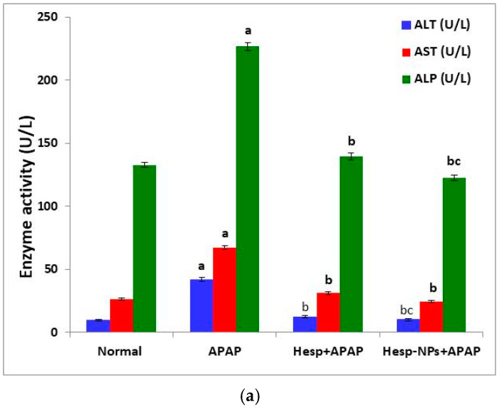
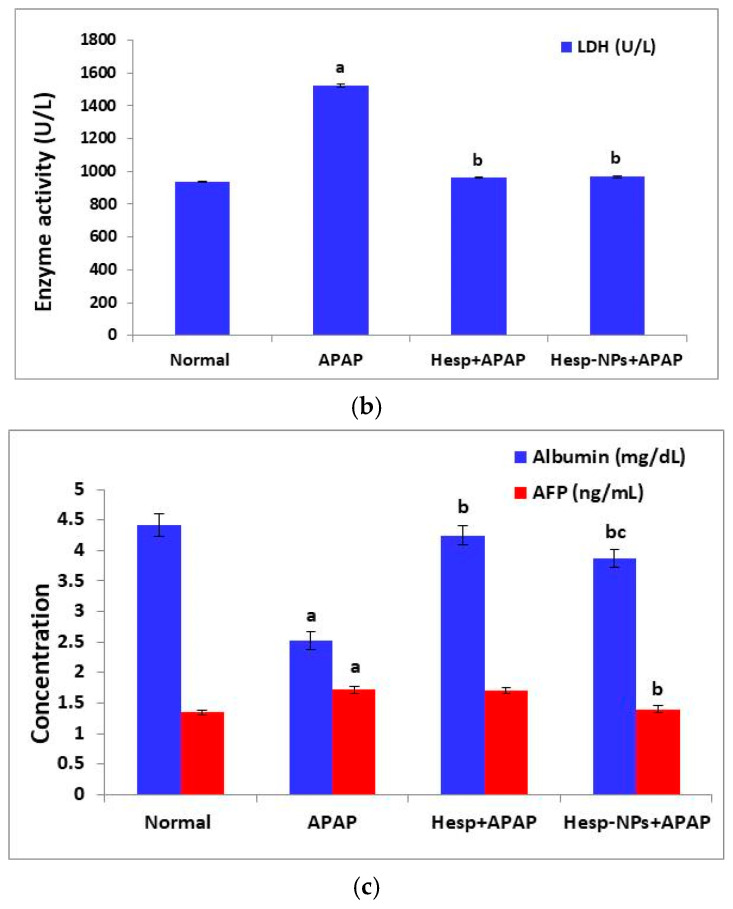
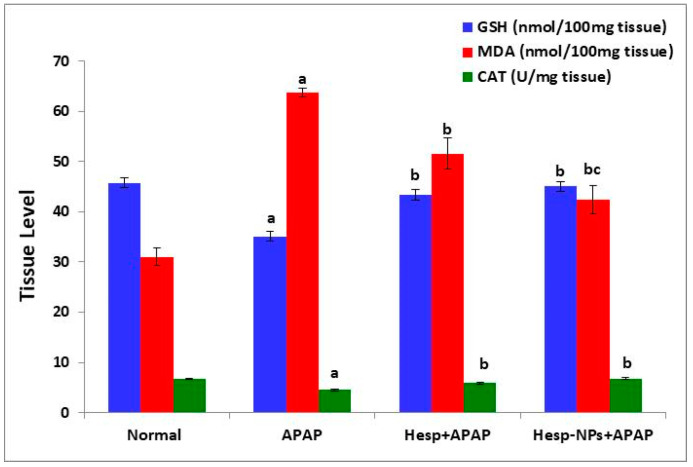
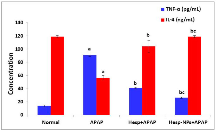
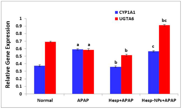
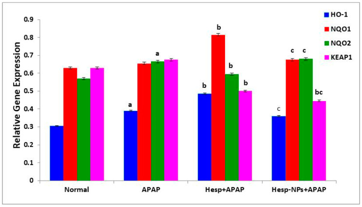
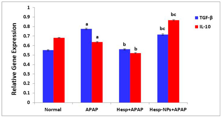
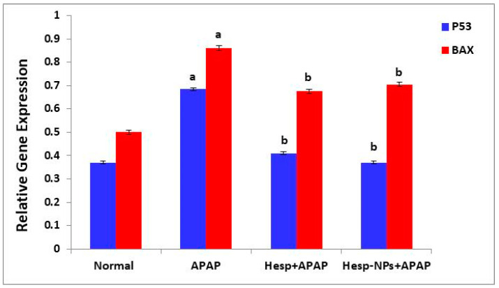
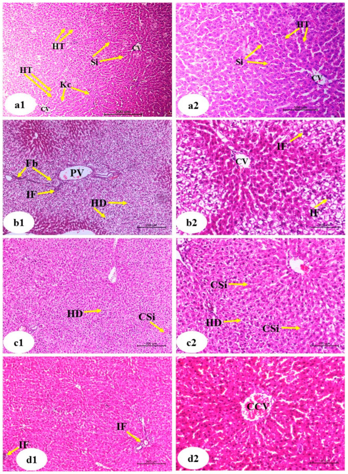
Background/Objectives: The most used antipyretic and pain relief treatment is paracetamol (acetaminophen), also known as N-acetyl-para-aminophenol (APAP). However, it is considered potentially hazardous if consumed repeatedly in large doses or over prolonged periods. This investigation explores the effectiveness of hesperidin (Hesp) and Hesp loaded on layered double hydroxide nanoparticles (Hesp-NPs) in inhibiting the progression of acute hepatotoxicity in rats induced by APAP. Methods: LDH-Hesp-NPs were prepared and characterized. Male Wistar rats were orally treated with Hesp and Hesp-NPs at the same adjusted dose (100 mg/kg) every other day for six weeks. After 2 h of the first doses of Hesp and Hesp-NPs, the rats received one oral dose of APAP (750 mg/kg). Results: Administering of Hesp and Hesp-NPs to APAP-treated rats significantly reduced oxidant parameter (malondialdehyde) and serum enzymes (ALT, AST, LDH, and ALP) associated with liver function. Antioxidant markers in the liver, such as catalase and glutathione, also increased notably. Moreover, Hesp and Hesp-NPs enhanced the mRNA expression of liver UGT1A6, IL-10, and HO-1. Conversely, the mRNA expressions of liver CYP1A1, KEAP1, TGF-β, P53, and BAX decreased. These improvements in biochemical and molecular markers were corroborated by liver histopathology. Conclusions: Hesp and Hesp-NPs protect significantly against APAP-induced hepatotoxicity in male Wistar rats. Hesp-NPs treatment was more potent. The protective effects may be mediated via modulation of APAP biotransformation, oxidative stress, inflammation and apoptosis.

Keywords: hepatotoxicity; hesperidin nanoparticles; layered double hydroxide; paracetamol.


Conflict of interest statement

The authors declare no conflict of interest.

Figures

Scheme 1
Scheme 1
Chemical structure of Hesp (A) and APAP (B).
Figure 1
Figure 1
MgAl LDH, MgAl LDH–hesperidin, and hesperidin powder FTIR spectroscopy.
Figure 2
Figure 2
HRTEM micrographs of (a) MgAl LDH (b) MgAl LDH–hesperidin nanopowder.
Figure 3
Figure 3
Measurements of (a) zeta sizer and (b) zeta potential MgAl LDH–hesperidin nanopowder.
Figure 4
Figure 4
Profile of in vitro release of hesperidin from MgAl LDH–hesperidin in PBS at 37 °C. Error bars represent the standard deviation from three replicates. The difference in release was significant (p < 0.05) after 2 h.
Figure 5
Figure 5
Effect of Hesp and Hesp-NPs on serum parameters measuring liver function, including ALT, AST, and ALP (a), LDH (b), albumin, and AFP (c) in rats given APAP. Data are presented as the mean ± SD. a,b and c indicate a significant change from control, APAP, and Hesp+APAP, respectively, at p < 0.05.
Figure 5
Figure 5
Effect of Hesp and Hesp-NPs on serum parameters measuring liver function, including ALT, AST, and ALP (a), LDH (b), albumin, and AFP (c) in rats given APAP. Data are presented as the mean ± SD. a,b and c indicate a significant change from control, APAP, and Hesp+APAP, respectively, at p < 0.05.
Figure 6
Figure 6
Effect of Hesp and Hesp-NPs on liver GSH, MDA, and CAT levels in rats given APAP. Data are presented as the mean ± SD. a,b and c indicate a significant change from control, APAP, and Hesp+APAP, respectively, at p < 0.05.
Figure 7
Figure 7
The effect of Hesp and Hesp-NPs on serum TNF-α and IL-4 levels in APAP-administered rats. Data are presented as the mean ± SD. a,b and c indicate a significant change from control, APAP, and Hesp+APAP, respectively, at p < 0.05. TNF-α: tumor necrosis factor-α.
Figure 8
Figure 8
Effect of Hesp and Hesp-NPs on mRNA expression of CYP1A1 and UGTA6 in liver of APAP-administered rats. Data are presented as the mean ± SD. a,b and c indicate a significant change from control, APAP, and Hesp+APAP, respectively, at p < 0.05.
Figure 9
Figure 9
Effect of Hesp and Hesp-NPs on mRNA expression of HO-1, NQO1, NQO2, and KEAP1 in liver of APAP-administered rats. Data are presented as the mean ± SD. a,b and c indicate a significant change from control, APAP, and Hesp+APAP, respectively, at p < 0.05 using ANOVA, followed by LSD test.
Figure 10
Figure 10
Effect of Hesp and Hesp-NPs on mRNA expression of TGF-β and IL-10. Data are presented as the mean ± SD. a,b and c indicate a significant change from control, APAP, and Hesp+APAP, respectively, at p < 0.05 using ANOVA followed by LSD test.
Figure 11
Figure 11
Effect of Hesp and Hesp-NPs on mRNA expression of markers related to apoptosis (P53 and BAX) in liver of APAP-treated rats. Data are presented as the mean ± SD. a,b indicate a significant change from control, APAP, and Hesp+APAP, respectively, at p < 0.05.
Figure 12
Figure 12
Photomicrographs of liver sections of normal ((a1), low magnification ×100; (a2), high magnification of a1 ×200), APAP ((b1), low magnification ×100; (b2), high magnification ×200), Hesp+APAP ((c1), low magnification ×100; (c2), high magnification ×200) and Hesp-NPs+APAP ((d1), low magnification ×100; (d2), high magnification ×200) groups. CV, central vein; HT, hepatic trabeculae; Si, sinusoids; HP, hepatic portal vein; HD, hydropic degeneration; CSi, congested sinusoids; Kc, Kupffer cells; Fb, fibroblast; IF, inflammatory infiltration; CCV, congested central vein.

Similar articles

References

    1. Mahmood N., Mamat S., Kamisan F., Yahya F., Kamarolzaman M., Nasir N., Mohtarrudin N., Tohid S., Zakaria Z. Amelioration of paracetamol-induced hepatotoxicity in rat by the administration of methanol extract of Muntingia calabura L. Leaves. BioMed Res. Int. 2014;2014:695678. - PMC - PubMed
    1. Halim A., Nur N.M., El-Agamy E.-S., Ibrahim A. Protective effect of hesperidin (HDN) on carbon tetrachloride (CCl4)-induced hepatic toxicity in male albino rats. AIJCR. 2017;8:20328–20338.
    1. Dart R.C., Bailey E. Does therapeutic use of acetaminophen cause acute liver failure? Pharmacotherapy. J. Hum. Pharmacol. Drug Ther. 2007;27:1219–1230. - PubMed
    1. Dar A., Saxena R., Bansal S. Hepatoprotection: A Hallmark of Citrullus colocynthis L. against Paracetamol Induced Hepatotoxicity in Swiss Albino Rats. Am. J. Plant Sci. 2012;3:1022–1027.
    1. Ahmed O.M. Natural Flavonoids: Chemistry, Therapeutic Potentials, Therapeutic Targets and Mechanisms of Actions. Curr. Pharm. Des. 2021;27:455. - PubMed

LinkOut - more resources