. 2025 Mar 28;17(4):434.
doi: 10.3390/pharmaceutics17040434.

Verteporfin Inhibits Severe Fever with Thrombocytopenia Syndrome Virus Infection via Inducing the Degradation of the Viral Gn Protein

Affiliations

Verteporfin Inhibits Severe Fever with Thrombocytopenia Syndrome Virus Infection via Inducing the Degradation of the Viral Gn Protein

Bingan Wu et al. Pharmaceutics. .

Abstract

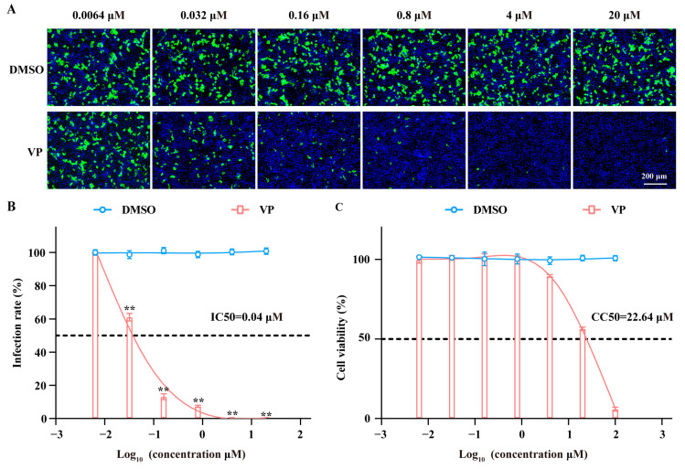
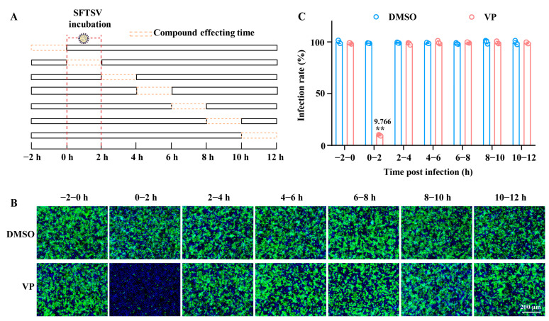
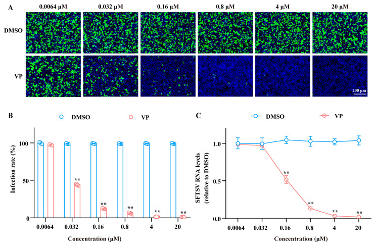
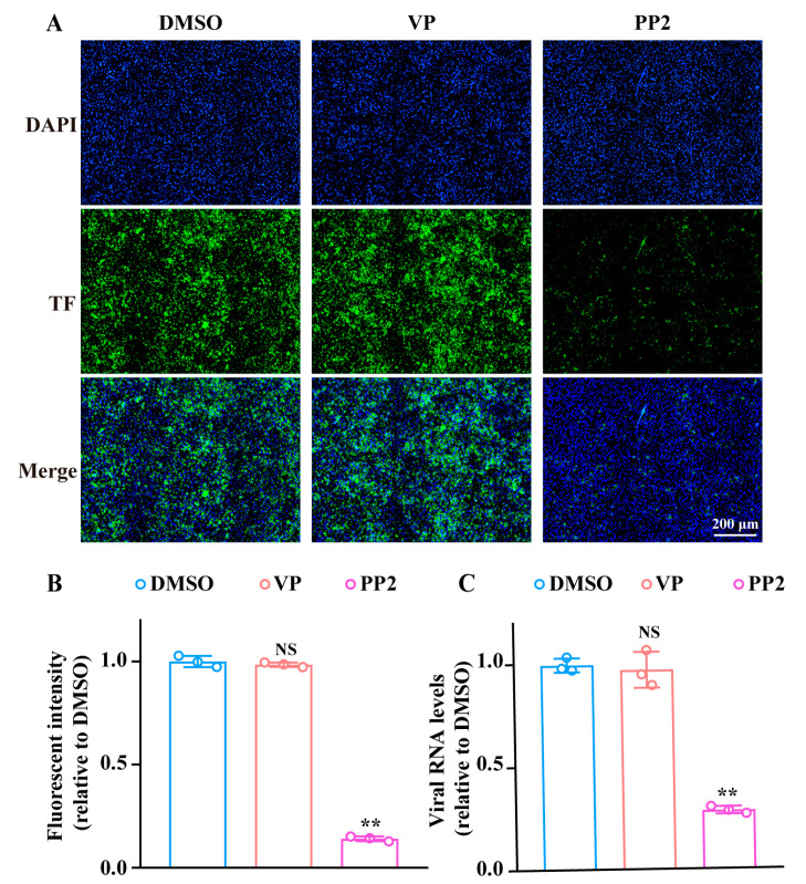
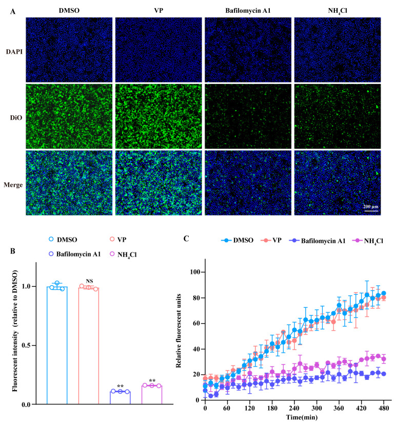
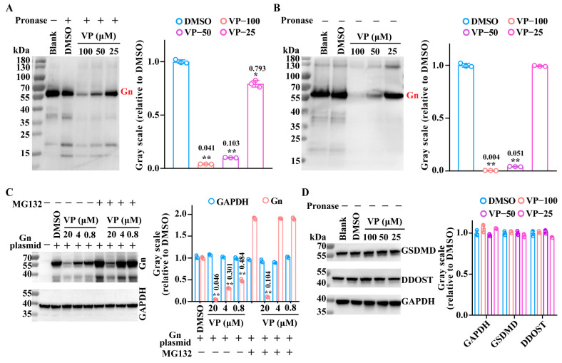
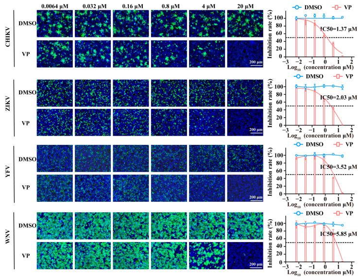
Background: Severe fever with thrombocytopenia syndrome virus (SFTSV) is a novel tick-borne bunyavirus, causing the hemorrhagic infectious disease of SFTS, with a case fatality rate up to 30% due to the absence of effective therapeutic interventions. Therefore, it is urgent to develop safe and effective therapeutic drugs to control this viral hemorrhagic fever. Methods: The activity of verteporfin (VP), screened from an FDA-approved drugs library, against SFTSV, was systematically evaluated in Huh7 cells in a wide range of concentrations. We performed time-of-addition experiments with VP, along with binding, endocytosis, and membrane fusion assays, to determine which part of the SFTSV life cycle VP has its effect on. The potential targets of VP were detected by a drug affinity responsive target stability (DARTS) assay. Results: VP exhibited a potent anti-SFTSV activity by blocking the initial viral binding to the target cells during viral entry via significantly inducing the degradation of the viral Gn protein. Conclusions: The VP-induced inhibition of SFTSV binding, the first step of viral invasion, suggested that VP might be an ideal and potent anti-SFTSV agent due to its prophylaxis and therapeutic effects on viral infection.

Keywords: Gn protein; antiviral agents; severe fever with thrombocytopenia syndrome virus; verteporfin; viral binding.


Conflict of interest statement

The authors declare no conflicts of interest.

Figures

Figure 1
Figure 1
VP displays dose-dependent anti-SFTSV activity and low cytotoxicity. (A,B) Huh7 cells were incubated with SFTSV (MOI = 1) in the presence of VP or DMSO at 37 °C for 24 h. The inhibition rate of VP was analyzed via an IF assay. The SFTSV was stained with the primary anti-SFTSV NP mAb and goat anti-mouse Alexa Fluor™ 488 secondary antibody (green). Nuclei were stained with DAPI (blue). (C) Huh7 cells were incubated with the indicated concentrations of VP at 37 °C for 24 h, and the cytotoxicity was detected by a CCK-8 assay. Data were shown as means with a SD of three independent experiments. Scale bar, 200 μm. ** p < 0.01 compared to the control group.
Figure 2
Figure 2
VP acts during the 0–2 h time window of the SFTSV infection. (A) A schematic diagram of the time window of the compound-affecting assay. (B,C) Huh7 cells were incubated with SFTSV (MOI = 1) at 37 °C for 2 h and treated with VP (4 μM) during −2–0, 0–2, 2–4, 4–6, 6–8, 8–10, and 10–12 h, respectively. The cells were detected by an IF assay 12 h after the virus inoculation. The SFTSV was stained with the primary anti-SFTSV NP mAb and goat anti-mouse Alexa Fluor™ 488 secondary antibody (green). Nuclei were stained with DAPI (blue). Data were shown as means with a SD of three independent experiments. Scale bar, 200 μm. ** p < 0.01 compared to the control group.
Figure 3
Figure 3
VP decreases SFTSV initial binding. Huh7 cells were co-incubated with SFTSV (MOI = 5) and the indicated concentrations (0.0064, 0.032, 0.16, 0.8, 4, and 20 μM) of VP or DMSO on ice for 1.5 h. A fraction of the infected and VP-treated cells were cultured at 37 °C for 24 h, with the SFTSV infection rate detected by IF. The SFTSV was stained with the primary anti-SFTSV NP mAb and goat anti-mouse Alexa Fluor™ 488 secondary antibody (green). Nuclei were stained with DAPI (blue) (A,B). The remaining cells were lysed and RT-qPCR was used to determine the amount of the bound virus (C). Data were shown as means with a SD of three independent experiments. Scale bar, 200 μm. ** p < 0.01 compared to the control group.
Figure 4
Figure 4
VP does not affect SFTSV endocytosis. (A,B) Huh7 cells were incubated with Alexa Fluor™ 488-labelled TF in the presence of VP (4 μM), PP2 (20 μM), or DMSO for 4 h. The fluorescent intensity of TF (green) was then examined by fluorescence microscopy. Nuclei were stained with DAPI (blue). (C) Huh7 cells were incubated with SFTSV on ice for 1.5 h. Then, the cellular supernatant was replaced with a fresh culture medium with VP (4 μM), PP2 (20 μM), or DMSO and the cells were incubated at 37 °C for 1 h. The intracellular SFTSV RNA levels were analyzed via RT-qPCR after being treated by proteinase K to remove the uninternalized SFTSV. Scale bar, 200 μm. ** p < 0.01 compared to the DMSO group. NS, no significance compared to the DMSO group.
Figure 5
Figure 5
VP does not affect SFTSV membrane fusion. (A,B) Huh7 cells were inoculated with DiO-labelled SFTSV (SFTSVDiO, MOI = 1) in the presence of VP (4 μM), bafilomycin A1 (20 nM), NH4Cl (20 mM), or DMSO at 37 °C for 2 h. Then, the fluorescent intensity of DiO (green) was detected by the BioTek Lionheart FX Imaging Reader. Nuclei were stained with DAPI (blue). (C) Huh7 cells were incubated with SFTSVDiO (MOI = 1) in the presence of VP (4 μM), bafilomycin A1 (20 nM), NH4Cl (20 mM), or DMSO. Then, the fluorescent units were collected every 15 min for a total of 33 cycles. Data were shown as means with a SD of three independent experiments. Scale bar, 200 μm. ** p < 0.01 compared to the DMSO group. NS, no significance compared to the DMSO group.
Figure 6
Figure 6
Degradation of the viral Gn protein by VP. (A) The Gn protein was pre-incubated with VP at the indicated concentrations (100 μM, 50 μM, and 25 μM) or DMSO for 1 h and subsequently digested using pronase for 30 min at room temperature. The influence of VP on Gn was analyzed by a Western blotting assay. (B) The Gn protein was incubated with VP at the indicated concentrations (100 μM, 50 μM, and 25 μM) or DMSO for 1.5 h, and the Gn was detected by a Western blotting assay. (C) The HeLa cells transfected with the Gn plasmid were treated with VP (20 μM, 4 μM, and 0.8 μM) and the proteasome inhibitor MG132 (10 μM) for 2 h. Then, the degradation effect was detected by a Western blotting assay. (D) The DDOST and GSDMD in cell lysates were incubated with the indicated concentrations of VP (100 μM, 50 μM, and 25 μM) for 1.5 h, and the influence of VP on DDOST and GSDMD was analyzed by a Western blotting assay. Data were shown as means with a SD of three independent experiments. ** p < 0.01 compared to the DMSO group. * p < 0.05 compared to the DMSO group.
Figure 7
Figure 7
The broad-spectrum antiviral activity of VP. Huh7 cells were infected with CHIKV, ZIKV, YFV, and WNV at an MOI of 1 in the presence of VP or DMSO at the indicated concentrations. Then, the inhibitory effect was quantified by an IF assay. The SFTSV was stained with the primary anti-SFTSV NP mAb and goat anti-mouse Alexa Fluor™ 488 secondary antibody (green). Nuclei were stained with DAPI (blue). Data were shown as means with a SD of three independent experiments. Scale bar, 200 μm.

Similar articles

References

    1. Li H., Zhang L.K., Li S.F., Zhang S.F., Wan W.W., Zhang Y.L., Xin Q.L., Dai K., Hu Y.Y., Wang Z.B., et al. Calcium channel blockers reduce severe fever with thrombocytopenia syndrome virus (SFTSV) related fatality. Cell Res. 2019;29:739–753. doi: 10.1038/s41422-019-0214-z. - DOI - PMC - PubMed
    1. Zhang W.K., Yan J.M., Chu M., Li B., Gu X.L., Jiang Z.Z., Li Z.M., Liu P.P., Yu T.M., Zhou C.M., et al. Bunyavirus SFTSV nucleoprotein exploits TUFM-mediated mitophagy to impair antiviral innate immunity. Autophagy. 2025;21:102–119. doi: 10.1080/15548627.2024.2393067. - DOI - PMC - PubMed
    1. Yu X.J., Liang M.F., Zhang S.Y., Liu Y., Li J.D., Sun Y.L., Zhang L., Zhang Q.F., Popov V.L., Li C., et al. Fever with thrombocytopenia associated with a novel bunyavirus in China. N. Engl. J. Med. 2011;364:1523–1532. doi: 10.1056/NEJMoa1010095. - DOI - PMC - PubMed
    1. Choi Y., Jiang Z., Shin W.J., Jung J.U. Severe Fever with Thrombocytopenia Syndrome Virus NSs Interacts with TRIM21 To Activate the p62-Keap1-Nrf2 Pathway. J. Virol. 2020;94:e01684-19. doi: 10.1128/JVI.01684-19. - DOI - PMC - PubMed
    1. Fang X., Hu J., Peng Z., Dai Q., Liu W., Liang S., Li Z., Zhang N., Bao C. Epidemiological and clinical characteristics of severe fever with thrombocytopenia syndrome bunyavirus human-to-human transmission. PLoS Negl. Trop. Dis. 2021;15:e0009037. doi: 10.1371/journal.pntd.0009037. - DOI - PMC - PubMed

Grants and funding

LinkOut - more resources