. 2025 Apr 1;17(4):453.
doi: 10.3390/pharmaceutics17040453.

Umbilical Cord Mesenchymal Stem Cell-Derived Apoptotic Extracellular Vesicles Improve 5-FU-Induced Delayed Wound Healing by Mitochondrial Transfer

Affiliations

Umbilical Cord Mesenchymal Stem Cell-Derived Apoptotic Extracellular Vesicles Improve 5-FU-Induced Delayed Wound Healing by Mitochondrial Transfer

Hongbin Lai et al. Pharmaceutics. .

Abstract

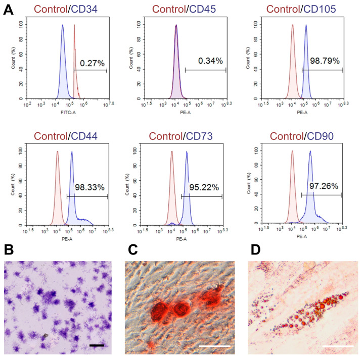
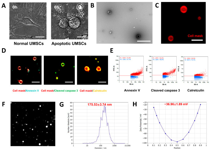
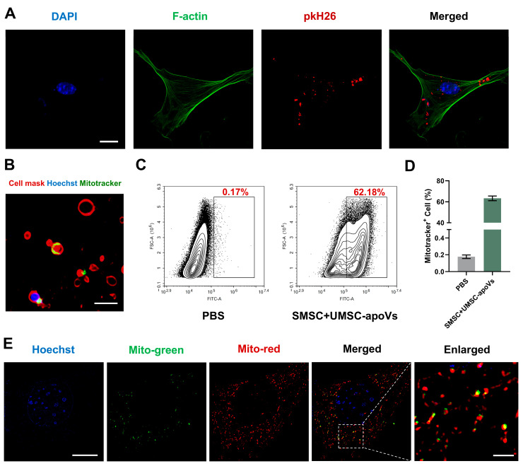
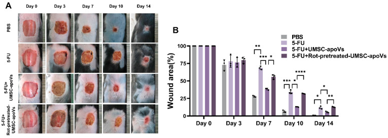
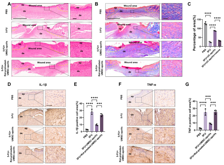
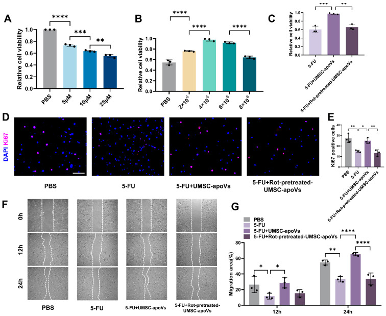
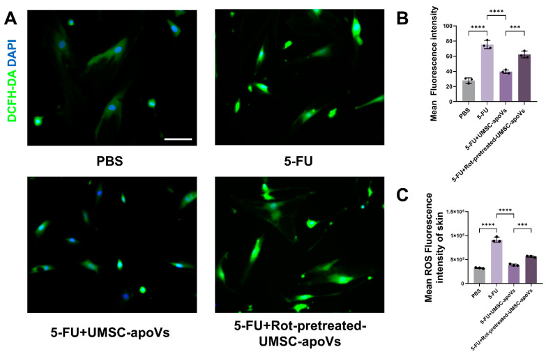
Background/Objectives: This study aimed to explore the therapeutic potential of umbilical mesenchymal stem cell-derived apoptotic vesicles (UMSC-apoVs) in a 5-Fluorouracil (5-FU)-induced impairment in skin wound healing. Methods: UMSC-apoVs were isolated from UMSCs using differential centrifugation after the induction of apoptosis. A murine model was established by administering 5-FU via intravenous tail injection, followed by full-thickness skin wound creation. Mice received local injections of UMSC-apoVs at the lesion site. Wound healing was evaluated based on wound closure rates, histological analysis, and in vivo/in vitro functional assays. Rotenone (Rot)-pretreated UMSC-apoVs were used to explore the role of mitochondrial transfer between skin mesenchymal stem cells (SMSCs) and UMSC-apoVs in wound healing. Results: UMSC-apoVs significantly accelerated wound healing in 5-FU-treated mice, as demonstrated by enhanced wound closure rates and histological findings of reduced inflammatory infiltration and increased collagen deposition. UMSC-apoVs transferred mitochondria to SMSCs, enhancing viability, proliferation, and migration while reducing reactive oxygen species (ROS) production in SMSCs. Rot pretreatment inhibited the therapeutic effects of UMSC-apoVs on wound healing by inducing mitochondrial dysfunction in UMSC-apoVs. Conclusions: Our findings indicate that UMSC-apoVs improve 5-FU-induced impaired skin wound healing by facilitating mitochondrial transfer, suggesting a novel therapeutic strategy for alleviating chemotherapy-induced impairment in wound healing.

Keywords: 5-fluorouracil; delayed wound healing; mitochondrial transfer; skin mesenchymal stem cells; umbilical cord mesenchymal stem cell-derived apoptotic vesicles.


Conflict of interest statement

The authors declare no conflicts of interest.

Figures

Figure 1
Figure 1
Characterization of UMSCs. (A) Flow cytometry analysis of UMSCs showing the expression of key surface markers, including CD34, CD45, CD44, CD73, CD90, and CD105; (B) CFU assay demonstrating the clonogenic potential of UMSCs (scale bar: 2000 μm); (C) Alizarin Red S staining indicating the osteogenic differentiation potential of UMSCs (red deposits indicate calcium accumulation, scale bar: 100 μm); (D) Oil Red O staining illustrating the adipogenic differentiation potential of UMSCs with lipid droplet accumulation (red staining marks intracellular lipid droplets, scale bar: 50 μm).
Figure 2
Figure 2
Characterization of UMSC-apoVs. (A) High-content imaging (40× magnification) showing the morphological changes in UMSCs at 0 and 6 h post-apoptosis induction; (B) TEM image of UMSC-apoVs (scale bar: 1 μm); (C) SIM image of PKH26-labeled apoVs (100× magnification; scale bar: 2 μm); (D) immunofluorescence staining of UMSC-apoVs markers (Annexin V, cleaved caspase 3, and calreticulin) (100× magnification; Scale bar: 2 μm); (E) nanoflow cytometry analysis showing the proportions of apoptotic markers (Annexin V, cleaved caspase 3, and calreticulin) on apoptotic vesicles. The red horizontal lines indicate gating thresholds used to distinguish marker-positive populations (P1, red) from marker-negative populations (P2, blue); (F) NTA image of apoptotic vesicles. The image was acquired using the ZetaView at a 10× objective magnification; (G) NTA analysis showing the mean size of UMSC-apoVs; (H) NTA analysis showing the mean zeta potential of apoptotic vesicles.
Figure 3
Figure 3
Mitochondrial transfer from UMSC-apoVs to SMSCs. (A) SIM image showing SMSCs internalizing UMSC-apoVs, labeled with PKH26 (100× magnification; scale bar: 20 μm); (B) immunofluorescent staining images showing mitochondrial staining in UMSC-apoVs (MitoTracker Green) (100× magnification; scale bar: 2 μm); (C,D) quantitative analysis by flow cytometry showing mitochondrial transfer from UMSC-apoVs to SMSCs; (E) co-culture of SMSCs with UMSC-apoVs, showing colocalization of mitochondria in both SMSCs and UMSC-apoVs as observed under Elyra 7 Lattice SIM (100× magnification; scale bar: 10 μm). Hoechst (blue) labels nuclei, MitoTracker Green (green) labels mitochondria in SMSCs, and MitoTracker Red (red) labels mitochondria in UMSC-apoVs. The dashed box in the merged image indicates the area shown at higher magnification (scale bar: 2 μm).
Figure 4
Figure 4
UMSC-apoVs transfer mitochondria to enhance wound healing of 5-FU skin wound model. (A) Representative images of dorsal wounds in different treatment groups on days 1, 3, 7, 10, and 14 post-wounding. (B) Quantification of wound area percentage at different time points. Data are presented as mean ± SEM (n = 3 per group). Statistical significance was determined by one-way ANOVA with Tukey’s post hoc test. * p < 0.05, ** p < 0.01, *** p < 0.001, **** p < 0.0001.
Figure 5
Figure 5
Histological analysis of 5-FU-induced delayed wound healing. (A) Hematoxylin and Eosin (H&E) staining of wound tissues on day 14, showing tissue architecture and inflammatory response across PBS, 5-FU, 5-FU + UMSC-apoVs, and 5-FU + Rot-pretreated-UMSC-apoVs groups. Yellow dashed lines delineate the wound margin. ep: epithelium, de: dermis (scale bar: 700 μm). Higher-magnification (scale bar: 50 μm). (B) Masson’s trichrome staining highlighting collagen deposition and dermal remodeling in the same groups. Yellow dashed lines indicate wound edges (scale bar: 700 μm). Higher-magnification insets (scale bar: 50 μm) illustrate cellular and extracellular matrix details. (C) Quantification of collagen deposition in the wound area based on Masson’s trichrome staining. (D) Immunohistochemical staining for IL-1β in wound tissues (scale bar: 200 μm). Higher-magnification insets (scale bar: 100 μm). (E) Quantification of IL-1β-positive staining area. (F) Immunohistochemical staining for TNF-α in wound tissues (scale bar: 200 μm). Higher-magnification insets (scale bar: 100 μm). (G) Quantification of TNF-α-positive staining area. Data are presented as mean ± SD (n = 3). *** p < 0.001, **** p < 0.0001, one-way ANOVA with Tukey’s post hoc test.
Figure 6
Figure 6
UMSC-apoVs promote the viability, proliferation, and migration of 5-FU-treated SMSCs by mitochondrial transfer. (A) CCK-8 assay showing SMSCs viability after treatment with 5-FU (0, 5, 10, 25 μM). (B) CCK-8 assay identifying the optimal UMSC-apoVs dose (0, 2 × 10⁶, 4 × 10⁶, 6 × 10⁶, 8 × 10⁶ particles/mL) after 24 h of co-incubation with 5-FU-pretreated SMSCs. (C) CCK-8 assay confirming mitochondrial involvement in UMSC-apoV-mediated protection against 5-FU toxicity, with Rot pretreatment (25 μM, 2 h). (D) Ki67 immunofluorescence staining of SMSCs across PBS, 5-FU, 5-FU + UMSC-apoVs, and 5-FU + Rot-pretreatment-UMSC-apoVs groups (Scale bar: 200 μm). (E) Quantification of Ki67-positive cells. (F) Scratch-wound assay showing SMSCs’ migration in different groups (scale bar: 400 μm). (G) Quantification of migration area at 12 and 24 h. Data are presented as mean ± SEM (n = 3 per group). Statistical significance was determined by one-way or two-way ANOVA with Tukey’s post hoc test. * p < 0.05, ** p < 0.01, *** p < 0.001, **** p < 0.0001.
Figure 7
Figure 7
UMSC-apoVs reduce 5-FU-induced oxidative stress in SMSCs and wounded skin via mitochondrial transfer. (A) Representative immunofluorescence images of SMSCs stained with DCFH-DA, showing ROS levels across PBS, 5-FU, 5-FU + UMSC-apoVs, and 5-FU +Rot-pretreated-UMSC-apoVs (scale bar: 100 μm). (B) Quantification of intracellular ROS fluorescence intensity in SMSCs. (C) Flow cytometry analysis of ROS levels in skin cells from wounded tissues in the same groups. Data are presented as mean ± SEM (n = 3 per group). Statistical significance was determined by one-way ANOVA with Tukey’s post hoc test. *** p < 0.001, **** p < 0.0001.

Similar articles

References

    1. Eming S.A., Martin P., Tomic-Canic M. Wound repair and regeneration: Mechanisms, signaling, and translation. Sci. Transl. Med. 2014;6:265sr266. doi: 10.1126/scitranslmed.3009337. - DOI - PMC - PubMed
    1. Yuan W., Ji G., Shi X., Sun Z., Liu C., Yu Y., Li W., Wang X., Hu H. The male reproductive toxicity after 5-Fluorouracil exposure: DNA damage, oxidative stress, and mitochondrial dysfunction in vitro and in vivo. Ecotoxicol. Env. Saf. 2024;278:116465. doi: 10.1016/j.ecoenv.2024.116465. - DOI - PubMed
    1. Sung H., Ferlay J., Siegel R.L., Laversanne M., Soerjomataram I., Jemal A., Bray F. Global Cancer Statistics 2020: GLOBOCAN Estimates of Incidence and Mortality Worldwide for 36 Cancers in 185 Countries. CA Cancer J. Clin. 2021;71:209–249. doi: 10.3322/caac.21660. - DOI - PubMed
    1. Zhao J., Cui H., Qu M., Xu Z., Zhang Y., Ma C. Impact of preoperative chemotherapy on cutaneous wound healing in lung cancer patients: A meta-analysis. Int. Wound J. 2024;21:e14518. doi: 10.1111/iwj.14518. - DOI - PMC - PubMed
    1. Słonimska P., Sachadyn P., Zieliński J., Skrzypski M., Pikuła M. Chemotherapy-Mediated Complications of Wound Healing: An Understudied Side Effect. Adv. Wound Care. 2024;13:187–199. doi: 10.1089/wound.2023.0097. - DOI - PMC - PubMed

LinkOut - more resources