. 2025 Apr 2;17(4):463.
doi: 10.3390/pharmaceutics17040463.

The Incorporation of CBD into Biodegradable DL-Lactide/Glycolide Copolymers Creates a Persistent Antibacterial Environment: An In Vitro Study on Streptococcus mutans and Staphylococcus aureus

Affiliations

The Incorporation of CBD into Biodegradable DL-Lactide/Glycolide Copolymers Creates a Persistent Antibacterial Environment: An In Vitro Study on Streptococcus mutans and Staphylococcus aureus

Ronit Vogt Sionov et al. Pharmaceutics. .

Abstract

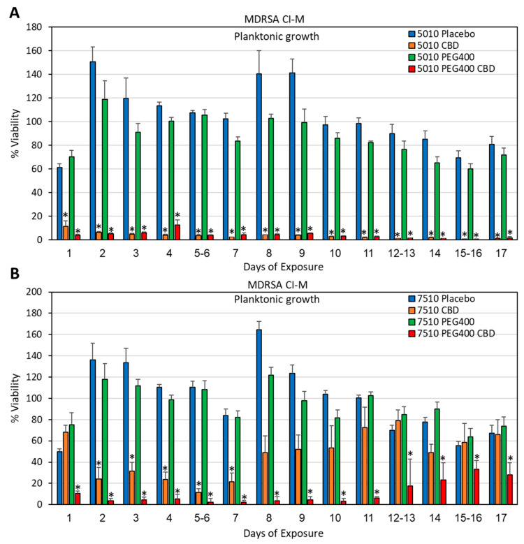
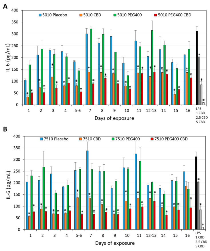
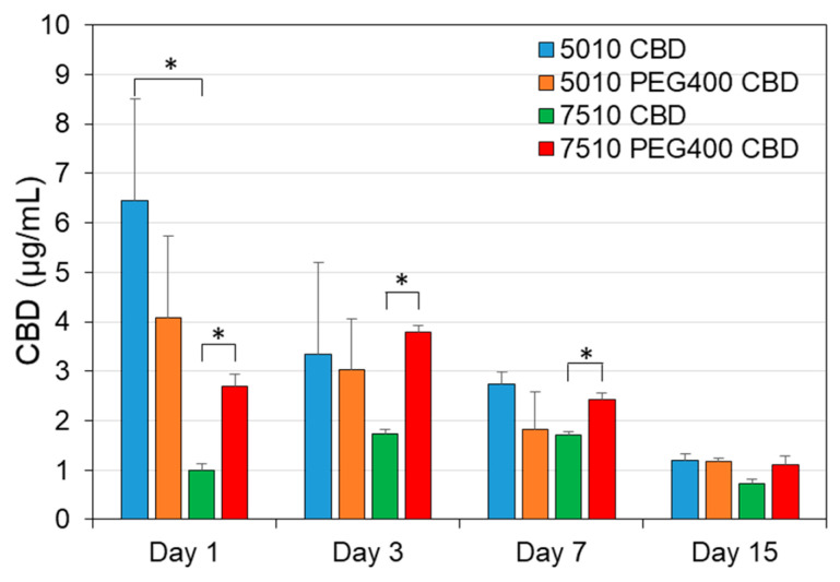
Background: Cannabidiol (CBD) is a natural compound from the Cannabis sativa L. plant, which has anti-inflammatory, anti-nociceptive, neuroprotective, and antibacterial activities. Objective: The aim of this study was to develop a sustained-release device of CBD that can provide an antibacterial effect against the Gram-positive bacteria Streptococcus mutans and Staphylococcus aureus for extended periods of time. Methods: CBD was incorporated into the biodegradable PURASORB 5010 or PURASORB 7510 DL-lactide/glycolide polymers using either dimethylsulfoxide (DMSO) or acetone as the solvent, and the dried polymer scaffolds were exposed daily to a fresh culture of bacteria. The bacterial growth was determined daily by optical density, and the metabolic activity of biofilms was determined using the MTT assay. Biofilm formation on the polymer scaffolds was visualized by HR-SEM. Its anti-inflammatory effect was determined by measuring the IL-6 release from LPS-stimulated RAW 264.7 macrophages by ELISA. Cell cytotoxicity on normal Vero epithelial cells was determined by the MTT assay. The daily release of CBD was determined by gas chromatography-mass spectrometry (GC-MS). Results: PURASORB 5010/CBD scaffolds had antibacterial activity against S. mutans UA159, S. aureus ATCC25923, and a clinical isolate of a multidrug-resistant S. aureus (MDRSA CI-M) strain for the tested period of up to 17 days. PURASORB 7510/CBD scaffolds also had antibacterial activity, but overall, it was less effective than PURASORB 5010/CBD over time. The addition of PEG400 to the copolymers significantly increased the antibacterial activity of PURASORB 7510/CBD but not of PURASORB 5010/CBD. The daily release of CBD from the polymer scaffolds was sufficient to reduce the LPS-induced IL-6 secretion from RAW 264.7 macrophages, and importantly, it was not cytotoxic to either RAW 264.7 macrophages or Vero epithelial cells. The daily release of CBD was found to be between 1.12 and 9.43 µg/mL, which is far below the cytotoxic dose of 25 µg/mL. Conclusions: The incorporation of CBD into the biodegradable PURASORB 5010 can be used to prepare sustained-release devices for medical purposes where combined antibacterial and anti-inflammatory activities are desirable.

Keywords: CBD; IL-6; PLGA scaffolds; Staphylococcus aureus; Streptococcus mutans; antibacterial; antibiofilm; cannabidiol; macrophages; sustained-release device.


Conflict of interest statement

The authors declare no conflicts of interest.

Figures

Figure 1
Figure 1
(A,B) Images of the PURASORB/CBD scaffolds formed using DMSO (A) or acetone (B) as the solvent. The scaffolds are flexible and can be made into different structures. The 5010 scaffolds were softer than the 7510 scaffolds, and the incorporation of CBD caused the scaffolds to become even softer.
Figure 2
Figure 2
(A,B) Antibacterial activity of the PURASORB 5010 (A) and 7510 (B) scaffolds containing two different ratios of polymer to CBD against S. mutans. Each scaffold contained 12.5 mg polymer alone or with either 2.5 or 12.5 mg CBD. DMSO was used as the solvent during the preparation of the scaffolds. The scaffolds were exposed daily to fresh S. mutans cultures in BHI broth, and the OD at 600 nm of the supernatant was measured after a 24 h incubation. N = 4. * p < 0.05 when compared to the placebo.
Figure 3
Figure 3
(AF) HR-SEM images of the PURASORB 5010 (AC) and PURASORB 7510 (DF) scaffolds with or without CBD after five times of exposure to fresh S. mutans cultures in BHI broth. Each scaffold contained 12.5 mg polymer alone (A,D) or with either 2.5 mg (ratio 5:1) (B,E) or 12.5 mg (ratio 1:1) CBD (C,F). DMSO was used as the solvent during the preparation of the scaffolds. The magnification is ×10,000, and the bars represent 5 μm, except for (D) where the bar is 10 μm. The live bacteria appear as smooth luminous 3D structures (green arrows), while the dead bacteria appear as smaller, dysmorphic bacteria that have lost the classical bacterial morphology and appear as darker structures (red arrows). Higher magnification is presented in Supplementary Figure S2.
Figure 4
Figure 4
(A,B) Long-term antibacterial (A) and antibiofilm (B) activity of the polymer/CBD scaffolds against MDRSA CI-M. The PURASORB 5010 and 7510 scaffolds with or without CBD at a polymer-to-CBD ratio of 2:1 (25 mg polymer and 12.5 mg CBD per scaffold) were exposed daily to a fresh culture of MDRSA CI-M in TSB for a 24 h incubation. The viability of bacteria in the supernatant was determined by measuring the OD at 600 nm (A), while the metabolic activity of biofilms formed on the well surface was determined by the MTT assay (B). The % viability and metabolic activity was calculated against control samples without scaffolds, which was set to 100%. DMSO was used as the solvent during the preparation of the scaffolds. N = 4. * p < 0.05 when compared to the placebo samples.
Figure 5
Figure 5
HR-SEM images of the PURASORB 5010 (A,B) and PURASORB 7510 (C,D) scaffolds with or without CBD after seven times of exposure to fresh MDRSA CI-M cultures in TSB. Each scaffold contained 25 mg polymer alone (A,C) or with 12.5 mg CBD (B,D). DMSO was used as the solvent during the preparation of the scaffolds. The magnification is ×10,000, and the bars represent 5 μm. The live bacteria appear as luminous, round 3D structures with smooth membranes (green arrows), while the dead bacteria appear shriveled with a lack of a defined surface and a lack of cell boundaries (red arrows). Higher magnification is presented in Supplementary Figure S3.
Figure 6
Figure 6
(A,B) Long-term antibacterial (A) and antibiofilm (B) activity of the polymer/CBD scaffolds against S. aureus ATCC25923. The PURASORB 5010 and 7510 scaffolds with or without CBD at a polymer-to-CBD ratio of 2:1 (25 mg polymer and 12.5 mg CBD per scaffold) were exposed daily to a fresh culture of S. aureus ATCC25923 in TSB for a 24 h incubation. The viability was determined by measuring the OD of the supernatant at 600 nm (A), while the metabolic activity of biofilms formed at the well surface was determined by the MTT assay (B). Acetone was used as the solvent during the preparation of the scaffolds. N = 4. * p < 0.05 compared to the placebo.
Figure 7
Figure 7
HR-SEM images of the PURASORB 5010 (A,B) and PURASORB 7510 (C,D) scaffolds with or without CBD after 13 times of exposure to fresh S. aureus ATCC25923 cultures in TSB. Each scaffold contained 25 mg polymer alone (A,C) or with 12.5 mg CBD (B,D). Acetone was used as the solvent during the preparation of the scaffolds. The magnification is ×10,000, and the bars represent 5 μm. The live bacteria appear as luminous, round 3D structures with smooth membranes (green arrows), while the dead bacteria appear darker with flat and shriveled structures (red arrows). Higher magnification is presented in Supplementary Figure S4.
Figure 8
Figure 8
(A,B) Long-term antibacterial activity of the polymer/CBD scaffolds against MDRSA CI-M. The PURASORB 5010 (A) and 7510 (B) scaffolds with or without CBD at a polymer-to-CBD ratio of 2:1 (25 mg polymer and 12.5 mg CBD per scaffold) and with or without 7% PEG400 were exposed daily to a fresh culture of S. aureus MDRSA CI-M in TSB for a 24 h incubation. The viability was determined by measuring the OD of the supernatant at 600 nm. Acetone was used as the solvent during the preparation of the scaffolds. Samples without any PURASORB scaffolds were used as controls and set to 100%. N = 4–5. * p < 0.05 compared to the placebo and control samples.
Figure 9
Figure 9
(A,B) Long-term antibiofilm activity of the polymer/CBD scaffolds against MDRSA CI-M. The PURASORB 5010 (A) and 7510 (B) scaffolds with or without CBD at a polymer-to-CBD ratio of 2:1 (25 mg polymer and 12.5 mg CBD per scaffold) and with or without 7% PEG400 were exposed daily to a fresh culture of MDRSA CI-M in TSB for a 24 h incubation. The metabolic activity of the biofilms that formed at the well surface was determined by the MTT assay. Acetone was used as the solvent during the preparation of the polymer scaffolds. Samples without any scaffolds were used as the control and set to 100%. N = 5, except for PURASORB 7510/CBD with N = 4. * p < 0.05 compared to the placebo and control samples.
Figure 10
Figure 10
HR-SEM images of the PURASORB 5010 (AD) and PURASORB 7510 (EH) scaffolds with or without CBD at a ratio of 2:1 and with or without 7% PEG400 after 17 days of exposure to fresh MDRSA CI-M cultures in TSB. Each scaffold contained 25 mg polymer alone (A,E) or with 12.5 mg CBD (B,D,F,H) and/or 7% PEG400 (C,D,G,H). Acetone was used as the solvent during the preparation of the scaffolds. The magnification is ×20,000, and the bars represent 4 μm. The appearance of the bacteria on the CBD-containing polymer scaffolds (B,F) differs from those on the placebo polymer scaffolds (A,E) (red and yellow arrows versus green arrows). The incorporation of PEG400 still caused the adherence of live bacteria (C,G) (green arrows), while the bacteria adhered to the polymer scaffolds containing both PEG400 and CBD showed distorted structures (D,H) (yellow arrows), suggesting that PEG400 enhances the antibacterial effect of CBD on the polymer surface.
Figure 11
Figure 11
(A,B) Long-term antibacterial activity of the polymer/CBD scaffolds against S. mutans UA159. The PURASORB 5010 (A) and 7510 (B) scaffolds with or without CBD at a polymer-to-CBD ratio of 2:1 (25 mg polymer and 12.5 mg CBD per scaffold) and with or without 7% PEG400 were exposed daily to a fresh culture of S. mutans in BHI broth for a 24 h incubation. The % viability was determined by measuring the OD of the supernatant at 600 nm and setting control samples without scaffolds to 100%. Acetone was used as the solvent during the preparation of the scaffolds. N = 4. * p < 0.05 compared to the placebo and control samples. PURASORB 7510/PEG400/CBD had significantly better antibacterial activity against S. mutans than the PURASORB 7510/CBD scaffolds (p < 0.05).
Figure 12
Figure 12
HR-SEM images of the PURASORB 5010 (AD) and PURASORB 7510 (EH) scaffolds with or without CBD at a ratio of 2:1 (B,D,F,H) and with or without 7% (w/w) PEG400 (C,D,G,H) after 17 days of exposure to fresh S. mutans UA159 cultures in BHI broth. Each scaffold contained 25 mg polymer alone (A,E) or with 12.5 mg CBD (B,D,F,H) and/or 7% (w/w) PEG400 (C,D,G,H). Acetone was used as the solvent during the preparation of the scaffolds. The magnification is ×10,000, and the bars represent 5 μm. Green arrows point to live bacteria with luminous 3D structures, while red arrows point to dead bacteria with a dysmorphic structure.
Figure 13
Figure 13
Long-term inhibition of the IL-6 secretion from LPS-stimulated macrophages. (A) The effect of the PURASORB 5010 and PURASORB 7510 scaffolds with or without CBD on the IL-6 secretion from LPS-stimulated RAW 264.7 macrophages. The gray-scaled columns at the very right present the IL-6 secretion from RAW 264.7 macrophages in the medium alone, in the presence of 10 ng/mL LPS alone or together with either 1 μg/mL or 5 μg/mL CBD after a 6 h incubation. The IL-6 concentration was determined by ELISA. (B) The metabolic activity of RAW 264.7 macrophages after 6 h incubation with the indicated treatments as indicated in (A). The polymer/CBD scaffolds were prepared by using DMSO as the solvent. N = 3. * p < 0.05 compared to the placebo and LPS-treated macrophages.
Figure 14
Figure 14
Long-term inhibition of the IL-6 secretion from LPS-stimulated macrophages. (A,B) The effect of the PURASORB 5010 (A) and PURASORB 7510 (B) scaffolds with or without CBD and/or PEG400 on the IL-6 secretion from LPS-stimulated RAW 264.7 macrophages. The gray-scaled columns at the very right presents the IL-6 secretion from RAW 264.7 macrophages in the presence of 10 ng/mL LPS alone or together with either 1 μg/mL, 2.5 μg/mL or 5 μg/mL CBD after a 6 h incubation. The IL-6 concentration was determined by ELISA. The polymer/CBD scaffolds were prepared by using acetone as the solvent. N = 3. * p < 0.05 compared to the placebo and LPS-treated macrophages.
Figure 15
Figure 15
Cytotoxicity assay using Vero epithelial cells. (A,B) Vero epithelial cells in monolayers were incubated with RPMI supernatants that had been exposed daily to the PURASORB 5010 or 7510 scaffolds with or without CBD at a polymer/drug ratio of 2:1 (25 mg polymer and 12.5 mg CBD). After a 24 h incubation, the total cell mass was determined by crystal violet (CV) staining (A), and the metabolic activity of the cells was determined by the MTT assay (B). The Vero cells receiving only RPMI were set to 100%. The scaffolds were prepared using DMSO as the solvent. * p < 0.05 compared to the placebo and control samples.
Figure 16
Figure 16
Quantification of CBD levels in the polymer-conditioned media by GC-MS. The PURASORB 5010/7510 scaffolds with or without CBD and/or PEG400 were incubated daily in 1 mL of 10 mM Tris-HCl pH 6.8, and the quantity of CBD in the supernatant was determined by GC-MS after lyophilizing the samples. The highest level was observed for a PURASORB 5010/CBD scaffold on day 1 (9.43 µg/mL), while the lowest level was observed for a PURASORB 7510/CBD scaffold on day 15 (0.575 µg/mL). The placebo scaffolds with or without PEG400 served as controls with no CBD detection. N = 3. * p ≤ 0.05.

Similar articles

References

    1. Amato M., Santonocito S., Polizzi A., Tartaglia G.M., Ronsivalle V., Viglianisi G., Grippaudo C., Isola G. Local delivery and controlled release drugs systems: A new approach for the clinical treatment of periodontitis therapy. Pharmaceutics. 2023;15:1312. doi: 10.3390/pharmaceutics15041312. - DOI - PMC - PubMed
    1. Steinberg D., Friedman M. Sustained-release delivery of antimicrobial drugs for the treatment of periodontal diseases: Fantasy or already reality? Periodontol. 2000. 2020;84:176–187. doi: 10.1111/prd.12341. - DOI - PubMed
    1. Vigata M., Meinert C., Hutmacher D.W., Bock N. Hydrogels as drug delivery systems: A review of current characterization and evaluation techniques. Pharmaceutics. 2020;12:1188. doi: 10.3390/pharmaceutics12121188. - DOI - PMC - PubMed
    1. Sabri A.H., Kim Y., Marlow M., Scurr D.J., Segal J., Banga A.K., Kagan L., Lee J.B. Intradermal and transdermal drug delivery using microneedles—Fabrication, performance evaluation and application to lymphatic delivery. Adv. Drug Deliv. Rev. 2020;153:195–215. doi: 10.1016/j.addr.2019.10.004. - DOI - PubMed
    1. Steinberg D., Friedman M. Sustained-release drug delivery of antimicrobials in controlling of supragingival oral biofilms. Expert. Opin. Drug Deliv. 2017;14:571–581. doi: 10.1080/17425247.2016.1213239. - DOI - PubMed

LinkOut - more resources