Review
. 2025 Apr 4;17(4):471.
doi: 10.3390/pharmaceutics17040471.

Targeting Regulatory Noncoding RNAs in Human Cancer: The State of the Art in Clinical Trials

Affiliations
Review

Targeting Regulatory Noncoding RNAs in Human Cancer: The State of the Art in Clinical Trials

Roberto Piergentili et al. Pharmaceutics. .

Abstract

Noncoding RNAs (ncRNAs) are a heterogeneous group of RNA molecules whose classification is mainly based on arbitrary criteria such as the molecule length, secondary structures, and cellular functions. A large fraction of these ncRNAs play a regulatory role regarding messenger RNAs (mRNAs) or other ncRNAs, creating an intracellular network of cross-interactions that allow the fine and complex regulation of gene expression. Altering the balance between these interactions may be sufficient to cause a transition from health to disease and vice versa. This leads to the possibility of intervening in these mechanisms to re-establish health in patients. The regulatory role of ncRNAs is associated with all cancer hallmarks, such as proliferation, apoptosis, invasion, metastasis, and genomic instability. Based on the function performed in carcinogenesis, ncRNAs may behave either as oncogenes or tumor suppressors. However, this distinction is not rigid; some ncRNAs can fall into both classes depending on the tissue considered or the target molecule. Furthermore, some of them are also involved in regulating the response to traditional cancer-therapeutic approaches. In general, the regulation of molecular mechanisms by ncRNAs is very complex and still largely unclear, but it has enormous potential both for the development of new therapies, especially in cases where traditional methods fail, and for their use as novel and more efficient biomarkers. Overall, this review will provide a brief overview of ncRNAs in human cancer biology, with a specific focus on describing the most recent ongoing clinical trials (CT) in which ncRNAs have been tested for their potential as therapeutic agents or evaluated as biomarkers.

Keywords: biomarker; cancer; clinical trial; noncoding RNA; oncological therapy.


Conflict of interest statement

The authors declare no conflicts of interest.

Figures
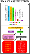
Figure 1
Figure 1
RNA origin and classification. (A) Approximately 95% of the genome is transcribed (transcriptome). Of this, around 90–95% is composed of noncoding RNAs (ncRNAs), mostly rRNAs and tRNAs; the remainder consists of mRNAs. (B) Broad classification of cellular RNAs; for each class, only representative RNAs are reported. The list is not intended to be comprehensive. Color codes are the same in both figure parts.
Figure 2
Figure 2
Main events in miRNA biology. In the nucleus, the gene containing the miR sequence is transcribed by RNA polymerase II, which produces a pri-miRNA. This molecule is then cleaved by Drosha to form a pre-miRNA, which is exported into the cytoplasm by Exportin-5. In the cytoplasm, the pre-miRNA is further processed by the DICER complex to produce a mature double-stranded miRNA. Upon loading into the RISC, one of the two RNA strand binds to its target mRNA, promoting either its translational repression (partial match, red X) or degradation (perfect match). Image partially built using freely available resources at NIH BioArt (https://bioart.niaid.nih.gov/).
Figure 3
Figure 3
Comparing different approaches in gene therapy. In the in vivo approach (red arrows), the starting material is incorporated into a vector (a viral or nanoparticle) and then injected into the patient. In the in situ approach (blue arrows), using appropriate vectors, the starting material is directly injected into the site of interest (e.g., a tumoral mass), where it exerts its effects. In the ex vivo procedure (yellow arrows), cells are explanted from the patient and cultured in vitro. Upon growth and selection, some cells are transformed using appropriate DNA vectors, such as a virus, to insert the sequence of interest into recipient cells, which are then transplanted back to the same donor. Image built using freely available resources at NIH BioArt (https://bioart.niaid.nih.gov/).
Figure 4
Figure 4
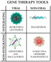
Main gene therapy tools. They can be broadly divided into viral and non-viral; in turn, each may or may not integrate into the host genome. Only representative examples are reported; the list is not intended to be comprehensive. Image partially built using freely available resources at NIH BioArt (https://bioart.niaid.nih.gov/).
Figure 5
Figure 5
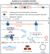
lncRNA biology. (A) Classification based on lncRNA gene position in the genome, which can be outside a coding region (intergenic lncRNA, sometimes named lincRNA) (1) or inside the intron of a coding gene (intragenic lncRNA) (2). Sometimes, the lncRNA is encoded on the complementary strand of a coding gene, resulting in an antisense lncRNA (3) with translation regulation functions. (B) Transcription and function of lncRNAs. Genes encoding lncRNAs are usually transcribed by Pol II and, in many cases, undergo maturation (i.e., capping, polyadenylation, and splicing) as their mRNA counterparts. Some transcripts can undergo a particular splicing mechanism called backsplicing, which generates circular RNAs (circRNA). lncRNAs may have roles either inside the nucleus or in the cytoplasm. In the figure, some examples of these roles are reported. Inside the nucleus, the lncRNA can modify the chromatin structure (e.g., modifying nucleosome positioning to achieve more compact chromatin, black arrows), or it can recruit proteins (green and pink elements), which can alter the gene expression profile (e.g., transcription factors or methylases), possibly causing chromatin modification and either enhancing (Pol II, top) or repressing (Pol II bottom, with the red X indicating the inhibition of transcription) target gene expression. Curved arrows indicate that the green protein interacting with the lncRNA can recruit the pink protein, which, in turn, interacts with Pol II to modify target gene expression. In the cytoplasm, lncRNAs (either linear or circular) may interact with target mRNAs (antisense) or with proteins (scaffolds) or may sponge microRNAs (either different or multiple copies of the same miRNA). Image partially built using freely available resources at NIH BioArt (https://bioart.niaid.nih.gov/).

Similar articles

References

    1. Ferlay J., Colombet M., Soerjomataram I., Parkin D.M., Piñeros M., Znaor A., Bray F. Cancer Statistics for the Year 2020: An Overview. Int. J. Cancer. 2021;149:778–789. doi: 10.1002/ijc.33588. - DOI - PubMed
    1. What Is Cancer. [(accessed on 22 December 2024)]; Available online: https://www.cancer.gov/about-cancer/understanding/what-is-cancer.
    1. Hatton I.A., Galbraith E.D., Merleau N.S.C., Miettinen T.P., Smith B.M., Shander J.A. The Human Cell Count and Size Distribution. Proc. Natl. Acad. Sci. USA. 2023;120:e2303077120. doi: 10.1073/pnas.2303077120. - DOI - PMC - PubMed
    1. Jiang Y., Lin W., Zhu L. Targeted Drug Delivery for the Treatment of Blood Cancers. Molecules. 2022;27:1310. doi: 10.3390/molecules27041310. - DOI - PMC - PubMed
    1. Sharma I., Son M.J., Motamedi S., Hoeft A., Teller C., Hamby T., Ray A. Utilization of Genomic Tumor Profiling in Pediatric Liquid Tumors: A Clinical Series. Hematol. Rep. 2023;15:256–265. doi: 10.3390/hematolrep15020026. - DOI - PMC - PubMed

LinkOut - more resources