. 2025 Apr 12;17(4):510.
doi: 10.3390/pharmaceutics17040510.

Delivery of PLGA-Loaded Influenza Vaccine Microparticles Using Dissolving Microneedles Induces a Robust Immune Response

Affiliations

Delivery of PLGA-Loaded Influenza Vaccine Microparticles Using Dissolving Microneedles Induces a Robust Immune Response

Emmanuel Adediran et al. Pharmaceutics. .

Abstract

Background: Influenza virus is one of the major respiratory virus infections that is a global health concern. Although there are already approved vaccines, most are administered via the intramuscular route, which is usually painful, leading to vaccine hesitancy. To this end, exploring the non-invasive, transdermal vaccination route using dissolving microneedles would significantly improve vaccine compliance. Research on innovative vaccine delivery systems, such as antigen-loaded PLGA microparticles, has the potential to pave the way for a broader range of vaccine candidates. Methods: In this proof-of-concept study, a combination of the inactivated influenza A H1N1 virus and inactivated influenza A H3N2 virus were encapsulated in a biodegradable poly (lactic-co-glycolic acid) (PLGA) polymeric matrix within microparticles, which enhanced antigen presentation. The antigen PLGA microparticles were prepared separately using a double emulsion (w/o/w), lyophilized, and characterized. Next, the vaccine microparticles were assessed in vitro in dendritic cells (DC 2.4) for immunogenicity. To explore pain-free transdermal vaccination, the vaccine microparticles were loaded into dissolving microneedles and administered in mice (n = 5). Results: Our vaccination study demonstrated that the microneedle-based vaccine elicited strong humoral responses as demonstrated by high antigen-specific IgA, IgG, IgG1, and IgG2a antibodies in serum samples and IgA in lung supernatant. Further, the vaccine also elicited a strong cellular response as evidenced by high levels of CD4+ and CD8a+ T cells in lymphoid organs such as the lymph nodes and spleen. Conclusion: The delivery of influenza vaccine-loaded PLGA microparticles using microneedles would be beneficial to individuals experiencing needle-phobia, as well as the geriatric and pediatric population.

Keywords: PLGA; influenza; microneedles; microparticles; polymer.


Conflict of interest statement

The authors declare no conflicts of interest.

Figures

Figure 1
Figure 1
Schematic of the experiment. The vaccine microparticles were formulated and loaded into dissolving microneedles using the spin-casting method. The vaccine microparticles were administered using transdermal delivery with a one prime and one booster dose vaccination strategy. The serum antibody levels were quantified using ELISA, and T cell responses were determined using flow cytometry.
Figure 2
Figure 2
Characterization of vaccine microparticles and dissolving microneedles using scanning electron microscope. (A) Height of microneedles was 486 µm imaged at a magnification of 130×. (B) Dissolving microneedles after administration onto the skin of the mice for 10 min, imaged at a magnification of 185×, (C) Dissolving microneedles form pores on the skin as determined using methylene staining.
Figure 3
Figure 3
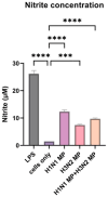
Nitrite oxide release from dendritic cells when pulsed with vaccine microparticles. The vaccine microparticles (MPs) induce a significant amount of nitrite oxide release. The release of nitrite oxide was assessed by the Griess assay in the supernatant. The cells were treated with the following groups: lipopolysaccharide (LPS), H1N1 microparticles, H3N2 microparticles, and H1N1 + H3N2 microparticles. Data expressed as Mean ± SEM n = 3, one-way ANOVA test, and post hoc Dunnett’s test were used for multiple comparisons. ns: non-significant, *** p ≤ 0.001, and **** p ≤ 0.0001.
Figure 4
Figure 4
At 24 h, there is no statistically significant difference between the cells only and the cells treated with vaccine microparticles. Two-fold serial dilution of (H1N1 + H3N2) microparticles was performed in complete DMEM; the concentration range was 31.25–1000 µg/mL at a volume of 100 µL/well. Dimethylsulfoxide (DMSO) was used as a positive control. Data expressed as Mean ± SEM n = 3, one-way ANOVA test, and post hoc Dunnett’s test were used for multiple comparisons. ns: non-significant, and **** p ≤ 0.0001.
Figure 5
Figure 5
H1N1-specific serum antibody levels in vaccinated mice: (A) total serum IgG levels in vaccinated mice. (B) Total serum IgA levels in vaccinated mice. (C) IgG1 serum subtypes in vaccinated mice. (D) IgG2a subtypes in vaccinated mice. The responses obtained were compared to the naïve group, and Fluzone was used as a positive control. Data expressed as Mean ± SEM n = 5, one-way ANOVA test, and post hoc Dunnett’s test were used for multiple comparisons. ns: non-significant, * p ≤ 0.05, ** p ≤ 0.01, *** p ≤ 0.001, and **** p ≤ 0.0001. Fluzone is the intramuscular marketed control group (1.5 µg), and the microneedle group is H1N1(20 µg) + H3N2(20 µg).
Figure 6
Figure 6
H3N2 specific serum antibody levels in vaccinated mice. (A) Total serum IgA levels in vaccinated mice. (B) Total serum IgG levels in vaccinated mice. (C) IgG1 serum subtypes in vaccinated mice. (D) IgG2a subtypes in vaccinated mice. The responses obtained were compared to the naïve group, and Fluzone was used as a positive control. Data expressed as Mean ± SEM n = 5, one-way ANOVA test, and post hoc Dunnett’s test were used for multiple comparisons. ns: non-significant, ** p ≤ 0.01, *** p ≤ 0.001, and **** p ≤ 0.0001. Fluzone is the intramuscular group (1.5 µg), and the microneedle group is H1N1(20 µg) + H3N2(20 µg).
Figure 7
Figure 7
Antigen-specific lung antibody levels in vaccinated mice. (A) H1N1-specific lung supernatant IgA levels in vaccinated mice. (B) H1N1-specific lung supernatant IgG levels in vaccinated mice. (C) H3N2-specific lung supernatant IgA levels in vaccinated mice. (D) H3N2-specific lung supernatant IgG levels in vaccinated mice. The responses obtained were compared to the naïve group, and Fluzone was used as a positive control. Data expressed as Mean ± SEM n = 5, one-way ANOVA test, and post hoc Dunnett’s test were used for multiple comparisons. ns: non-significant, ** p ≤ 0.01, and **** p ≤ 0.0001. Fluzone is the intramuscular marketed control group (1.5 µg), and the microneedle group is H1N1(20 µg) + H3N2(20 µg).
Figure 8
Figure 8
H1N1 and H3N2 cellular responses in vaccinated mice. (A) CD4 cells in the spleen of vaccinated mice. (B) CD8 cells in the spleen of vaccinated mice. (C) CD4 cells in the lymph node of vaccinated mice. (D) CD8 cells in the lymph node of vaccinated mice. The responses obtained are compared to the naïve group. Data expressed as Mean ± SEM n = 5 and two-way ANOVA test. ns: non-significant, ** p ≤ 0.01, and **** p ≤ 0.0001.

Similar articles

References

    1. Deng L., Wang B.Z. A Perspective on Nanoparticle Universal Influenza Vaccines. ACS Infect. Dis. 2018;4:1656–1665. doi: 10.1021/acsinfecdis.8b00206. - DOI - PMC - PubMed
    1. Dou D., Revol R., Östbye H., Wang H., Daniels R. Influenza A Virus Cell Entry, Replication, Virion Assembly and Movement. Front. Immunol. 2018;9:1581. doi: 10.3389/fimmu.2018.01581. - DOI - PMC - PubMed
    1. Krammer F. Emerging influenza viruses and the prospect of a universal influenza virus vaccine. Biotechnol. J. 2015;10:690–701. doi: 10.1002/biot.201400393. - DOI - PubMed
    1. Brankston G., Gitterman L., Hirji Z., Lemieux C., Gardam M. Transmission of influenza A in human beings. Lancet Infect. Dis. 2007;7:257–265. doi: 10.1016/S1473-3099(07)70029-4. - DOI - PubMed
    1. Bischoff W.E., Swett K., Leng I., Peters T.R. Exposure to Influenza Virus Aerosols During Routine Patient Care. J. Infect. Dis. 2013;207:1037–1046. doi: 10.1093/infdis/jis773. - DOI - PubMed

LinkOut - more resources