. 2025 Apr 15;17(4):518.
doi: 10.3390/pharmaceutics17040518.

Intracellular Protein Binding of Zr-89 Oxine Cell Labeling for PET Cell Tracking Studies

Affiliations

Intracellular Protein Binding of Zr-89 Oxine Cell Labeling for PET Cell Tracking Studies

Emmanuel Nyong et al. Pharmaceutics. .

Abstract

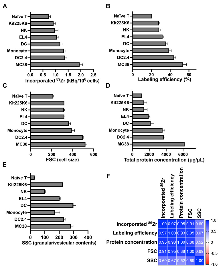
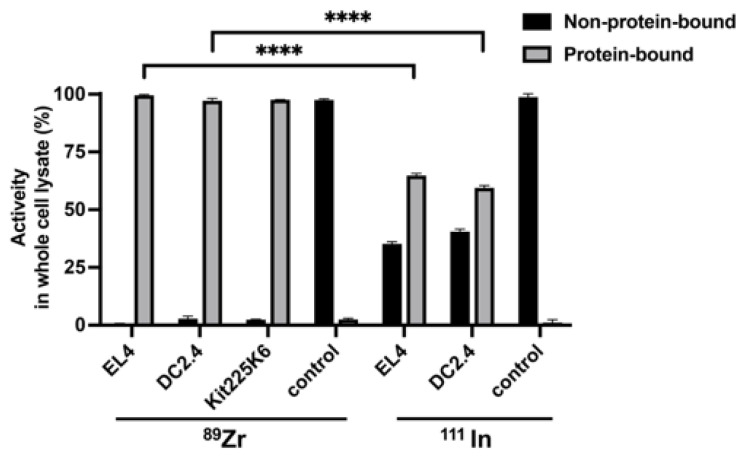
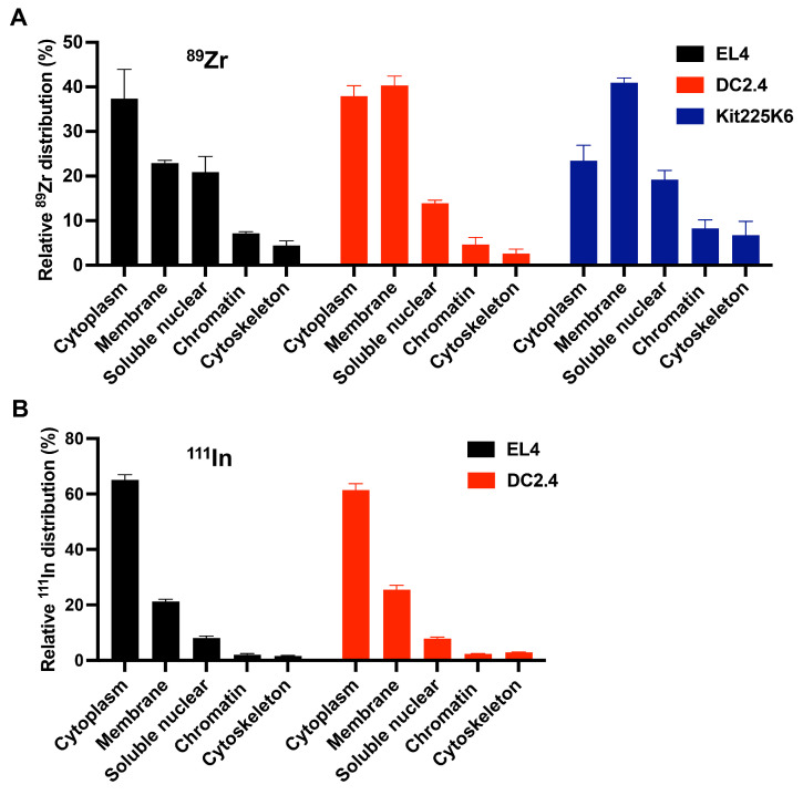
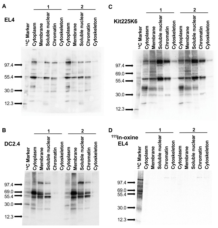
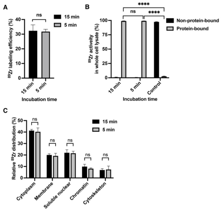
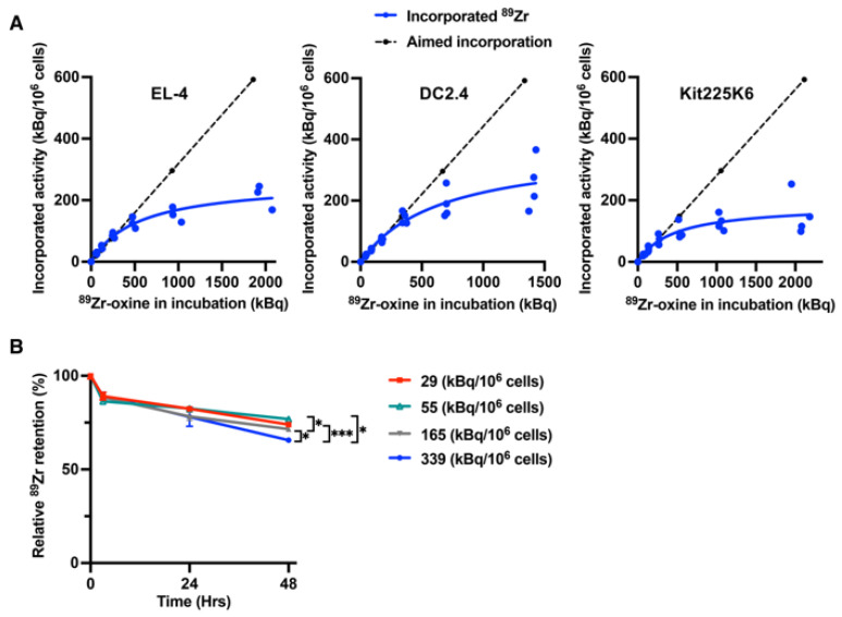
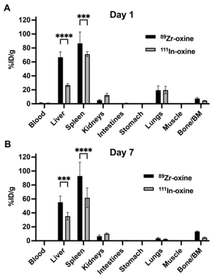
Background/Objectives: 89Zr-oxine is an ex vivo cell labeling agent that enables cells to be tracked in vivo by positron emission tomography (PET) over a period of up to two weeks. To better understand where 89Zr-oxine binds within cellular components, factors affecting labeling and intracellular distribution of 89Zr were examined. Methods: Mouse primary T cells, natural killer cells, dendritic cells, and monocytes, and cell lines EL4 (mouse lymphoma), DC2.4 (mouse dendritic cell), Kit225K6 (human T cell leukemia) and MC38 (mouse colon adenocarcinoma) were labeled with 89Zr-oxine or 111In-oxine and protein binding within the cellular compartments, the labeling thresholds, and radioactivity retention were subsequently determined. Results: Cell incorporation of 89Zr-oxine (27.8-71.8 kBq/106 cells) positively correlated with cellular size and protein mass. Most (>97%) 89Zr was protein-bound and primarily localized in the cytoplasm, membrane, and nuclear fractions (>81%) with distribution patterns varying by cell type. By contrast, 111In-oxine showed lower protein-binding activity of approximately 59-65%, with 62-65% of 111In localized in the cytoplasm. Autoradiography of electrophoresed subcellular fractionated cell samples indicated stable binding by 89Zr-oxine to proteins in all subcellular fractions but unstable protein binding by 111In. Saturation studies showed that 89Zr-oxine labeling was saturable, and further labeling reduced cellular retention. Biodistribution of dendritic cells labeled with either 89Zr-oxine or 111In-oxine indicated greater retention of 89Zr in the labeled cells in vivo than 111In. Conclusions: 89Zr-oxine stably binds many intracellular proteins and shows much higher and more stable protein binding than 111In-oxine. Intracellular protein binding of 89Zr accounts for the ability of 89Zr-oxine labeling to successfully track cells in vivo long-term on PET.

Keywords: cell labeling; cell tracking; positron emission tomography; protein labeling; radiolabeling; zirconium-89 oxine.


Conflict of interest statement

The authors declare no conflicts of interest.

Figures

Figure 1
Figure 1
89Zr-oxine cell labeling positively correlates with the protein mass and cell size. (A) Various cell types incubated with 37 kBq/106 cells of 89Zr-oxine incorporated different amounts of activity (n = 3–6). (B) Labeling efficiency (percentage of incorporated activity vs. incubation activity) was calculated for each cell type (n = 3–6). (C) Flow cytometry was performed to determine the forward scatter (FSC) values that reflect cellular size (n = 3–6). (D) Various cell types were lysed at 100 µL/106 cells, and total protein concentration of each lysate was measured as a readout for protein mass of a cell (n = 3–6). (E) Using flow cytometry, the side scatter (SSC) value reflecting vesicular contents was obtained for each cell type (n = 3–6). All data in (AE) are represented as mean ± standard deviation. (F) Correlation analysis using the values shown in (AE) indicated strong positive correlation between 89Zr activity incorporation or labeling efficiency with protein concentration and cell size (FSC). Pearson r values are shown.
Figure 2
Figure 2
89Zr incorporated into cells is protein-bound. Trichloroacetic acid (TCA) protein precipitation of lysates generated from 2 × 10⁶ 89Zr-oxine labeled EL4, DC2.4, and Kit225K6 cells demonstrated that virtually all activity was protein-bound (n = 3). By contrast, more than one-third of the activity was non-protein-bound in the cells labeled with 111In-oxine (n = 4), demonstrating significant differences in % protein binding with 89Zr-oxine labeling. PBS containing either 89Zr-oxine alone or 111In-oxine alone was used as a control in the relevant assays, and the addition of TCA to these control samples did not precipitate the activity. Data are represented as mean ± standard deviation. ****: p < 0.0001 by two-way ANOVA. Radioactivity of the protein-bound fraction in each cell lysate was significantly higher than that in the relevant control (p < 0.0001, not indicated in the graph).
Figure 3
Figure 3
89Zr-oxine cell labeling leads to distinct intracellular distribution of 89Zr by cell type, predominantly in the cytoplasm, membranes, and nucleus, whereas 111In-oxine labeling results in the activity limited to the cytoplasm. (A) 89Zr-oxine labeled EL4, DC2.4, and Kit225K6 cells were fractionated into the subcellular compartments indicated, and 89Zr activity in each fraction was measured. 89Zr primarily localized in the cytoplasm, membrane, and soluble nuclear fractions, showing different distribution patterns among the cell types (n = 4 for EL4 and DC2.4 cells, n = 3 for Kit225K6 cells). (B) Subcellular fractionation of 111In-oxine labeled EL4 and DC2.4 cells was performed, followed by activity measurement for each fraction. 111In primarily localized in the cytoplasm, membrane, and soluble nuclear fractions, showing different distribution patterns among the cell types (n = 3). All data are represented as mean ± standard deviation.
Figure 4
Figure 4
89Zr-oxine labels multiple cellular proteins of different sizes in various subcellular compartments. (AC) Autoradiography of the membranes obtained from western blotting of subcellular fractions of 89Zr-oxine labeled EL4 (A), DC2.4 (B), and Kit225K6 (C) cells. Multiple bands in each fraction indicate the binding of 89Zr to multiple proteins of different sizes. The pattern of protein binding of 89Zr differed by cell type. Representative results of more than three independent experiments showing duplicated sample preparations for each cell type. (D) Autoradiography of 111In-oxine labeled EL4 cell subcellular fractions only showed extremely faint bands in the cytoplasmic, chromatin-bound, and cytoskeletal fractions. Representative results of two independent experiments, showing duplicated sample preparations.
Figure 5
Figure 5
Intracellular protein binding of 89Zr-oxine occurs rapidly. (A) EL4 cells incubated with 89Zr-oxine for 15 min and 5 min showed similar labeling efficiency with no statistical difference (ns: not significant by Student’s two-tailed t-test, n = 3). (B) EL4 cells incubated with 89Zr-oxine for 5 min showed virtually all activity bound to protein(s) similar to the cells incubated for 15 min, as indicated by the TCA protein precipitation of whole cell lysates. PBS containing 89Zr-oxine alone was used as a control, which did not precipitate the activity (n = 3, ns: not significant, ****: p < 0.0001, by one-way ANOVA). (C) Five min incubation was sufficient to similarly label intracellular proteins as 15 min incubation in EL4 cells (n = 3, ns: not significant, by two-way ANOVA). All data are represented as mean ± standard deviation.
Figure 6
Figure 6
89Zr-oxine incorporation demonstrates a cell type-dependent saturation, and an activity lower than the saturation shows better retention. (A) Saturation assay shows the maximum of 89Zr-oxine incorporation in EL4, DC2.4, and Kit225K6 cells that were incubated with increasing doses of 89Zr-oxine calculated from the labeling efficiency shown in Figure 1B for the following targeted incorporation doses: 18.5 KBq/106, 37KBq/106, 74 kBq/106, 148 kBq/106, 296 kBq/106, and 592 kBq/106 cells (broken line). Incorporated activity plateaued at higher doses, indicating the presences of saturation thresholds. EL4 and DC2.4 showed a threshold point around 90 kBq/106 cells, while that of Kit225K6 was around 60 kBq/106 cells (n = 4 for EL4 and Kit225K6 cells, n = 3 for DC2.4 cells). (B) Kit225K6 cells labeled with 89Zr-oxine at 29, 55, 165, and 339 kBq/106 cells were cultured, and decay-corrected cell-associated activity was examined over 48 h (also see Figure S2A). Relative 89Zr retention compared to the activity immediately after the labeling (0 h) is plotted for each labeling dose (n = 3, *: p < 0.05, ***: p < 0.001, by repeated measure two-way ANOVA). Data are represented as mean ± standard deviation.
Figure 7
Figure 7
Biodistribution of 89Zr-oxine labeled cells showed higher activity retention compared to 111In-oxine labeled cells. (A,B) The graphs show %ID/g values of intravenously infused DCs (6 × 10⁶ cells) labeled with 89Zr-oxine (29.6 kBq/106 cells) or 111In-oxine (76.0 kBq/106 cells) in mice 1 day (A) and 7 days (B) after infusion. The cells primarily migrated to the liver and spleen, regardless of the labeling method. However, the %ID/g values of 89Zr were significantly higher than those of 111In in both organs, indicating greater activity retention in 89Zr-oxine labeled cells (n = 4, ***: p < 0.001, ****: p < 0.0001, by two-way ANOVA). Data are decay corrected, normalized to a 20-g mouse, and represented as mean ± standard deviation. BM: bone marrow.

Similar articles

References

    1. June C.H., O’Connor R.S., Kawalekar O.U., Ghassemi S., Milone M.C. CAR T cell immunotherapy for human cancer. Science. 2018;359:1361–1365. doi: 10.1126/science.aar6711. - DOI - PubMed
    1. El-Kadiry A.E., Rafei M., Shammaa R. Cell Therapy: Types, Regulation, and Clinical Benefits. Front. Med. 2021;8:756029. doi: 10.3389/fmed.2021.756029. - DOI - PMC - PubMed
    1. Buzhor E., Leshansky L., Blumenthal J., Barash H., Warshawsky D., Mazor Y., Shtrichman R. Cell-based therapy approaches: The hope for incurable diseases. Regen. Med. 2014;9:649–672. doi: 10.2217/rme.14.35. - DOI - PubMed
    1. Perrin J., Capitao M., Mougin-Degraef M., Guerard F., Faivre-Chauvet A., Rbah-Vidal L., Gaschet J., Guilloux Y., Kraeber-Bodere F., Cherel M., et al. Cell Tracking in Cancer Immunotherapy. Front. Med. 2020;7:34. doi: 10.3389/fmed.2020.00034. - DOI - PMC - PubMed
    1. Kurebayashi Y., Choyke P.L., Sato N. Imaging of cell-based therapy using (89)Zr-oxine ex vivo cell labeling for positron emission tomography. Nanotheranostics. 2021;5:27–35. doi: 10.7150/ntno.51391. - DOI - PMC - PubMed

LinkOut - more resources