. 2025 Apr 15;17(4):519.
doi: 10.3390/pharmaceutics17040519.

Osteogenic and Antibacterial Response of Levofloxacin-Loaded Mesoporous Nanoparticles Functionalized with N-Acetylcysteine

Affiliations

Osteogenic and Antibacterial Response of Levofloxacin-Loaded Mesoporous Nanoparticles Functionalized with N-Acetylcysteine

Alberto Polo-Montalvo et al. Pharmaceutics. .

Abstract

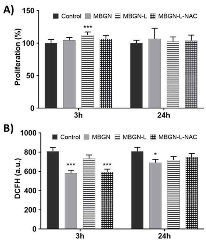
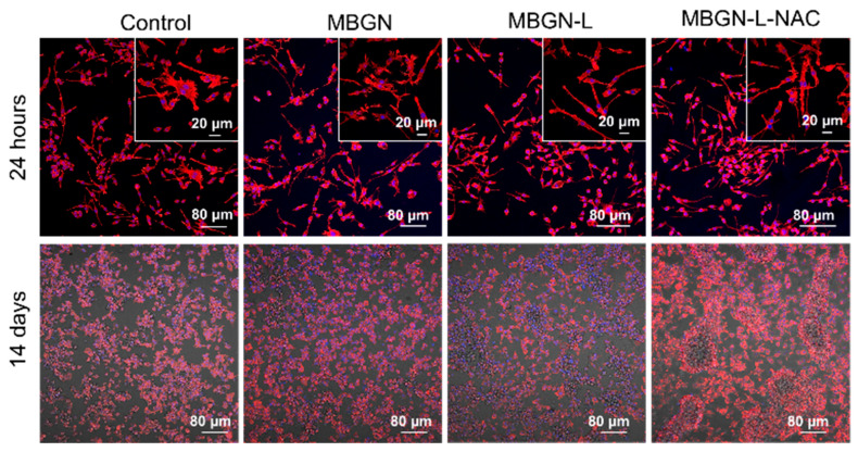
Background/Objectives: Bone infection is one of the most prevalent complications in orthopedic surgery. This pathology is mostly due to bacterial pathogens, among which S. aureus stands out. The formation of a bacterial biofilm makes systemic treatment with antibiotics ineffective. Herein we propose a nanosystem composed of mesoporous bioactive glass nanoparticles (MBGN) loaded with levofloxacin and functionalized with N-acetylcysteine (NAC), aiming to offer an alternative to current treatments. These nanoparticles would present antibacterial activity able to disintegrate the biofilm and regenerate the peri-implantar osseous tissue. Methods: MBGN of composition 82.5 SiO2-17.5 CaO have been synthesized, loaded with levofloxacin, and functionalized with NAC (MBGN-L-NAC). The antimicrobial activity against mature S. aureus biofilms and bioactivity of the nanosystem have been evaluated, as well as its biocompatibility and ability to promote murine pre-osteoblastic MC3T3-E1 differentiation. Results: MBGNs exhibited high surface areas and radial mesoporosity, allowing up to 23.1% (% w/w) of levofloxacin loading. NAC was covalently bound keeping the mucolytic thiol group, SH, available. NAC and levofloxacin combination enhances the activity against S. aureus by disrupting mature biofilm integrity. This nanosystem was biocompatible with pre-osteoblasts, enhanced their differentiation towards a mature osteoblast phenotype, and promoted bio-mimetic mineralization under in vitro conditions. MBGN-L-NAC nanoparticles induced greater osteogenic response of osteoprogenitor cells through increased alkaline phosphatase expression, increased mineralization, and stimulation of pre-osteoblast nodule formation. Conclusions: MBGN-L-NAC exhibits a more efficient antibacterial activity due to the biofilm disaggregation exerted by NAC, which also contributes to enhance the osteoinductive properties of MBGNs, providing a potential alternative to conventional strategies for the management of bone infections.

Keywords: N-acetylcysteine; bone infection; bone regeneration; levofloxacin; mesoporous bioactive nanoparticles.


Conflict of interest statement

The authors declare no conflicts of interest.

Figures

Scheme 1
Scheme 1
Schematic representation of the synthesis of the different nanomaterials prepared in this work. (A) Synthesis of MBGN and levofloxacin loading to obtain MBGN-L. (B) Functionalization of MBGN-L with APTES for NAC anchoring to obtain MBGN-L-NAC.
Figure 1
Figure 1
Physicochemical characterization of the different materials synthesized. TEM images of (A) MBGN, (B) MBGN-L, and (C) MBGN-L-NAC samples. (D) FTIR spectra of the obtained materials. “&” symbol indicates the bands assigned to silica mesoporous framework; “#” symbol indicates the levofloxacin bands, and “*” symbol corresponds to the N-acetylcysteine anchoring via amide bond.
Figure 2
Figure 2
Atom concentrations of Ca, Si, and P in SBF from a suspension of MBGN during 168 h.
Figure 3
Figure 3
In vitro bioactivity testing of MBGN-L-NAC after being soaked in SBF for 0, 3, and 7 days. (A) FTIR spectra and (B) TEM images corresponding to MBGN-L-NAC after soaking in SBF for different times. The white arrows show the needle-like calcium phosphate crystals formed.
Figure 4
Figure 4
“In vial” cumulative levofloxacin release profiles from MBGN-L and MBGN-L-NAC nanosystems at pH 6.0 in PBS during 72 h (A). QT refers to the amount of released levofloxacin versus time, and Q0 refers to the total amount of loaded levofloxacin into the material. (B) Amount of levofloxacin released from both materials during the first 24 h.
Figure 5
Figure 5
Viability of mature bacterial biofilm after 24 h of treatment with the different synthesized nanosystems. Statistical significance: + p < 0.05 vs. Control, +++ p < 0.005 vs. Control, *** p < 0.005 vs. Levo, ### p < 0.005 vs. MBGN, ^^^ p < 0.005 vs. MBGN-L.
Figure 6
Figure 6
Confocal studies after treating the biofilms for 24 h with the different nanosystems. Confocal images of the (A) Control, (B) MBGN-L, and (C) MBGN-L-NAC-treated biofilms. (D) Ratio of Alive vs. Dead bacteria. (** p < 0.01 vs. Levo, *** p < 0.005 vs. Levo, ## p < 0.05 vs. MBGN, ### p < 0.005 vs. MBGN).
Figure 7
Figure 7
In vitro biocompatibility of different obtained materials in MC3T3-E1 murine pre osteoblasts after different time periods (3 and 24 h). (A) Cell proliferation measured with CCK-8 and (B) Intracellular ROS content analysed by flow cytometry. Statistical significance: * p < 0.05 vs. Control, *** p < 0.005 vs. Control.
Figure 8
Figure 8
In vitro cell-differentiation studies on MC3T3-E1 murine pre-osteoblasts in contact with the different synthesized nanomaterials. (A) ALP activity measured at different times (8 and 14 days of culture), after 24 h of contact with the nanomaterials and (B) Mineralization of calcium deposits at 21 days of culture, after 24 h of contact with the nanomaterials. Statistical significance: ** p < 0.01 vs. Control, *** p < 0.005 vs. Control, ## p < 0.01 vs. MBGN, ### p < 0.005 vs. MBGN.
Figure 9
Figure 9
Confocal and phase contrast microscopy images of MC3T3-E1 cells treated with MBGN, MBGN-L, and MBGN-L-NAC for 24 h and after 14 days of culture post-treatment. Staining: Nuclei (DAPI, blue), Cytoskeleton (Phalloidin, red). The selected confocal plane is the closest to the coverglass.

References

    1. Patel R. Periprosthetic Joint Infection. N. Engl. J. Med. 2023;388:251–262. doi: 10.1056/NEJMra2203477. - DOI - PubMed
    1. Dufour D., Leung V., Lévesque C.M. Bacterial Biofilm: Structure, Function, and Antimicrobial Resistance. Endod. Top. 2010;22:2–16. doi: 10.1111/j.1601-1546.2012.00277.x. - DOI
    1. Flemming H.-C., Wingender J., Szewzyk U., Steinberg P., Rice S.A., Kjelleberg S. Biofilms: An Emergent Form of Bacterial Life. Nat. Rev. Microbiol. 2016;14:563–575. doi: 10.1038/nrmicro.2016.94. - DOI - PubMed
    1. Lee S.F., Davey L. Disulfide Bonds: A Key Modification in Bacterial Extracytoplasmic Proteins. J. Dent. Res. 2017;96:1465–1473. doi: 10.1177/0022034517725059. - DOI - PubMed
    1. Davis R., Bryson H.M. Levofloxacin. Drugs. 1994;47:677–700. doi: 10.2165/00003495-199447040-00008. - DOI - PubMed

LinkOut - more resources