. 2025 Apr 1;13(4):803.
doi: 10.3390/microorganisms13040803.

The Role of Membrane-Bound Extracellular Vesicles During Co-Stimulation and Conjugation in the Ciliate Tetrahymena thermophila

Affiliations

The Role of Membrane-Bound Extracellular Vesicles During Co-Stimulation and Conjugation in the Ciliate Tetrahymena thermophila

Eric S Cole et al. Microorganisms. .

Abstract

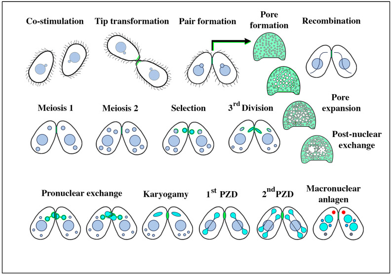
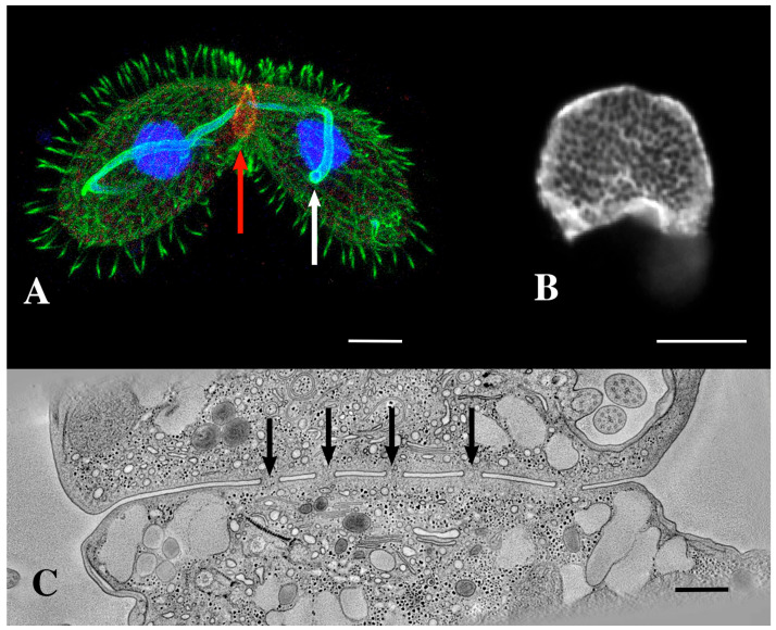
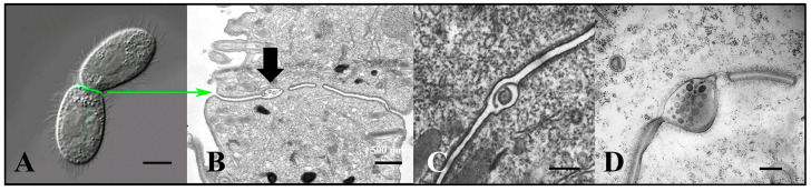
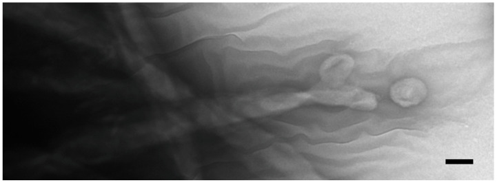
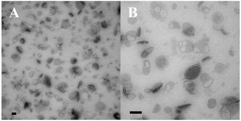
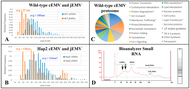
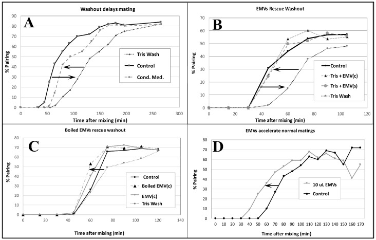
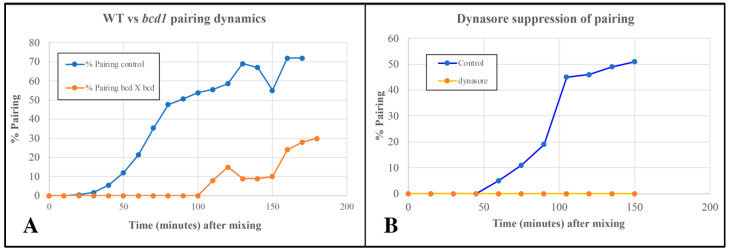
During sexual reproduction, the freshwater ciliate Tetrahymena thermophila sheds membrane-bound vesicles into the extracellular environment (cEMVs: ciliary extracellular micro-vesicles). We provide evidence that 100 nm vesicles shed from the cilia of starved cells promote mating between cells of complementary mating types. A proteomic analysis revealed that these EMVs are decorated with mating-type proteins expressed from the MAT locus, proteins that define a cell's sex (one of seven). Once the mating junction is established between cells, smaller 60 nm vesicles (junction vesicles) appear within the extracellular gap that separates mating partners. Junction vesicles (jEMVs) may play a role in remodeling the mating junction through which gametic pronuclei are exchanged. Evidence is presented demonstrating that cells must be able to internalize extracellular signals via some form of endocytosis in order to trigger conjugation. Finally, an evolutionarily conserved fusogen (Hap2) implicated in pore formation also appears necessary for jEMV processing. This system offers an excellent opportunity for studies on ectosome shedding, intercellular signaling and shed vesicle uptake by macro-pinocytosis, as they relate to sexual reproduction in the ciliate Tetrahymena thermophila.

Keywords: EMVs; Tetrahymena; ciliates; co-stimulation; conjugation; ectosomes; pheromones.


Conflict of interest statement

The authors declare no conflict of interest.

Figures

Figure 1
Figure 1
Stages of conjugation in the ciliate Tetrahymena thermophila. The mating junctions (pale green) are enlarged and en face to highlight details of pore formation and expansion, and the disruption following pronuclear exchange that must be repaired to restore cellular integrity. Turquoise nuclei represent the haploid, the ‘selected’ micronucleus within each partner just following meiosis, and their mitotic products (the migratory and stationary pronuclei) during nuclear exchange and fertilization. Following fertilization, the postzygotic nuclei are also turquoise. During macronuclear anlagen formation (differentiation of the newly recombinant genome into a polyploid pair of macronuclei), one can observe a non-membrane-bound organelle in the anterior cytoplasm (shown in red) named the ‘conjusome’ [28].
Figure 2
Figure 2
(A) A pair of mating Tetrahymena cells decorated with anti-acetylated tubulin (green) and expressing LF4B-GFP, an RCK Kinase involved in determining the cilia length that decorates the mating junction, analyzed by immunofluorescence using anti-GFP antibodies (red) 12G10 anti-α-tubulin (green) and DAPI (blue) (from Jiang et al., 2019 [33]) Scale bar = 10 µM. The white arrow indicates the crescent elongation of the pre-meiotic chromosomes. The red arrow indicates the mating junction. (Photo courtesy of Jacek Gaertig). (B) An en-face view of an isolated mating junction decorated with fenestrin:GFP (photo courtesy of Benjamin Reister) Scale bar = 5 µM. One can see over a hundred fusion pores. (C) A transmission electron micrograph cross section of the mating junction. Arrows indicate fusion pores in profile. (Image taken as a single frame from a 3-D tomogram first published by Cole, et al. [34]. Scale bar = 400 nm.
Figure 3
Figure 3
Extracellular vesicles (jEMVs) being shed into the mating junction. (A) Intact pair with DIC and fluorescence microscopy highlighting conA label. (Scale bar = 10 µM). (B) TEM image of mating junction. Arrow highlights extracellular pocket within mating junction with jEMVs (scale bar = 500 nm). (C) Close-up TEM of wild-type mating junction capturing what appears to be a macro-pinocytotic membrane fold enveloping a jEMV (scale bar = 250 nm) (Cole et al., 2015 [34]). (D) A bcd1 × bcd1 mutant mating junction showing excess accumulation of jEMVs (scale bar = 250 nm).
Figure 4
Figure 4
Negative-stained cilia from an intact, starved, co-stimulating Tetrahymena cell showing what appears to be concave vesicles shedding from their tips. Scale bar = 100 nm.
Figure 5
Figure 5
Two samples of ciliary EMVs (cEMVs) collected by differential ultra-centrifugation from co-stimulating Tetrahymena cells and prepared for TEM using special methyl-cellulose technique to protect their 3D structure. (A) Low mag image, (B) High mag image. Scale bars = 100 nm.
Figure 6
Figure 6
Analysis of both cEMVs and jEMVs collected from mutant and wild-type Tetrahymena during co-stimulation and mating. (A,B) Distribution plots of shed vesicle diameters measured from TEM images of wild-type and HAP2Δ mutant matings. Blue cEMVs (ciliary EMVs) were measured from material collected by differential ultra-centrifugation from 4 h mating cells of either (A) wild-type partners or (B) HAP2Δ deletion partners. For the HAP2Δ matings, the cEMVs are no doubt contaminated with jEMVs, (junction vesicles) since mating pairs fall apart during centrifugation, releasing vesicles from the mating junction and into the supernatant. This is far less likely from the tightly bound wild-type partners. Junction EMVs (jEMVs) were measured in situ from thin sections of 4 h mating pairs. jEMVs imaged from HAP2Δ mating junctions exhibit twice the average size of wild-type jEMVs with significant variance. (C) Proteomic analysis of shed vesicles (cEMVs) collected 4 h into mating. This diagram shows proteins clustered by functional annotation. The complete list appears in the Supplementary Materials. (D) Spectrophotmetric analysis of small RNAs present in shed vesicles. Note, these EMVs were collected from disrupted HAP2Δ matings taking advantage of the mutant pair fragility. Centrifugation disrupts pairing, liberating the jEMVs into the medium for differential centrifugation. This also means we are not analyzing wild-type jEMVs. Asterisks indicate proteins of special interest described in the text.
Figure 7
Figure 7
Pairing assays demonstrate the ‘mating activity’ present in conditioned medium collected from starved single mating-type culture (EMVs) and from mating co-stimulating cultures (EMVc). (A) Demonstration that washing cells in fresh starvation medium delays pairing by up to an hour. Pairing dynamics are restored by adding back conditioned medium following centrifugation. (B) Shed vesicles collected from either starved cells of a single mating type, EMV(s) or from cultures 40 min into co-stimulation EMV(c) can restore pairing when added to fresh, unconditioned starvation medium. (C) Shed vesicles that have been briefly boiled can restore pairing delayed by washing out. (D) Shed vesicles collected from 40 min co-stimulating cells accelerate pairing in cells mating in normal ‘conditioned’ medium. Pairing assays were performed by gently sampling cells from a mating mixture under the microscope, and counting the number of ‘objects’ (out of a sample of 100) representing pairs vs. single, unmated cells. (# pairs/total # objects) × 100 = % pairs.
Figure 8
Figure 8
(A) Delayed pairing in matings involving cells homozygous for the bcd1 mutant defective in endocytosis. (B) Inhibition of pairing in wild-type cells exposed to 70 µM Dynasore (endocytosis inhibitor).
Figure 9
Figure 9
A 3-D electron tomogram of a cilium from a co-stimulated cell with its ciliary pocket and parasomal sac. (A) TEM image of wildtype cilium, ciliary pocket and parasomal sac (ps) (from Richard Allen image archive at the University of Hawaii, Manoa. (https://www6.pbrc.hawaii.edu/allen/ch18/, accessed on 1 March 2015). Note: blue arrow indicates previously unidentified vesicle in a membrane pocket within the ciliary pocket. Scale bar = 200 nm. (B) A section of a tomogram from a wild-type cell highlighting the cilium, parasomal sac (ps) and clathrin-coated vesicles (white arrows). Scale bar = 400 nm. (C) A tomographic slice from (B) highlighting parasomal sac and abundant coated vesicles (golden vesicles, white arrows). Note the absence of extracellular micro-vesicles.
Figure 10
Figure 10
(A) cilium and its ciliary pocket from a mating bcd1 mutant cell with vesicles of varying sizes, filling the ciliary pocket. (A) Thick section view used for tomography in (BD). (B) A tomographic slice of the thick section in (A) showing the clathrin-coated parasomal sac (green arrow) adjacent to the cilium. (C) A tomographic slice from deeper in the volume showing the continuity of the vesicle-filled pocket with the ciliary pocket (blue arrow highlights a massive EMV). (D) The same cytoplasmic volume modeled to reveal the parasomal sac (green arrow), and extracellular vesicles in a variety of sizes (blue) occupying an enlarged ciliary pocket (turquoise arrow). Note the absence of coated vesicles issuing from the parasomal sac (pink vesicles are endosomes without clathrin coats). Scale bars = 250 nm.
Figure 11
Figure 11
(AD) Serial TEM section through a pocket of shed vesicles (jEMVs, red bracket) showing that it is flanked by developing conjugal fusion pores (blue arrows), (Scale bar = 250 nm). (E,F) Tomogram of bcd1 × bcd1 mating junction showing presence of junction pores, and over-abundance of jEMVs. Scale bar = 500 nm. (G) TEM image of mating junction in HAP2Δ × HAP2Δ mutant pair (Modified from Pinello, et al. [37]). Note absence of junction pores and hypertrophied, multi-membrane shed vesicles (jEMVs). Scale bar = 100 nm.
Figure 12
Figure 12
Summary of shed vesicle activity during Tetrahymena conjugation. (A) cEMVs (ciliary EMVs) are shed from cilia of cells expressing complementary mating types (indicated by red and blue) and transferred between cells during ‘brush-by encounters’. Lower diagram highlights red vesicles being shed from the cilium, and blue vesicles (from the mating partner) being internalized by endocytosis at the ciliary pocket and near sites of pinocytosis (the parasomal sacs). The turquoise cylinder represents the basal body subtending the cilium. (B) After pairs have joined, jEMVs (junction EMVs) are shed into the intercellular space within the mating junction (green). The lower diagram illustrates consecutive views of the mating junction over time. jEMVs (green) appear to be internalized by macro-pinocytosis, likely for degradation by autophagy. Red double arrows suggest pore expansion coincident with jEMV shedding and internalization (membrane excavation).

Similar articles

References

    1. Guttery D.S., Zeeshan M., Holder A.A., Tromer E.C., Tewari R. Meiosis in Plasmodium: How does it work? Trends Parasitol. 2023;39:812–821. doi: 10.1016/j.pt.2023.07.002. - DOI - PubMed
    1. Figueroa R.I., Howe-Kerr L.I., Correa A.M.S. Direct evidence of sex and a hypothesis about meiosis in Symbiodiniaceae. Sci. Rep. 2021;11:18838. doi: 10.1038/s41598-021-98148-9. - DOI - PMC - PubMed
    1. Orias E. Ciliate conjugation. In: Gall J.G., editor. The Molecular Biology of Ciliated Protozoa. Academic Press; London, UK: 1986. pp. 45–84.
    1. Cole E., Sugai T. Developmental progression of Tetrahymena through the cell cycle and conjugation. Methods Cell Biol. 2012;109:177–236. - PubMed
    1. Ruehle M.D., Orias E., Pearson C.G. Tetrahymena as a unicellular model eukaryote: Genetic and genomic tools. Genetics. 2016;203:649–665. doi: 10.1534/genetics.114.169748. - DOI - PMC - PubMed

Grants and funding

LinkOut - more resources