. 2025 Apr 21;13(4):955.
doi: 10.3390/microorganisms13040955.

The Vaginally Exposed Extracellular Vesicle of Gardnerella vaginalis Induces RANK/RANKL-Involved Systemic Inflammation in Mice

Affiliations

The Vaginally Exposed Extracellular Vesicle of Gardnerella vaginalis Induces RANK/RANKL-Involved Systemic Inflammation in Mice

Yoon-Jung Shin et al. Microorganisms. .

Abstract

Gardnerella vaginalis (GV), an opportunistic pathogen excessively proliferated in vaginal dysbiosis, causes systemic inflammation including vaginitis, neuroinflammation, and osteitis. To understand its systemic inflammation-triggering factor, we purified extracellular vesicles isolated from GV (gEVs) and examined their effect on the occurrence of vaginitis, osteitis, and neuroinflammation in mice with and without ovariectomy (Ov). The gEVs consisted of lipopolysaccharide, proteins, and nucleic acid and induced TNF-α and RANKL expression in macrophage cells. When the gEVs were vaginally exposed in mice without Ov, they significantly induced RANK, RANKL, and TNF-α expression and NF-κB+ cell numbers in the vagina, femur, hypothalamus, and hippocampus, as observed in GV infection. The gEVs decreased time spent in the open field (OT) in the elevated plus maze test by 47.3%, as well as the distance traveled in the central area (DC) by 28.6%. In the open field test, they also decreased the time spent in the central area (TC) by 39.3%. Additionally, gEVs decreased spontaneous alteration (SA) in the Y-maze test by 33.8% and the recognition index (RI) in the novel object recognition test by 26.5%, while increasing the immobility time (IT) in the tail suspension test by 36.7%. In mice with OV (Ov), the gEVs also induced RANK, RANKL, and TNF-α expression and increased NF-κB+ cell numbers in the vagina, femur, hypothalamus, and hippocampus compared to vehicle-treated mice. When gEVs were exposed to mice with Ov, gEVs also reduced the DC, TC, OT, SA, and RI to 62.1%, 62.7%, 28.2%, 90.7%, and 85.4% of mice with Ov, respectively, and increased IT to 122.9% of mice with Ov. Vaginally exposed fluorescein-isothiocyanate-tagged gEVs were detected in the blood, femur, and hippocampus. These findings indicate that GV-derived gEVs may induce systemic inflammation through the activation of RANK/RANKL-involved NF-κB signaling, leading to systemic disorders including vaginitis, osteoporosis, depression, and cognitive impairment. Therefore, gEVs may be an important risk factor for vaginitis, osteoporosis, depression, and cognitive impairment in women.

Keywords: Gardnerella vaginalis; extracellular vesicle; osteoporosis; psychiatric disorder; vaginitis.


Conflict of interest statement

The authors declare no conflicts of interest.

Figures

Figure 1
Figure 1
Effects of GV and gEVs on TNF-α, IL-6, RANKL, and RANK expression in macrophage cells. (a) Transmission electron microscope image of gEVs. One bar is 0.5 μm. (b) SDS-PAGE of gEVs. M, molecular weight markers; GV, treated with GV; EV, treated with gEVs. (c) Identified gEV components in (b): A, type I polyketide synthase; B, partial hypothetical protein of Klebsiella oxytoca. Effect of gEVs on TNF-α (d), IL-6 (e), RANKL (f), and RANK expression (g) in macrophage cells. Vh, GV, and EV were treated with saline, GV, and gEVs, respectively. Statistical significance is indicated by different letters (p < 0.05).
Figure 2
Figure 2
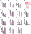
Effects of vaginally exposed GV and gEVs on the occurrence of vaginitis in mice. Effect on uterine weight (a). Effect on TNF-α (b), IL-6 (c), and RANKL expression (d), and TNF-α+ and NF-κB+ cell number (e) in the vagina. Effects on TNF-α (f), and NF-κB intensities (g) in the (e). Effect on TNF-α (h) and RANKL levels (i) in the blood. Bars in (e) indicate 0.5 mm. NF-κB/DAPI and TNF-α/DAPI staining were performed, as described in Materials and Methods. Nc, Gv, and Ev were treated with saline, GV, and gEVs, respectively. Statistical significance is indicated by different letters (p < 0.05).
Figure 3
Figure 3
Effects of vaginally exposed GV and gEVs on osteoporosis-like symptoms in mice. Effect on femur weight (a), TNF-α (b), RANK (c), RANKL (d), osteoprotegerin (e), MMP2 (f), and TRAP expression (g), and TRAP+ALP+, RANK+, and TNF-α+ cell number (h) in the femur. Effects on RANKL (i) and TNF-α intensities (j) in the (h). Bars in (h) indicate 0.5 mm. TRAP/ALP (arrows indicate positive cells), H&E, RANK/DAPI, and TNF-α/DAPI staining were performed, as described in Materials and Methods. Nc, Gv, and Ev were treated with saline GV, and gEVs, respectively. Statistical significance is indicated by different letters (p < 0.05).
Figure 4
Figure 4
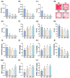
Effects of vaginally exposed GV and gEVs on DCi-like symptoms in mice. Effect on TD (a), TC (b), and DC (c), and movement track path (d) in the OFT, OT in the EMPT (e), IT in the TST (f), SA (g) in the YMT, and RI (h) in NORT. Effect on serotonin levels (i), TNF-α (j), and IL-6 expression (k) in the hypothalamus, and serotonin levels (l), TNF-α (m), IL-6 (n), and RANKL expression (o) in the hippocampus. Nc, Gv, and Ev were treated with saline GV and gEVs, respectively. Statistical significance is indicated by different letters (p < 0.05).
Figure 5
Figure 5
Effects of vaginal GV and gEV exposure on vaginitis development in ovariectomized mice. Effect on uterine weight (a). Effect on TNF-α (b), IL-6 (c), and RANKL expression (d), and TNF-α+ and NF-κB+ cell number (e) in the vagina. Effects on TNF-α (f) and NF-κB intensities (g) in the (e). Bars in (e) indicate 0.5 mm. NF-κB/DAPI and TNF-α/DAPI staining were performed, as described in Materials and Methods. Effect on TNF-α (h), RANKL (i), and estradiol levels (j) in the blood. Bars in (h) indicate scale size. Sh, saline in sham mice; Ov, saline in ovariectomized mice; oGv, GV in ovariectomized mice; oEv, gEVs in ovariectomized mice. Statistical significance is indicated by different letters (p < 0.05).
Figure 6
Figure 6
Effects of vaginally exposed GV and gEVs on osteoporosis-like symptoms in ovariectomized mice. Effect on femur weight (a), TNF-α (b), IL-6 (c), RANK (d), RANKL (e), osteoprotegerin (f), MMP2 (g), and TRAP expression (h) and TRAP+ALP+, RANK+, and TNF-α+ cell number (i) in the femur. Effects on RANKL (j) and TNF-α intensities (k) in the (i). Bars in (i) indicate 0.5 mm. TRAP/ALP (arrows indicate positive cells), H&E, RANK/DAPI, and TNF-α/DAPI staining were performed, as described in Materials and Methods. Sh, saline in sham mice; Ov, saline in ovariectomized mice; oGv, GV in ovariectomized mice; oEv, gEVs in ovariectomized mice. Statistical significance is indicated by different letters (p < 0.05).
Figure 7
Figure 7
Effects of vaginally exposed GV and gEVs on DCi-like symptoms in ovariectomized mice. Effect on DC (a), TD (b), and TC (c), and movement track path (d) in the OFT, OT in the EMPT (e), IT in the TST (f), SA (g) in the YMT, and RI (h) in NORT. Effect on serotonin (i), TNF-α (j), and IL-6 expression (k) in the hypothalamus, and serotonin (l), TNF-α (m), IL-6 (n), and RANKL expression (o) in the hippocampus. Sh, saline in sham mice; Ov, saline in ovariectomized mice; oGv, GV in ovariectomized mice; oEv, gEV in ovariectomized mice. Statistical significance is indicated by different letters (p < 0.05).
Figure 8
Figure 8
Vaginally exposed FITC-labeled gEVs (F-gEVs) were translocated into the blood (a), hippocampal CA1 and CA3 regions (b), and femur (c). Bars in (ac) indicate 0.1 mm. N, treated with saline; F, treated with FITC alone; F-gEV, treated with F-gEV. Arrows indicate FITC-positive.
Scheme 1
Scheme 1
Vaginally infected Gardnerella vaginalis (GV) and its extracellular vesicles (gEVs) can cause vaginitis, osteoporosis, and depression/cognitive impairment with systemic inflammation by activating RNAK/RANKL-involved NF-κB signaling. Thick red arrows, induced; thick blue arrows, suppressed; thin arrows, supported process pathway; dotted arrows, proposed signaling pathway processed through several molecules.

Similar articles

References

    1. Holdcroft A.M., Ireland D.J., Payne M.S. The Vaginal Microbiome in Health and Disease-What Role Do Common Intimate Hygiene Practices Play? Microorganisms. 2023;11:298. doi: 10.3390/microorganisms11020298. - DOI - PMC - PubMed
    1. Chen X., Lu Y., Chen T., Li R. The Female Vaginal Microbiome in Health and Bacterial Vaginosis. Front. Cell Infect. Microbiol. 2021;11:631972. doi: 10.3389/fcimb.2021.631972. - DOI - PMC - PubMed
    1. Kalia N., Singh J., Kaur M. Microbiota in vaginal health and pathogenesis of recurrent vulvovaginal infections: A critical review. Ann. Clin. Microbiol. Antimicrob. 2020;19:5. doi: 10.1186/s12941-020-0347-4. - DOI - PMC - PubMed
    1. Almeida C., Oliveira R., Soares R., Barata P. Influence of gut microbiota dysbiosis on brain function: A systematic review. Porto Biomed. J. 2020;5:1. doi: 10.1097/j.pbj.0000000000000059. - DOI - PMC - PubMed
    1. Jemimah S., Chabib C.M.M., Hadjileontiadis L., AlShehhi A. Gut microbiome dysbiosis in Alzheimer’s disease and mild cognitive impairment: A systematic review and meta-analysis. PLoS ONE. 2023;18:e0285346. doi: 10.1371/journal.pone.0285346. - DOI - PMC - PubMed

LinkOut - more resources