. 2025 Apr 15;12(4):368.
doi: 10.3390/vetsci12040368.

Mitofusin-Mediated Mitochondrial Fusion Inhibits Pseudorabies Virus Infection in Porcine Cells

Affiliations

Mitofusin-Mediated Mitochondrial Fusion Inhibits Pseudorabies Virus Infection in Porcine Cells

Xiuhan Xu et al. Vet Sci. .

Abstract

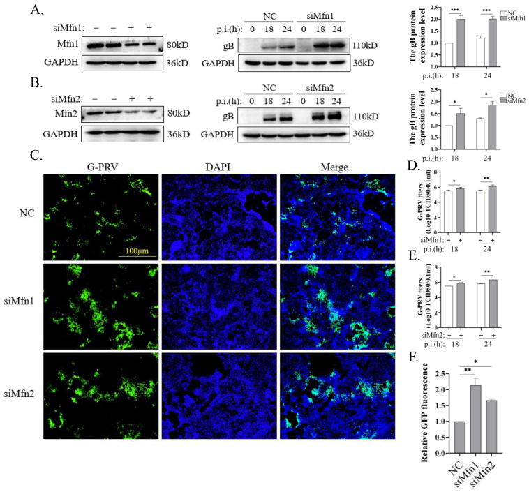
Background: Mitochondria are highly dynamic organelles that undergo fusion/fission dynamics, and emerging evidence has established that mitochondrial dynamics plays a crucial regulatory role in the process of viral infection. Nevertheless, the function of mitochondria dynamics during pseudorabies (PRV) infection remains uncertain. Methods: Our investigation commenced with examining PRV-induced alterations in mitochondrial dynamics, focusing on morphological changes and the expression levels of fusion/fission proteins. We then restored mitochondrial dynamics through Mfn1 (Mitofusin 1)/Mfn2 (Mitofusin 2) overexpression and mdivi-1 (mitochondrial division inhibitor-1) treatment to assess their impact on PRV replication and mitochondrial damage. Results: We found a downregulation of the mitochondrial fusion proteins Mfn1, Mfn2, and OPA1 (optic atrophy 1), along with the activation of the fission protein Drp-1 (dynamin-related protein 1) upon PRV infection. Restoring the function of mitochondrial fusion inhibited PRV infection. Furthermore, elevated mitochondrial membrane potential (MMP), decreased reactive oxygen species (ROS) levels, and an increased mitochondrial number were observed after overexpressing Mfns or treatment with mdivi-1. Conclusions: PRV infection impairs mitochondrial dynamics by altering mitochondrial fusion and fission proteins, and the promotion of Mfn-mediated mitochondrial fusion inhibits PRV replication.

Keywords: mitochondrial dynamics; mitochondrial fusion; mitofusin proteins; pseudorabies virus.


Conflict of interest statement

The authors declare no conflicts of interest.

Figures

Figure 1
Figure 1
PRV infection impairs mitochondrial dynamics (Western Blot original figures see Supplementary Materials). (A) Confocal microscopy of PK-15 cells infected with W-PRV (moi 1) for 12, 18, and 24 h. The mitochondria network was analyzed by Image J. Green: anti-gB; red: anti-TOMM20; blue: DAPI. Scale bar: 10 μm. The yellow boxes indicated these areas were enlarged on the right. (B) Western blot analysis and quantification of expression levels of PK-15 cells infected with G-PRV (moi 1) for 12, 18, and 24 h. (C) Confocal microscopy of PK-15 cells infected with W-PRV (moi 1) for 12, 18, and 24 h. Green: anti-p-Drp1; red: anti-TOMM20; blue: DAPI. Scale bar: 10 μm. The yellow boxes indicated these areas were enlarged on the right. (D) Western blot analysis and quantification of expression levels of PK-15 cells infected with G-PRV (moi 0.1, 1, and 5) for 24 h. (E) Western blot analysis of PK-15 cells infected with G-PRV (moi 1) for 12, 18, and 24 h. G-PRV was pre-treated with UV or natural light (control) for 30 s. (FH) The mRNA levels of indicated genes were detected by RT-qPCR. Data are mean ± SD values (n = 3 per group). *, **, and *** indicate statistically significant differences with p < 0.05, p < 0.01, and p < 0.001, respectively. ns indicates not statistically significant.
Figure 2
Figure 2
The promotion of mitochondrial fusion inhibited PRV replication (Western Blot original figures see Supplementary Materials). (A,B) Western blot analysis and quantification of expression levels of PK-15 cells. Cells were transfected with EV, Mfn1 or Mfn2 plasmids for 24 h and then infected with G-PRV (1 moi) for (A) 24 h, (B) 18, 24, 36, or 48 h. (C) Analysis of the culture supernatants derived from panel A to B for PRV titer. (DG) The levels of G-PRV replication (green) were visualized by an inverted fluorescence microscope upon G-PRV (1 moi) infection at 24 h. The GFP/DAPI fluorescence intensity ratios of each group were compared to quantify the data. Scale bar: 100 μm. (H,I) Western blot analysis and quantification of gB expression levels. Cells were infected with G-PRV (1 moi) for 24 h in the absence or presence of mdivi-1 (25 µM). (J) Analysis of the culture supernatants derived from panel H for PRV virus titer. Data are mean ± SD values (n = 3 per group). *, **, and *** indicate statistically significant differences with p < 0.05, p < 0.01, and p < 0.001, respectively. ns indicates not statistically significant.
Figure 3
Figure 3
Blocking mitochondrial fusion promoted PRV replication (Western Blot original figures see Supplementary Materials). (A,B) Western blot analysis and quantification of different protein expression levels of PK-15 cells. Cells were transfected with NC or siMfn1 or siMfn2 siRNAs for 24 h and then infected with G-PRV (moi 1) for 18 and 24 h. (D,E) Analysis of the culture supernatants derived from panel A to B for PRV titer. (CF) The levels of G-PRV replication (green) were visualized by an inverted fluorescence microscope (OLYMPUS, DP74) upon G-PRV (1 moi) infection at 24 h. The GFP/DAPI fluorescence intensity ratios of each group were compared to quantify the data. Scale bar: 100 μm. Data are mean ± SD values (n = 3 per group). *, **, and *** indicate statistically significant differences with p < 0.05, p < 0.01, and p < 0.001, respectively. ns indicates not statistically significant.
Figure 4
Figure 4
The promotion of mitochondrial fusion alleviated mitochondrial damage and decreased the mitochondrial number caused by PRV infection (Western Blot original figures see Supplementary Materials). (A,B) Flow cytometry with JC-1 (A) or DCFH-DA (B) staining of PK-15 cells. Cells were transfected with EV or Mfn1 or Mfn2 plasmids for 24 h and then infected with W-PRV (1 moi) for 24 h. Decreased red/green fluorescence ratio of JC-1 represents disrupted MMP. (C) Western blot analysis and quantification of the expression levels of PK-15 cells. Cells were infected with G-PRV (1 moi) for 24 h in the absence or presence of CQ (50 μM), MG132 (10 μM), or Z-VAD (10 μM). (D,E) Western blot analysis and quantification of TOMM20 and COX IV expression levels of PK-15 cells. Cells were transfected with EV or Mfn1 or Mfn2 plasmids for 24 h and then infected with G-PRV (moi 1) for 24 h. (F) Western blot analysis and quantification of TOMM20 and COX IV expression levels of PK-15. Cells were infected with G-PRV (1 moi) for 24 h in the absence or presence of mdivi-1(25 μM). *, **, and *** indicate statistically significant differences with p < 0.05, p < 0.01, and p < 0.001, respectively. ns indicates not statistically significant.
Figure 5
Figure 5
Blocking mitochondrial fusion enhanced PRV-induced mitochondrial damage and mitophagy (Western Blot original figures see Supplementary Materials). (A) Flow cytometry with DCFH-DA staining of PK-15 cells. Cells were transfected with NC, siMfn1, or siMfn2 siRNAs for 24 h and then infected with W-PRV (1 moi) for 24 h. (B,C) Western blot analysis and quantification of Mfn1, Mfn2, TOMM20, and COX IV expression levels in PK-15 cells. Cells were transfected with NC or siMfn1 or siMfn2 siRNAs for 24 h and then infected with G-PRV (1 moi) for 24 h. *, **, and *** indicate statistically significant differences with p < 0.05, p < 0.01, and p < 0.001, respectively.

Similar articles

Cited by

References

    1. Laval K., Enquist L.W. The Neuropathic Itch Caused by Pseudorabies Virus. Pathogens. 2020;9:254. doi: 10.3390/pathogens9040254. - DOI - PMC - PubMed
    1. Muller T.F., Teuffert J., Zellmer R., Conraths F.J. Experimental infection of European wild boars and domestic pigs with pseudorabies viruses with differing virulence. Am. J. Vet. Res. 2001;62:252–258. doi: 10.2460/ajvr.2001.62.252. - DOI - PubMed
    1. Ezura K., Usami Y., Tajima K., Komaniwa H., Nagai S., Narita M., Kawashima K. Gastrointestinal and skin lesions in piglets naturally infected with pseudorabies virus. J. Vet. Diagn. Investig. 1995;7:451–455. doi: 10.1177/104063879500700405. - DOI - PubMed
    1. Freuling C.M., Muller T.F., Mettenleiter T.C. Vaccines against pseudorabies virus (PrV) Vet. Microbiol. 2017;206:3–9. doi: 10.1016/j.vetmic.2016.11.019. - DOI - PubMed
    1. Wu R., Bai C., Sun J., Chang S., Zhang X. Emergence of virulent pseudorabies virus infection in northern China. J. Vet. Sci. 2013;14:363–365. doi: 10.4142/jvs.2013.14.3.363. - DOI - PMC - PubMed

LinkOut - more resources