. 2025 Mar 23;17(4):458.
doi: 10.3390/v17040458.

The Virus Entry Pathway Determines Sensitivity to the Antiviral Peptide TAT-I24

Affiliations

The Virus Entry Pathway Determines Sensitivity to the Antiviral Peptide TAT-I24

Eva Kicker et al. Viruses. .

Abstract

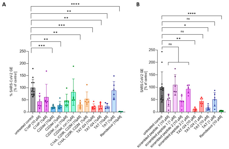
The peptide TAT-I24, a fusion of the TAT peptide (amino acids 48-60) and the 9-mer peptide I24, has been previously shown to neutralize several double-stranded (ds) DNA viruses in vitro. We have now extended the testing to potentially sensitive RNA viruses and analyzed the antiviral effect of the peptide against Severe Acute Respiratory Syndrome Coronavirus-2 (SARS-CoV-2). In Vero E6 cells, TAT-I24 neutralized the human 2019-nCoV isolate (Wuhan variant) in a dose-dependent manner, while it was unable to neutralize two SARS-CoV-2 variants of concern, Delta and Omicron. Moreover, TAT-I24 could not significantly neutralize any of the SARS-CoV-2 variants in the human lung carcinoma cell line Calu-3, which provides an alternative entry route for SARS-CoV-2 by direct membrane fusion. Therefore, a possible dependence on virus uptake by endocytosis was investigated by exposing Vero E6 cells to chloroquine (CQ), an inhibitor of endosomal acidification. The Wuhan variant was highly sensitive to inhibition by CQ, an effect which was further enhanced by TAT-I24, while the Delta variant was less sensitive to inhibition by higher concentrations of CQ compared to the Wuhan variant. The microscopic analysis of COS-7 cells using a rhodamine-labeled TAT-I24 (Rho-TAT-I24) showed the endosomal localization of fluorescent TAT-I24 and co-localization with transfected GFP-Rab14 but not GFP-Rab5. As these proteins are found in distinct endosomal pathways, our results indicate that the virus entry pathway determines sensitivity to the peptide.

Keywords: Rab14; SARS-CoV-2; antiviral peptide; endocytosis; virus entry.


Conflict of interest statement

Hanna Harant owns 100% of the shares of Pivaris BioScience GmbH. Kurt Zatloukal is the co-founder and CEO of Zatloukal-Innovations GmbH. The other authors declare no conflicts of interest.

Figures

Figure 1
Figure 1
RNA binding of TAT-I24 or TAT and effect of peptides on replication of SARS-CoV-2 (Wuhan variant). (A) Binding of TAT or TAT-I24 to synthetic ssRNA. Mean ± SD of relative fluorescence units are shown (n = 6). (B) Relative viral genome equivalents (GE) in supernatants of Vero E6 cells infected with original Wuhan variant in presence of increasing concentrations of TAT-I24 (24 h p.i.). IC50 values were calculated by non-linear curve fit analysis. Data points show mean ± 95% confidence interval (CI) of n ≥ 9 from three independent experiments. (C) Relative SARS-CoV-2 GE in supernatants of Vero E6 cells infected with original Wuhan variant, untreated or treated with 10 µM of TAT-I24 or 10 µM of TAT for 24 h. Significant differences were detected using one-way ANOVA (Kruskal–Wallis multiple comparison of means). p-values: p ≥ 0.05 (ns); p ≤ 0.01 (**), and p ≤ 0.0001 (****); n = 12, mean ± SD are shown. (D) Corresponding wells (one representative experiment) from (B) with immunohistochemical staining against SARS-CoV-2 nucleocapsid. Images were quantified for red-positive area signals and normalized to untreated control (infected, untreated cells); representative sections of wells are shown. Scale bars indicate 200 µm.
Figure 2
Figure 2
The effect of TAT-I24 analogs (A) and scrambled peptides (B) on the replication of SARS-CoV-2 (Wuhan variant). The relative viral genome equivalent (GE) in the supernatants of Vero E6 cells infected with the original Wuhan variant in the presence of 1 µM or 10 µM of TAT-analogs, TAT-I24, TAT, and scrambled peptides (24 h p.i.). The data points with the mean ± SD of n ≥ 6 from two independent experiments are shown. Significant differences were detected using one-way ANOVA (Kruskal–Wallis multiple comparison of means). p-values: p ≥ 0.05 (ns), p ≤ 0.05 (*), p ≤ 0.01 (**), p ≤ 0.001 (***), and p ≤ 0.0001 (****).
Figure 3
Figure 3
Differential inhibitory effect of TAT-I24 against SARS-CoV-2 virus variants. Relative viral GE in the supernatants of Vero E6 cells infected with the Wuhan variant (A), the Delta variant B.1.617.2 (B), and the Omicron variant B.1.1.529 (C) in untreated cells (gray), in the presence of various concentrations of TAT-I24 (red), 10 µM of Remdesivir (green), or 10 µM of TAT (blue) 24 h p.i.; significant differences were detected using one-way ANOVA (Kruskal–Wallis multiple comparison of means). p-values: p ≤ 0.05 (*); p ≤ 0.001 (***); and p ≤ 0.0001 (****), n ≥ 9, means ± SD from four independent experiments are shown for TAT-I24 with the Wuhan, Delta, and Omicron variants; for TAT, n ≥ 6 with the Wuhan, Delta, and Omicron variants from two independent experiments. Heatmap of the genomic (D) and subgenomic (E) SARS-CoV-2 RNA levels in the presence of 10 µM of TAT-I24 compared to untreated and infected controls; a 2 log-fold change at the time points 0.5 h, 4 h, 8 h, and 24 h p.i. for the Wuhan, Delta, and Omicron virus variants is shown.
Figure 4
Figure 4
Effect of peptides on dsRNA levels in infected Vero E6 cells. (A) Cells were infected with Wuhan variant (500 pfu/well) or (B) Delta variant (500 pfu/well) for 2 h and 24 h in absence (untreated) and presence of TAT-I24 (10 µM) or TAT (10 µM), followed by staining for dsRNA (FITC/green); nuclei were stained with DAPI (blue), and one representative section is shown. Scale bars indicate 50 µm. (C) Quantification of dsRNA positive signals (FITC/green) from three randomly chosen sections normalized against DAPI (blue) signals and expressed as % of control; mean + SD from dsRNA relative to untreated controls are shown. Significant differences were detected using one-way ANOVA (Kruskal–Wallis multiple comparison of means); p-value: p ≤ 0.05 (*).
Figure 5
Figure 5
SARS-CoV-2 is not sensitive to TAT-I24 in Calu-3 cells. Relative viral GE in the supernatants of Calu-3 cells infected with the original Wuhan variant (A), the Delta variant B.1.617.2 (B), and the Omicron B.1.1.529 variant (C) in untreated cells (gray), in the presence of various concentrations of TAT-I24 (red), 10 µM of Remdesivir (green), or 10 µM of TAT (blue) 24 h p.i.; significant differences were detected using one-way ANOVA (Kruskal–Wallis multiple comparison of means). p-values: p ≤ 0.001 (***), p ≤ 0.0001 (****), n ≥ 6, the means ± SD from three independent experiments are shown for TAT-I24 and n = 3 for TAT from one experiment. (D) Staining of Calu-3 cells for SARS-CoV-2 nucleocapsid 24 h p.i. with the Wuhan variant, Delta variant, and Omicron variant. Images were quantified for positive area signals and normalized to the untreated control; representative sections of the wells are shown. Scale bars indicate 1 mm.
Figure 6
Figure 6
Differential sensitivity of the Wuhan variant and Delta variant to high concentrations of CQ. Vero E6 cells were infected with the SARS-CoV-2 Wuhan variant (A) or the Delta variant (B) in the presence of increasing concentrations of CQ (magenta) or combinations of CQ with 10 µM of TAT-I24 (pink). The mean ± SD of GE 24 h p.i. relative to the untreated control is shown. Significant differences were detected using one-way ANOVA (Kruskal–Wallis multiple comparison of means) from three independent experiments. p-values: p ≤ 0.05 (*); p ≤ 0.01 (**); p ≤ 0.001 (***); and p ≤ 0.0001 (****), n ≥ 6, mean ± SD are shown. (C) Luciferase levels in COS-7 cells infected with baculovirus-Luc in the presence of increasing concentrations of CQ (light green) and combinations of CQ with 10 µM of TAT-I24 (dark green). The data shown are the mean ± SD from the luciferase levels relative to the untreated control from three independent experiments (n = 9). Multiple t-test was used for statistical analysis; **** statistically significant at p ≤ 0.0001.
Figure 7
Figure 7
Localization of TAT-I24 in endosomal compartments. (A) Localization of Rho-TAT-I24 (red) in COS-7 cells after 15, 90, and 180 min incubation or (B) in Vero E6 cells after 30 and 120 min incubation. (C) Localization of Rho-TAT-I24 after 120 min either alone or in presence of 30 µM of CQ. (D) Staining of COS-7 cells for LAMP1 (green) after 30 min of incubation with Rho-TAT-I24 (red). Arrows indicate co-localization of Rho-TAT-I24 and LAMP1. (E) Overexpression of GFP-Rab5 (green) and lack of co-localization with Rho-TAT-I24 (red), or (F) overexpression of GFP-Rab14 (green) and incubation with Rho-TAT-I24 (red). Arrows indicate co-localization of GFP-Rab14 and Rho-TAT-I24 in enlarged vesicles. Nuclei are stained with DAPI (blue). Scale bars indicate 40 µm. (G) Co-localization of GFP-Rab5 or GFP-Rab14 with Rho-TAT-I24, expressed as percent (%) of total GFP signal, was determined from four areas of two independent experiments using color threshold tool in ImageJ.

Similar articles

References

    1. Lytras S., Xia W., Hughes J., Jiang X., Robertson D.L. The Animal Origin of SARS-CoV-2. Science. 2021;373:968–970. - PubMed
    1. Hao Y.J., Wang Y.L., Wang M.Y., Zhou L., Shi J.Y., Cao J.M., Wang D.P. The Origins of COVID-19 Pandemic: A Brief Overview. Transbound. Emerg. Dis. 2022;69:3181–3197. doi: 10.1111/tbed.14732. - DOI - PMC - PubMed
    1. Hillary V.E., Ceasar S.A. An Update on COVID-19: SARS-CoV-2 Variants, Antiviral Drugs, and Vaccines. Heliyon. 2023;9:e13952. doi: 10.1016/j.heliyon.2023.e13952. - DOI - PMC - PubMed
    1. Markov P.V., Ghafari M., Beer M., Lythgoe K., Simmonds P., Stilianakis N.I., Katzourakis A. The Evolution of SARS-CoV-2. Nat. Rev. Microbiol. 2023;21:361–379. doi: 10.1038/s41579-023-00878-2. - DOI - PubMed
    1. Carabelli A.M., Peacock T.P., Thorne L.G., Harvey W.T., Hughes J., de Silva T.I., Peacock S.J., Barclay W.S., de Silva T.I., Towers G.J., et al. SARS-CoV-2 Variant Biology: Immune Escape, Transmission and Fitness. Nat. Rev. Microbiol. 2023;21:162–177. doi: 10.1038/s41579-022-00841-7. - DOI - PMC - PubMed

Publication types

Supplementary concepts

LinkOut - more resources