. 2025 Mar 24;17(4):464.
doi: 10.3390/v17040464.

Identification of B-Cell Epitopes Located on the Surface of the S1 Protein of Infectious Bronchitis Virus M41 Strains

Affiliations

Identification of B-Cell Epitopes Located on the Surface of the S1 Protein of Infectious Bronchitis Virus M41 Strains

Zichen Gao et al. Viruses. .

Abstract

Avian infectious bronchitis is caused by the avian infectious bronchitis virus (IBV), which poses a significant threat to the poultry industry and public health. The S1 protein of IBV plays a crucial role in the process of the virus invading host cells. To investigate the significant antigenic targets within the S1 protein, in this study, the truncated S1 sequence of the IBV M41 strain was cloned with approximately 660 bp and expressed. After purification and renaturation, the recombinant S1 protein was immunized into BALB/c mice. Then, following fusion with lymphocytes and SP2/0 cells, the indirect ELISA and Western blotting techniques were employed to screen hybridoma cell lines secreting monoclonal antibodies (mAbs) targeting the S1 protein. Antigenic epitopes of the mAbs were identified using truncated S1 fragments and peptide scanning. The results indicated that three hybridoma cell lines stably secreting S1 protein-specific mAbs (2A10, 4E9, and 5E12) were screened. The heavy chains of the three mAbs were IgG1, and all three mAbs contained kappa light chains. The identified minimal B-cell epitopes were 132RVSAMK137 and 142FYNLTV147. Homology analysis showed these both epitopes were conserved across IBV subtypes and located on the S1 protein surface. The conserved β-sheet epitope 132RVSAMK137 and the surface-exposed, flexible loop epitope 142FYNLTV147 serve as ideal targets for broad-spectrum diagnostics and early infection detection, respectively. These epitopes provide unique structural advantages for antibody binding, enabling the design of multivalent epitope vaccines or the development of immunomodulatory drugs. They offer novel biomaterials and targets for antibody-based drug development and rapid detection methods for avian infectious bronchitis virus (IBV), holding significant potential for the prevention and control of IBV.

Keywords: B-cell epitope; S1 antigenic determinants; avian infectious bronchitis virus; monoclonal antibody; overlapping fragments; peptide scanning; prokaryotic expression.


Conflict of interest statement

The authors declare no conflicts of interest.

Figures

Figure 1
Figure 1
Selection, expression, and purification of the truncated S1 protein. (A) Structural characteristics of the S1 protein. The software Protean was employed to analyze the hydrophilicity, antigenic index, and surface probability of the S1 protein, with the region highlighted in the red box representing the selected fragment (38–257 AA). (B) PCR amplification of the truncated S1 gene. Lane 1 and 2, agarose electrophoresis of S1 gene amplification. (C) Identification of recombinant plasmid pCold I-S1 by double enzyme digestion. Lane 1 and 2, electrophoresis of pCold I-S1 after enzyme digestion. (D) S1 protein expression. The recombinant plasmid pColdI-S1 was transformed into BL21, and induced by IPTG, which was analyzed on SDS-PAGE. Lane 1, pCold I-S1 recombinant strain; Lane 2, supernatant after sonication of pCold I-S1 recombinant strain; Lane 3, precipitated after sonication of pCold I-S1 recombinant strain; Lane 4, pCold I recombinant strain control. (E) Specific reaction of recombinant S1 protein reacted with positive serum. Lane 1 and 2 pCold I-S1 (27.1 kDa); Lane 3, pCold I vector expression control. (F) Purification of recombinant S1 protein. Lane 1, pCold I-S1 recombinant strain; Lane 2, effluent after column hanging; Lane 3-12, effluent after elution with different imidazole concentrations. The imidazole concentrations were 50, 80, 100, 100, 120, 120, 150, 150, 150 and 250 mM.
Figure 2
Figure 2
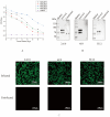
Screening and identification of monoclonal antibodies. (A) Antibody levels of the immunized mice. One week after the third immunization, the potency of antibodies from all mice was measured by indirect ELISA, and the results are presented as the absorbance value at OD450. (B) Recognition between mAbs 2A10, 4E9, and 5E12 and S1 protein. CEK cells were infected with IBV, and the protein samples were collected at 36 h to detect recognition between S1 protein and mAbs by Western blotting. (C) Reactivity of three mAbs detected by IFA. CEK cells were infected with IBV and subsequently incubated with selected mAbs to evaluate the reactivity of these mAbs against the S1 protein of IBV.
Figure 3
Figure 3
Identification of B cell epitopes of mAbs specific to S1 protein. (A) Schematic diagram of S1 epitope identification. The red-labeled regions represent the final identified antigenic epitope fragments. (B) Differentiation among truncated S1-38, S1-114, and S1-190 with three screened mAbs. (C) Differentiation between truncated S1-107 and S1-128 with three screened mAbs. (D) Identification of the coupled peptides with mAbs following Western blot analysis. The reactivities of mAbs with different truncated S1 proteins are shown in Table 5.
Figure 4
Figure 4
Comparison of antigenic epitopes in S1 protein. Homology analysis of the selected sequences was performed in 56 strains by the software MEGA-11. The antigenic epitope 132RVSAMK137 is marked in the red box and the antigenic epitope 142FYNLTV147 is marked in the blue box.
Figure 5
Figure 5
Localization of the identified S1 protein-specific epitopes. (A) Epitopes identified 132RVSAMK137 by 2A10 mAb are marked in pale green on the surface of the S1 protein. (B) Epitopes 142FYNLTV147 identified by 4E9 and 5E12 mAbs are marked in light blue on the surface of the S1 protein. (C) Overall structure of S1-NTD, displayed as a cartoon. α-helices are shown in pale cyan, β-sheets in smudge, and loop regions in light teal. The epitope 132RVSAMK137 located within a β-sheet region, is highlighted in pale green. The epitope 142FYNLTV147, located within a loop region, is highlighted in light blue. Boxed regions indicate the areas magnified in the insets, highlighting the secondary structures where the epitopes are located. Insets: Close-up views of the boxed regions, showing the detailed structures of the epitopes 132RVSAMK137 (β-sheet) and 142FYNLTV147 (loop).

Similar articles

Cited by

References

    1. Cavanagh D. Coronavirus avian infectious bronchitis virus. Vet. Res. 2007;38:281–297. - PubMed
    1. Abolnik C., Phiri T.P., van der Zel G., Anthony J., Daniell N., de Boni L. Wild bird surveillance in the Gauteng Province of South Africa during the high-risk period for highly pathogenic avian influenza virus introduction. Viruses. 2022;14:2027. doi: 10.3390/v14092027. - DOI - PMC - PubMed
    1. Andreychev A., Boyarova E., Brandler O., Tukhbatullin A., Kapustina S. Terrestrial and subterranean mammals as reservoirsof zoonotic diseases in the central part of European Russia. Diversity. 2023;15:39.
    1. Schalk A.F., Hawn M.C. An apparently new respiratory disease of baby chicks. J. Am. Vet. Med. Assoc. 1931;78:19.
    1. Beach J.R., Schalm O.W. A filterable virus, distinct from that of laryngotracheitis, the cause of a respiratory disease of chicks. Poult. Sci. 1936;15:199–206.

Publication types

MeSH terms

Substances

LinkOut - more resources