. 2025 Mar 29;17(4):495.
doi: 10.3390/v17040495.

Experimental Infection of Rats with Influenza A Viruses: Implications for Murine Rodents in Influenza A Virus Ecology

Affiliations

Experimental Infection of Rats with Influenza A Viruses: Implications for Murine Rodents in Influenza A Virus Ecology

Long Li et al. Viruses. .

Abstract

Rattus norvegicus (brown rat), a widely distributed rodent and common biomedical model, is a known reservoir for many zoonotic pathogens but has not been traditionally recognized as a host for influenza A virus (IAV). To evaluate their susceptibility, we intranasally inoculated Sprague-Dawley rats with various IAV subtypes, including H5Nx, H7N9, H9N2, H10N8 and the 2009 pandemic H1N1. All strains productively infected the rats, inducing seroconversion without overt clinical signs. While replication efficiency varied, all viruses caused significant lung injury with a preferential tropism for the upper respiratory tract. Investigation of receptor distribution revealed a predominance of α2,3-linked sialic acid (SA) in the nasal turbinates and trachea, whereas α2,6-linked SA was more abundant in the lungs. Notably, both receptor types coexisted throughout the respiratory tract, aligning with the observed tissue-specific replication patterns and broad viral infectivity. These findings demonstrate that rats are permissive hosts for multiple IAV subtypes, challenging their exclusion from IAV ecology. The asymptomatic yet pathogenic nature of infection, combined with the global synanthropy of rats, underscores their potential role as cryptic reservoirs in viral maintenance and transmission. This study highlights the need for expanded surveillance of rodents in influenza ecology to mitigate zoonotic risks.

Keywords: host range; interspecies transmission; sialic acid receptor; surveillance; zoonotic influenza.


Conflict of interest statement

The authors declare no conflicts of interest.

Figures

Figure 1
Figure 1
Changes in body weight of Sprague-Dawley (SD) rats inoculated with selected influenza A viruses (see Table 1). To evaluate the pathogenicity of different influenza viruses in rats, 8- to 9-week-old SD rats were inoculated with 106 TCID50 of the indicated viruses (AK). Body weight was monitored daily for 14 days post-inoculation (dpi). No deaths occurred in any group during the observation period. Data present mean changes in body weight ± standard deviation for five rats in the control group (grey) and five rats in each virus-inoculated group (blue). The area under the curve (AUC) for each group was calculated using GraphPad Prism 10.1.0 (L). One-way analysis of variance (ANOVA) of the group-specific AUC and pairwise t-tests between the virus-inoculated groups and the control group revealed no statistically significant changes in body weight (p > 0.05).
Figure 2
Figure 2
Replication of selected influenza A viruses in the respiratory tract of SD rats. To evaluate the infectivity of different influenza viruses (see Table 1) in rats, 8- to 9-week-old SD rats were inoculated with doses of 106 (A), 104 (B), and 102 (C) TCID50 of the indicated viruses. Animals were humanely euthanized, and tissues harvested for virus titration 4 days post-inoculation. Virus titers in the nasal turbinate (blue), trachea (green), and lung (red) were determined by titration on MDCK cells, and TCID50 was calculated using the Reed-Muench method. Log10TCID50/mL is shown, with error bars representing standard deviations for three individual rats in each virus group. No viable virus was detected in the control group.
Figure 3
Figure 3
Serological antibody titers in rats challenged with selected influenza A viruses. Blood samples from rats inoculated with 106 TCID50 of each virus (see Table 1) were collected for serological analysis at the end of the 14-day experiment (14 dpi). Antibodies to influenza A viruses were measured using the hemagglutination inhibition (HAI) test. Data from seropositive rats are presented here, with each red dot representing one rat; seronegative (HAI titer < 10) and control group data are provided in Table S1.
Figure 4
Figure 4
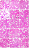
Histopathological analysis of the lungs of SD rats inoculated with different subtypes of influenza A virus. Three rats per virus group were inoculated at the indicated doses, and lungs were harvested at 4 dpi. Hematoxylin-eosin (H&E) staining was performed on 3 μm sections. Whole sections were used for analysis, but only representative images are shown, as pathological changes were comparable among the different virus groups at the same dose. Dose-dependent effects are illustrated with representative lung sections from rats inoculated with SH2 (H7N9) at 102 TCID50 (A), 104 TCID50 (B), and 106 TCID50 (C). Pathological changes in the lungs of mock-infected SD rats (D) and those inoculated with all tested influenza A viruses are shown with representative lung sections from the 104 TCID50 group ((E), CA7, pandemic 2009 H1N1; (F), VN1203, H5N1; (G), GY5096, H5N1; (H), SZ2400, H5N6; (I), SP378, H5N6; (J), HZ4258, H5N8; (K), AH1, H7N9; (L), SH2, H7N9; (M), SP440, H7N9; (N), WZ598, H9N2; (O), JX346, H10N8). Scale bars indicate 200 μm.
Figure 5
Figure 5
Immunohistochemical analysis of influenza virus nucleoprotein (NP) in the respiratory tract of SD rats. Airway tissues were harvested at 4 dpi and 3 μm sections were used for immunohistochemical analysis. A representative image was selected for viruses with similar virus distribution patterns. Influenza NP antigens (brown) are shown in the nasal turbinate ((A), CA7, pandemic 2009 H1N1, 106 TCID50; (B), HZ4258, H5N8, 106 TCID50, with arrows indicating the positively stained cells; (C), AH1, H7N9, 102 TCID50; (D), AH1, H7N9, 104 TCID50; (E), SH2, H7N9, 102 TCID50; (F), SH2, H7N9, 104 TCID50; (G), SH2, H7N9, 106 TCID50; (H), SP440, H7N9, 102 TCID50; (I), SP440, H7N9, 104 TCID50; (J), SP440, H7N9, 106 TCID50; (K), WZ598, H9N2, 106 TCID50; (L), JX346, H10N8, 104 TCID50) and lungs ((M), VN1203, H5N1, 104 TCID50; (N), AH1, H7N9, 102 TCID50; (O), WZ598, H9N2, 104 TCID50). Scale bars indicate 200 μm.
Figure 6
Figure 6
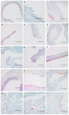
Distribution of α2,3- and α2,6-linked sialosides in the respiratory tracts of SD rats. The presence of α2,3- and α2,6-linked sialosides in the nasal turbinate (AC), trachea (DF) and lung (GI) of naïve rats were detected using biotinylated Maackia amurensis agglutinin I or II (MAA I, MAA II) for α2,3-linked sialosides or Sambucus nigra agglutinin (SNA) for α2,6-linked sialosides. Both α2,3- and α2,6-linked sialosides are shown in brown. Scale bars indicate 200 μm.

Similar articles

Cited by

References

    1. Smith G.J., Vijaykrishna D., Bahl J., Lycett S.J., Worobey M., Pybus O.G., Ma S.K., Cheung C.L., Raghwani J., Bhatt S., et al. Origins and evolutionary genomics of the 2009 swine-origin H1N1 influenza A epidemic. Nature. 2009;459:1122–1125. doi: 10.1038/nature08182. - DOI - PubMed
    1. Gao R., Cao B., Hu Y., Feng Z., Wang D., Hu W., Chen J., Jie Z., Qiu H., Xu K., et al. Human infection with a novel avian-origin influenza A (H7N9) virus. N. Engl. J. Med. 2013;368:1888–1897. doi: 10.1056/NEJMoa1304459. - DOI - PubMed
    1. World Health Organization (WHO) Global Influenza Programme. [(accessed on 27 February 2025)]. Available online: https://www.who.int/teams/global-influenza-programme/avian-influenza/mon....
    1. Webster R.G., Bean W.J., Gorman O.T., Chambers T.M., Kawaoka Y. Evolution and ecology of influenza A viruses. Microbiol. Rev. 1992;56:152–179. doi: 10.1128/mr.56.1.152-179.1992. - DOI - PMC - PubMed
    1. Yoon S.W., Webby R.J., Webster R.G. Evolution and ecology of influenza A viruses. Curr. Top. Microbiol. Immunol. 2014;385:359–375. doi: 10.1007/82_2014_396. - DOI - PubMed

Publication types

LinkOut - more resources