. 2025 Apr 1;17(4):514.
doi: 10.3390/v17040514.

Design and Characterization of Inhibitors of Cell-Mediated Degradation of APOBEC3G That Decrease HIV-1 Infectivity

Affiliations

Design and Characterization of Inhibitors of Cell-Mediated Degradation of APOBEC3G That Decrease HIV-1 Infectivity

Aubrey M Sawyer et al. Viruses. .

Abstract

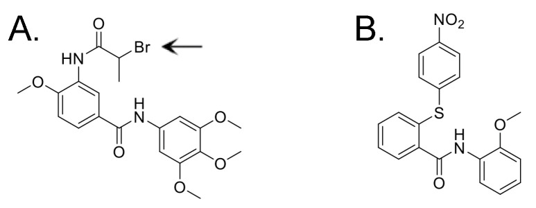
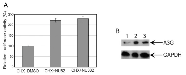
The cytoplasmic human Apolipoprotein B mRNA-editing enzyme catalytic polypeptide-like 3 (APOBEC3 or A3) cytidine deaminases G and F (A3G and A3F) can block the spread of human immunodeficiency virus (HIV). HIV counteracts this cell-intrinsic defense through a viral protein called viral infectivity factor (Vif). Vif causes proteasomal degradation of A3G and A3F proteins (A3G/F) in HIV-producing cells to ensure infectivity of virions subsequently released from these cells. Here, we optimized a lead compound reported previously to boost cellular levels of A3G. The modified analogs designed, synthesized, and evaluated here inhibit cell-mediated post-translational degradation of A3G/F, whether Vif is present or not. This increases A3G/F incorporation into Vif-positive virions to decrease viral infectivity. The compounds and processes described here can facilitate the development of new anti-HIV therapeutics whose host-targeted effect may not be evaded by resistance-conferring mutations in HIV Vif.

Keywords: APOBEC3F; APOBEC3G; HIV; HIV cure research; HIV infectivity; HIV-1; Vif.


Conflict of interest statement

The authors declare no conflicts of interest. The funders had no role in the design of the study; in the collection, analyses, or interpretation of data; in the writing of the manuscript; or in the decision to publish the results.

Figures

Figure 1
Figure 1
Structural formulas of compounds previously identified to inhibit Vif-mediated A3G degradation. Methyl groups indicated by a line. (A) IMB-26. Arrow indicates R2 site. (B) RN-18.
Figure 2
Figure 2
Characterization of puromycin-resistant 293T-cell lines stably expressing A3G-Luc and Vif/A3G-Luc. (A) Relative luciferase activities (RLUs) of two different clones (#5 and #6) transduced by pseudotyped viral particles to stably express different levels of A3G-Luc. A similarly transduced control clonal line lacks A3G-Luc expression. (B) Immunoblots of these three clones probed with an anti-A3G antibody are shown. Lane 1 is A3G-Luc #5. Lane 2 is A3G-Luc #6. Lane 3 is the control line. (C) RLU of clone #5 or clone #6, each with or without stable expression of Vif using the FUGW-Vif vector (indicated as Vif+) and the corresponding empty vector (indicated as EV) control. (D) Immunoblots for A3G, Vif, and β-actin proteins from C are shown. Lane 1 is EV/A3G-Luc #5. Lane 2 is Vif/A3G-Luc #5. Lane 3 is EV/A3G-Luc #6. Lane 4 is Vif/A3G-Luc #6. (E) Luciferase activities produced by A3G-Luc cell clone #5 following transfection with the EV control, the Vif-null NL4.3 plasmid, or the Vif+ NL4.3 plasmid are shown. (F) Luciferase activities produced by A3G-Luc cell clone #6 following transfection with the EV control, the Vif-null NL4.3 plasmid, or the Vif+ NL4.3 plasmid are shown. (G) Relative infectivity of viral particles produced from transfection of Vif+ NL4.3 expression plasmid (indicated as Vif+) into A3G-Luc cell clones #5 and 6, as well as the control line, are shown. Supernatants of Vif+ NL4.3-transfected cultures of each cell clone were normalized by p24 value prior to infection of TZM-bl cells to ensure the same viral input. RLUs were read 24 h after infection of TZM-bl cells. (H) Relative infectivity of viral particles produced from transfection of Vif-null NL4.3 expression plasmid (indicated as Vif-null) into A3G-Luc cell clones #5 and 6, as well as the control line, are shown. Infectivity was assessed as in G.
Figure 3
Figure 3
Structural formulas of compounds selected for further study. The R2 site is circled in NU-52. Note that methyl groups are shown here, rather than being depicted only as lines, as in Figure 1 and Supplementary Table S1.
Figure 4
Figure 4
NU compounds boost A3G protein levels in uninfected CD4+ T cells and A3F protein levels in A3F-transfected 293T cells. (A) Immunoblot shows resting, uninfected CD4+ T cells treated with NU-52, -302, and -611 (10 μM each). (B) Quantification of band densities in A. (C) Immunoblot shows 293T cells transiently transfected with A3F expression plasmid and treated with DMSO, IMB-26, or NU-52. (D) Quantification of band densities in C.
Figure 5
Figure 5
NU compounds increase incorporation of A3G or A3F into Vif+ virions and decrease Vif+ virion infectivity. (A) Vif+ NL4.3 was co-transfected with an A3G expression plasmid into 293T cells. DMSO or NU compounds (10 μM each) were added 24 h after transfection. One day later, immunoblots of A3G and p24 of pelleted supernatant viral particles were performed from cultures transfected with Vif+ NL4.3 alone (Lane 1) or co-transfected with both Vif+ NL4.3 and A3G followed by treatment with either DMSO (Lane 2), NU-52 (Lane 3), or NU-302 (Lane 4). (B) Relative infectivity of supernatant viruses from the different conditions described in A was determined. Vif+ HIV infectivity was measured by luciferase activity after infection of TZM-bl cells. (C) Immunoblots of A3F and p24 in pelleted supernatant viral particles from 293T cells transfected with Vif+ NL4.3 alone (Lane 1) or co-transfected with both Vif+ NL4-3 and A3F followed by treatment with either DMSO (Lane 2), NU-52 (Lane 3), or NU-302 (Lane 4) are shown. (D) Relative infectivity of supernatant viruses from 293T cells from the different conditions described in C was determined. Vif+ HIV infectivity was measured as in B. (E) Immunoblots of A3G and p24 in pelleted supernatant viral particles from 293T cells stably expressing A3G that were transiently transfected with Vif+ NL4.3 and treated with either DMSO (Lane 1), NU-52 (Lane 2), or NU-302 (Lane 3) are shown. (F) Relative infectivity of supernatant viruses from 293T cells stably expressing A3G from the different conditions described in E was determined. (G) Immunoblots of A3F and p24 in pelleted supernatant viral particles from 293T cells stably expressing A3F that were transiently transfected with Vif+ NL4.3 and treated with either DMSO (Lane 1), NU-52 (Lane 2), or NU-302 (Lane 3) are shown. (H) Relative infectivity of supernatant viruses from 293T cells stably expressing A3F from the conditions described in G was determined. All data shown as means ± SD from ≥3 independent experiments; ANOVA was used to analyze the differences (* p < 0.05, ** p < 0.01, **** p < 0.0001, ns p > 0.05).
Figure 6
Figure 6
NU-611 enhances A3G-mediated reduction in HIV virion infectivity more than NU-52. 293T cells stably expressing A3G (293T IP/G cells) were transfected with Vif+ NL4.3 and then treated with either DMSO control or 15 μM each of either NU-52 or -611. DMSO was plotted as 100% relative infectivity. Data from 8 independent experiments are expressed as means ± SD; ANOVA was used to analyze the differences, **** p < 0.0001.
Figure 7
Figure 7
NU compounds decrease infectivity of Vif+ HIV produced by H9 cells expressing endogenous A3G and A3F after infection. After Vif+ NL4.3 infection, H9 cells were treated with a compound (NU-52, -302, or -611, each at 10 μM) or control (DMSO). TZM-bl cells were infected with p24-normalized H9 culture supernatants and relative infectivity quantified by luciferase activity of TZM-bl cell lysates. Infectivity of virus from DMSO control is plotted as 100%. All data expressed as means ± SD from ≥3 independent experiments; ANOVA was used to analyze the differences (*** p < 0.001, **** p < 0.0001, ns p > 0.05).
Figure 8
Figure 8
Stability of A3G and A3F in the presence of protein synthesis-blocking cycloheximide (CHX) increases with treatment with NU-52 or -302 at 10 μM concentration. (A) Relative luciferase activities are shown from the cell line stably expressing intermediate levels of A3G-Luc (A3G-Luc #6) after treatment with either CHX+DMSO, CHX+NU-52, or CHX+NU-302. (B) Immunoblots from 293T cells transfected with A3G expression plasmid are shown after treatment with either CHX and DMSO (lane 1), CHX plus NU-52 (lane 2), or CHX plus NU-302 (lane 3).
Figure 9
Figure 9
NU-52 and -611 diminish pVHL-mediated degradation of A3G or -F. (A) Immunoblots from cell lysates of 293T cells transfected with pVHL and A3G are shown. (B) Band densities of immunoblots depicted in A are quantified. (C) Immunoblots from cell lysates of 293T cells transfected with pVHL along with A3F are shown. (D) Band densities of immunoblots depicted in C are quantified. Mean ± SD for at least 2 independent immunoblots are shown. ANOVA was used to analyze the differences (* p < 0.05, ** p < 0.01, *** p < 0.001).

Similar articles

References

    1. Ito J., Gifford R.J., Sato K. Retroviruses drive the rapid evolution of mammalian APOBEC3 genes. Proc. Natl. Acad. Sci. USA. 2020;117:610–618. - PMC - PubMed
    1. Conticello S.G., Thomas C.J., Petersen-Mahrt S.K., Neuberger M.S. Evolution of the AID/APOBEC family of polynucleotide (deoxy)cytidine deaminases. Mol. Biol. Evol. 2005;22:367–377. - PubMed
    1. Poulain F., Lejeune N., Willemart K., Gillet N.A. Footprint of the host restriction factors APOBEC3 on the genome of human viruses. PLoS Pathog. 2020;16:e1008718. - PMC - PubMed
    1. Refsland E.W., Harris R.S. The APOBEC3 family of retroelement restriction factors. Curr. Top. Microbiol. Immunol. 2013;371:1–27. doi: 10.1007/978-3-642-37765-5_1. - DOI - PMC - PubMed
    1. Burns M.B., Lackey L., Carpenter M.A., Rathore A., Land A.M., Leonard B., Refsland E.W., Kotandeniya D., Tretyakova N., Nikas J.B., et al. APOBEC3B is an enzymatic source of mutation in breast cancer. Nature. 2013;494:366–370. doi: 10.1038/nature11881. - DOI - PMC - PubMed

Publication types

MeSH terms

Substances

LinkOut - more resources