. 2025 Apr 5;17(4):534.
doi: 10.3390/v17040534.

Genomic and Epidemiological Investigations Reveal Chromosomal Integration of the Acipenserid Herpesvirus 3 Genome in Lake Sturgeon Acipenser fulvescens

Affiliations

Genomic and Epidemiological Investigations Reveal Chromosomal Integration of the Acipenserid Herpesvirus 3 Genome in Lake Sturgeon Acipenser fulvescens

Sharon Clouthier et al. Viruses. .

Abstract

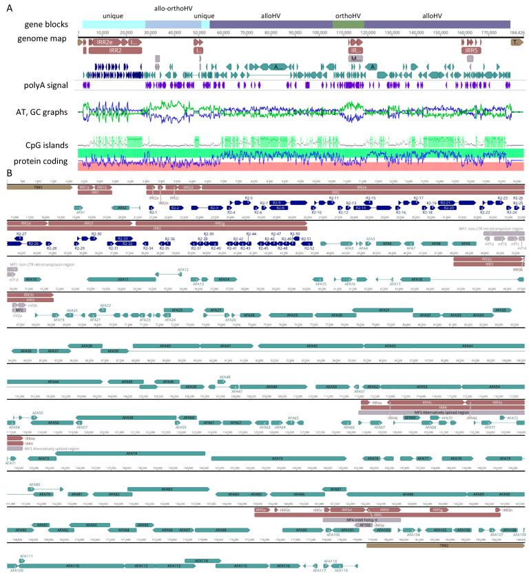
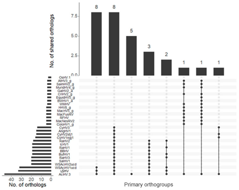
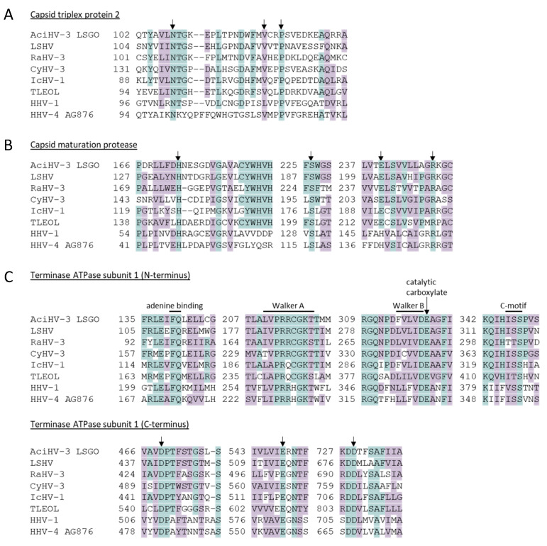
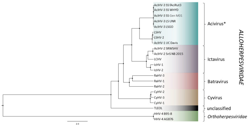
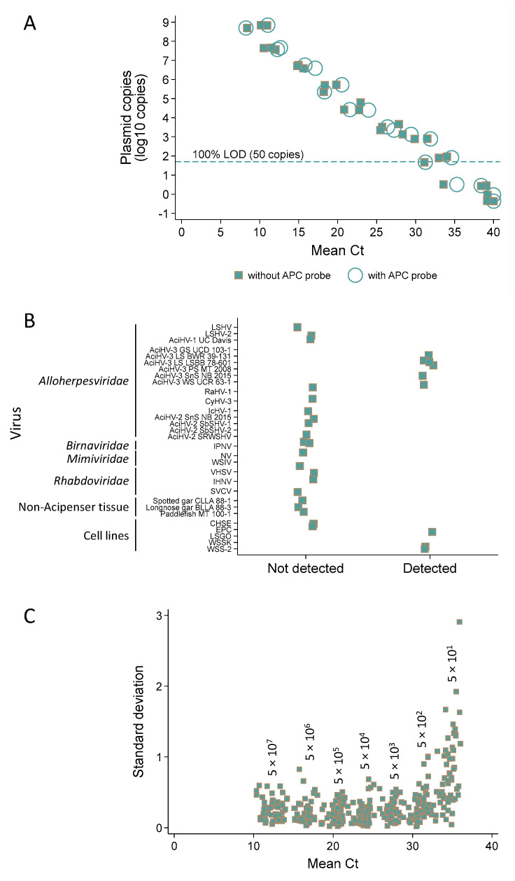
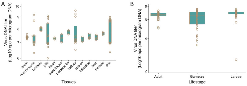
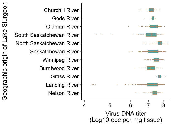
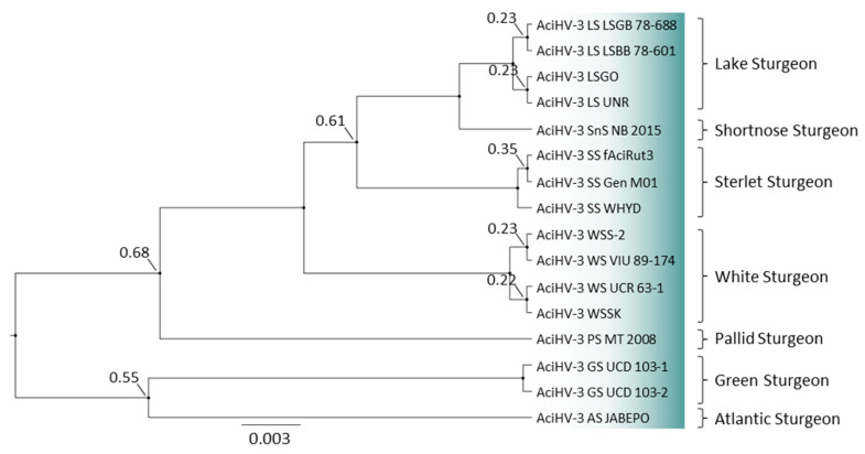
DNA sequence from a new alloherpesvirus named acipenserid herpesvirus 3 (AciHV-3) was found in sturgeon species that are vulnerable to decline globally. A study was undertaken to develop a better understanding of the virus genome and to develop diagnostic tools to support an epidemiological investigation. A 184,426 bp genome was assembled from PacBio HiFi sequences generated with DNA from a Lake Sturgeon Acipenser fulvescens gonad cell line. The AciHV-3 genome was contiguous with host chromosomal DNA and was structured with telomere-like terminal direct repeat regions, five internal direct repeat regions and a U region that included intact open reading frames encoding alloherpesvirus core proteins. Diagnostic testing conducted with a newly developed and analytically validated qPCR assay established the ubiquitous presence and high titer of AciHV-3 DNA in somatic and germline tissues from wild Lake Sturgeon in the Hudson Bay drainage basin. Phylogenetic reconstructions confirm that the monophyletic AciHV-3 lineage shares a common ancestor with AciHV-1 and that AciHV-3 taxa cluster according to their sturgeon host. The same genotype of AciHV-3 is found in disjunctive Lake Sturgeon populations within and among drainage basins. The results support the hypotheses that AciHV-3 has established latency through germline chromosomal integration, is vertically transmitted via a Mendelian pattern of inheritance, is evolving in a manner consistent with a replication competent virus and has co-evolved with its host reaching genetic fixation in Lake Sturgeon populations in central Canada.

Keywords: Alloherpesviridae; Lake Sturgeon; endogenous; germline; herpesvirus.


Conflict of interest statement

The authors declare no conflicts of interest.

Figures

Figure 1
Figure 1
Study area map (2010–2021) showing geographic distribution of acipenserid herpesvirus 3 (AciHV-3) qPCR-positive Lake Sturgeon Acipenser fulvescens in the Hudson Bay drainage basin (HBDB). A map inset of North America provides the relative location of the HBDB study area (black) in Canada (stipple) with the red dot marking the location where samples were collected in 2019 from Lake Sturgeon in Lake Superior. The map was created using ESRI [44] ArcGIS 10.8.1 software with publicly available CANVec Series shapefiles from Natural Resources Canada [45]. AciHV-3 DNA prevalence levels and number of fish tested annually from each river across the HBDB are available in Table S1.
Figure 2
Figure 2
Schematics of contig sequences (5′–3′) illustrating the location of the acipenserid herpesvirus 3 (AciHV-3) genome relative to Lake Sturgeon Acipenser fulvescens chromosomal DNA. Features of the virus–host junction are provided on the left side of the figure. Contig id and length in base pairs (bps) are provided to the left of each rectangle in which the AciHV-3 genome unique (U) region is shown in blue, its terminal repeat region(s) (TRR) in brown and host sequence in grey. The dark grey box within the host sequence illustrates regions rich in transposable elements (TEs) including Penelope-like elements (PLEs), (non)long terminal repeat retrotransposons (nLTRs, LTRs) and various DNA TEs, some of which encode the CRISPR-associated protein Csa3 (CRISPR). The orientation and integrity of the U region is provided within each blue box. The number of base pairs within each colored box denotes the length of each region within the contig sequence. Additional details are provided in Table S3. Those contigs encoding TRR1 and TRR2 contain the complete virus genome. nt, nucleotide.
Figure 3
Figure 3
Schematics of contig sequences (5′–3’) in which the acipenserid herpesvirus 3 (AciHV-3) genome sequence is present in the absence of the host sequence. A description of the genome arrangement is provided on the left side of the figure. Contig id and length in base pairs (bps) are provided to the left of each rectangle in which the AciHV-3 genome unique (U) region is shown in blue and its terminal repeat region(s) (TRRs) in brown. The orientation and integrity of the U region is provided within each blue box. The number of base pairs within each colored box denotes the length of each region within the contig sequence. Additional details are provided in Table S3. nt, nucleotide.
Figure 4
Figure 4
Genome map for acipenserid herpesvirus 3 from the Lake Sturgeon Acipenser fulvescens gonad cell line. The genome map represents an informed interpretation of bioinformatic-based predictions of ORFs, splice sites and repeat sequences. (A) Genome-wide mini-maps are presented for the gene blocks (Alloherpesviridae (alloHV), Orthoherpesviridae (orthoHV) or neither family (unique)), poly(A) signal sequence (i.e., aataaa), AT (green) or GC (blue) content of the nucleotide sequence, CpG islands and predicted protein coding regions. (B) Enlarged genome map in which the terminal repeat regions (TRR1 and 2; brown), internal repeat regions (IRR1 to 5; red), ORFs R2-1 to R2-52 in IRR2 (dark blue), unique region with predicted functional ORFs AFA1 to 119 (blue) and miscellaneous features (MF1 to 4 and AFA102, grey) are presented for the 184,426 bp genome. Introns are displayed as a thin line between coding regions. Image created in Geneious v2023.2.
Figure 5
Figure 5
Orthology analyses of acipenserid herpesvirus 3 (AciHV-3). The UpSet plot displays the distribution of orthogroups between AciHV-3 and 29 members of the Herpesvirales order. The number of per-species orthologs are shown on the x-axis, whereas the shared orthologs are enumerated using vertical bars (annotated with number) for each orthogroup. The black dots represent the occurrence of a specific orthogroup in each species.
Figure 6
Figure 6
Location of conserved amino acids in universal Herpesvirales proteins involved in viral morphogenesis. The amino acid sequence alignments display functional signatures within the (A) capsid triplex protein 2 (critical amino acids involved in protein–protein interactions between capsid triplex proteins 1 and 2); (B) capsid maturation protease (key amino acids of the protease catalytic triad (H, S, E/H) and the oxyanion-binding site (R) conserved across the order Herpesvirales); (C) terminase ATPase subunit 1 (functionally significant amino acids within 4 regions of the N-terminal ATPase catalytic center and the catalytic metal-binding triad (D, E, D) of the C-terminal nuclease center). Conserved residues are highlighted in blue, and similar amino acids are highlighted in purple. Arrows show the location of conserved residues. The numbers correspond to the relative location of the first amino acid in the protein sequence that follows. The corresponding full protein sequence alignments are provided in Figure S5. Taxa are described in Table S2.
Figure 7
Figure 7
Bayesian phylogeny of concatenated amino acid sequences of 5 core alloherpesvirus proteins (capsid maturation protease, capsid triplex subunit 2, DNA polymerase catalytic subunit, helicase-primase helicase subunit and the major capsid protein). The tree was constructed under the LG amino acid substitution model [88] combined with a gamma-invariant site heterogeneity model with 4 rate categories, a strict clock and the Yule Process tree prior [89,90]. Full length protein sequences were used to generate the tree. Nodes are marked with a black circle of fixed size, and all node posterior probabilities are 1.0. The taxa are described in Table S2, and the Alloherpesviridae genera are provided to the right of the tree. * Acivirus is the genus name proposed in Clouthier et al. [25] for the unclassified AciHV-1 and AciHV-3 taxa.
Figure 8
Figure 8
Analytical validation of the Q1mcp qPCR assay for detection of acipenserid herpesvirus 3 DNA. Unless noted otherwise, the assay was duplexed with the artificial positive control (APC) probe. (A) Analytical sensitivity—for each dilution, the mean cycle threshold (Ct) value for 3 replicates is reported for 2–3 runs of pLSAciHV3-APC plasmid DNA 10-fold serially diluted from 5 × 10⁸ to 0.5 copies. The dashed line marks the 100% limit of detection (LOD). Circles, with APC probe; squares, no APC probe. (B) Analytical specificity—binary test results of detected (<40 Ct) or not detected (40 Ct) with select viruses, tissues and naïve cell lines. (C) Analytical repeatability—for each dilution, the standard deviation and mean Ct value obtained for 3 replicates are plotted for pLSAciHV3-APC plasmid 10-fold serially diluted from 5 × 10⁷ to 50 copies (n = 58 runs).
Figure 9
Figure 9
Acipenserid herpesvirus 3 DNA titer in tissues from Lake Sturgeon Acipenser fulvescens at different life stages. Virus DNA titer was determined using the Q1mcp qPCR test and expressed as equivalent plasmid copies (epcs) per µg DNA for (A) somatic tissues collected from juvenile Landing River Lake Sturgeon (age—0, 2017 year class (n = 10); age—1, 2019 year class (n = 3)) housed at the Grand Rapids Hatchery in Manitoba and (B) adult fin tissue, gametes (eggs, milt) and larvae (whole; 46–74 days post-fertilization) from select mating crosses of wild Lake Sturgeon broodstock from the Winnipeg, Landing or Burntwood Rivers. Further details are available in Table 2. The horizontal line within each box is the median virus DNA titer, the individual boxes represent 50% of the observations and the circles denote virus DNA titer values.
Figure 10
Figure 10
Acipenserid herpesvirus 3 DNA titer in fin tissue of wild Lake Sturgeon Acipenser fulvescens in the Hudson Bay drainage basin. The DNA titer was quantified as equivalent plasmid copies (epcs) per mg tissue using the Q1mcp qPCR test and the standard curve method. The range of values obtained for each population is presented according to their river of origin. The vertical line within each box is the median virus DNA titer, the individual boxes represent 50% of the observations, and the circles denote virus DNA titer values.
Figure 11
Figure 11
Phylogenetic tree of acipenserid herpesvirus 3 genotypes. A 493 bp sequence of the major capsid protein gene from each sample was aligned in ClustalX2 and the Bayesian inference tree was reconstructed under the HKY model [91]. Ancestral nodes are fixed in size and labelled if the posterior probability of their existence was <0.997. Taxa are described in Table S2, and their host of origin is provided to the right of the tree.

Similar articles

References

    1. ICTV 2024 (International Committee on Taxonomy of Viruses). 2023 Taxonomy Release. [(accessed on 20 September 2024)]. Available online: https://ictv.global/taxonomy.
    1. McGeoch D.J., Rixon F.J., Davison A.J. Topics in herpesvirus genomics and evolution. Virus Res. 2006;117:90–104. - PubMed
    1. McGeoch D.J., Davison A.J., Dolan A., Gatherer D., Sevilla-Reyes E.E. Molecular evolution of the Herpesvirales. In: Domingo E., Parrish C.R., Holland J.J., editors. Origin and Evolution of Viruses. 2nd ed. Academic Press; London, UK: 2008. pp. 447–475.
    1. Roizman B., Pellett P.E. The family of Herpesviridae: A brief introduction. In: Knipe D.M., Howley P.M., editors. Fields Virology. Lippincott Williams & Wilkins; Philadelphia, PA, USA: 2001. pp. 2381–2397.
    1. Roizman B., Knipe D.M., Whitley R.J. Herpes simplex viruses. In: Knipe D.M., Howley P.M., editors. Fields Virology. 5th ed. Lippincott Williams & Wilkins; Philadelphia, PA, USA: 2007. pp. 2501–2601.

Publication types

Grants and funding

LinkOut - more resources