. 2025 Apr 12;17(4):559.
doi: 10.3390/v17040559.

Screening of Neutralizing Antibodies Targeting Gc Protein of RVFV

Affiliations

Screening of Neutralizing Antibodies Targeting Gc Protein of RVFV

Chuanyi Zhao et al. Viruses. .

Abstract

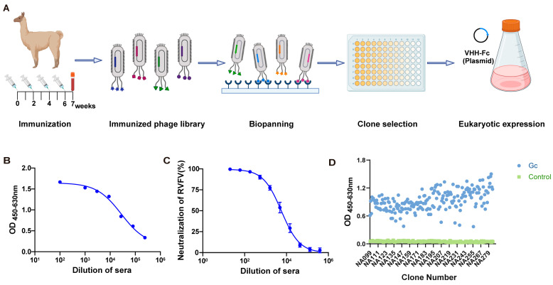
Rift Valley fever virus (RVFV) is a mosquito-transmitted bunyavirus that can cause substantial morbidity and mortality in livestock and humans, for which there are no currently available licensed human therapeutics or vaccines. Therefore, the development of safe and effective antivirals is both necessary and urgent. The Gc protein is the primary target of the neutralizing antibody response related to Rift Valley fever virus. Here, we report one Gc-specific neutralizing antibody (NA137) isolated from an alpaca and one bispecific antibody (E2-NA137), the protective efficacies of which we evaluated in A129 mice. In this prophylactic study, the survival rates of the NA137 and E2-NA137 groups were both 80%, and in the treatment study, the survival rates were 20% and 60%, respectively. Altogether, our results emphasize that NA137 and E2-NA137 provide a potential approach for treating RVFV either prophylactically or therapeutically.

Keywords: Gc protein; Rift Valley fever virus (RVFV); VHH; bispecific antibody (bsAb); nanobody (Nb); neutralizing antibody (NAb).


Conflict of interest statement

The authors declare no conflicts of interest.

Figures

Figure 1
Figure 1
Construction and panning of the Gc-specific Phage Display Nanobody Library. (A) Schematic of phage-displayed VHH (variable domain of the heavy chain of heavy-chain antibodies) antibody screening and expression. (B) Detection of Gc-specific immunoglobulin G (IgG) titer in alpaca serum using an enzyme-linked immunosorbent assay (ELISA). (C) Detection of serum-neutralizing activity in immunized alpaca. (D) Monoclonal antibody identification from the fourth- and fifth-round screenings.
Figure 2
Figure 2
Screening for Gc-specific neutralizing antibodies and antibody sequence analysis targeting Gc protein. (A) Schematic comparison of nanobodies (Nbs) from heavy-chain antibodies and single-chain variable fragments (scFv) from conventional antibodies. (B) Binding activity of antibodies to Gc protein analyzed via ELISA. (C) seqLogo plot by entropy. The y-axis represents the entropy of amino acids, and the x-axis represents the position in the variable domain. The larger the letter of the amino acid, the greater the entropy value. (D) Protection efficacy of monoclonal antibodies (NA137) against RVFV infection in Vero E6 cells. (E) Determination of affinity between NA137 and Gc protein using surface plasmon resonance (SPR).
Figure 3
Figure 3
Design and enhanced neutralizing potency for IgG-VHH. (A) Schematic diagram of bispecific antibody. (B) Reduced SDS-PAGE analysis of bispecific antibody. (C) Binding of E2-NA19, E2-NA137, and E2-NA220 to Gc protein analyzed via ELISA. 1A8, a monoclonal antibody to the Marburg virus GP protein, was used as a negative control. (D) Protection efficacies of bispecific antibodies against RVFV infection in Vero E6 cells. (E) Determination of affinity between E2-NA19 and Gc protein using surface plasmon resonance (SPR). (F) Determination of affinity between E2-NA137 and Gc protein using surface plasmon resonance (SPR). (G) Determination of affinity between E2-NA220 and Gc protein using surface plasmon resonance (SPR).
Figure 4
Figure 4
NA137 and E2-NA137 prevented infection when administered prior to the challenge. (A) Schematic diagram of antibody delivery, virus challenge, sample collection, and immunological assays. (B) Mouse mortality and survival curves. (C) Body weight changes in mice after RVFV challenge. The tissues from the PBS and E2 groups (at the time of death) and antibody-administrated groups (14 days after challenge) were collected and analyzed with histopathological and immunohistochemical assays. The Blank group consisted of female A129 mice that had not been challenged with RVFV and that were injected with an equal volume of PBS. (D) Hepatocyte necrosis is indicated by black arrows, excessive cell necrosis in the red pulp is indicated by red arrows, neuronal necrosis is indicated by green arrows, gliocyte proliferation is indicated by blue arrows, inflammatory cell infiltration is indicated by orange arrows, and necrosis of renal tubular epithelial cells is indicated by gray arrows. Representative histopathology (H&E) of hearts, livers, spleens, lungs, kidneys, and brains in RVFV-infected mice. Scale bar, 200 μm. (E) Representative immunohistochemistry (IHC) of brains, livers, and spleens with RVFV Gc-specific monoclonal antibodies. Red arrows indicate RVFV-positive signals. Scale bar, 200 μm. Viral loads in brains (F), livers (G), and spleens (H) of challenged mice were determined via qRT-PCR at the time of death (PBS and E2 groups) or 14 days after the challenge (Blank, NA137, and E2-NA137 groups). Data are displayed as means ± SD. Comparisons based on one-way ANOVA with Dunnett’s multiple comparisons test with * p < 0.1, *** p < 0.001, and **** p < 0.0001.
Figure 5
Figure 5
NA137 and E2-NA137 prevented infection when delivered post-challenge. (A) Schematic diagram of antibody delivery, virus challenge, sample collection, and immunological assays. (B) Mouse mortality and survival curves. (C) Body weight changes in mice after RVFV challenge. The tissues from the PBS and E2 groups (at the time of death) and antibody-administrated groups (14 days after the challenge) were collected and analyzed with histopathological and immunohistochemical assays. The Blank group consisted of female A129 mice that had not been challenged with RVFV and that were injected with an equal volume of PBS. (D) Hepatocyte necrosis is indicated by black arrows, inflammatory cell infiltration is indicated by yellow arrows, a small amount of cell necrosis in the red pulp is indicated by red arrows, neuronal necrosis is indicated by green arrows, gliocyte proliferation is indicated by blue arrows, alveolar wall capillary congestion is indicated by brown arrows, and necrosis of renal tubular epithelial cells is indicated by gray arrows. Representative histopathology (H&E) of hearts, livers, spleens, lungs, kidneys, and brains in RVFV-infected mice. Scale bar, 200 μm. (E) Representative immunohistochemistry (IHC) of brains, livers, and spleens with RVFV Gc-specific monoclonal antibodies. Red arrows indicate RVFV-positive signals. Scale bar, 200 μm. Viral loads in brains (F), livers (G), and spleens (H) of challenged mice were determined via qRT-PCR at the time of death (PBS and E2 groups) or 14 days after the challenge (Blank, NA137, and E2-NA137 groups). Data are displayed as means ± SD. Comparisons based on one-way ANOVA with Dunnett’s multiple comparisons test with * p < 0.1, ** p < 0.01, *** p < 0.001, and **** p < 0.0001.
Figure 6
Figure 6
Epitope analysis of E2-NA137. (A) Prediction of the interaction between NA137 and Gc using AlphaFold 3. The structure of NA137 is shown in orange, and that of Gc is shown in gray. (B) Prediction of the interaction between E2 and Gn using AlphaFold 3. The heavy chain of the E2 antibody is represented in orange, the light chain of the E2 antibody is shown in blue, and the Gn protein is shown in gray.

Similar articles

References

    1. Wright D., Kortekaas J., Bowden T.A., Warimwe G.M. Rift Valley fever: Biology and epidemiology. J. Gen. Virol. 2019;100:1187–1199. doi: 10.1099/jgv.0.001296. - DOI - PMC - PubMed
    1. Bird B.H., Githinji J.W., Macharia J.M., Kasiiti J.L., Muriithi R.M., Gacheru S.G., Musaa J.O., Towner J.S., Reeder S.A., Oliver J.B., et al. Multiple virus lineages sharing recent common ancestry were associated with a Large Rift Valley fever outbreak among livestock in Kenya during 2006–2007. J. Virol. 2008;82:11152–11166. doi: 10.1128/JVI.01519-08. - DOI - PMC - PubMed
    1. Ikegami T., Makino S. The pathogenesis of Rift Valley fever. Viruses. 2011;3:493–519. doi: 10.3390/v3050493. - DOI - PMC - PubMed
    1. Jin Y., Lei C., Hu D., Dimitrov D.S., Ying T. Human monoclonal antibodies as candidate therapeutics against emerging viruses. Front. Med. 2017;11:462–470. doi: 10.1007/s11684-017-0596-6. - DOI - PMC - PubMed
    1. Murithi R.M., Munyua P., Ithondeka P.M., Macharia J.M., Hightower A., Luman E.T., Breiman R.F., Njenga M.K. Rift Valley fever in Kenya: History of epizootics and identification of vulnerable districts. Epidemiol. Infect. 2011;139:372–380. doi: 10.1017/S0950268810001020. - DOI - PubMed

MeSH terms

Associated data

LinkOut - more resources