. 2025 Apr 12;25(8):2441.
doi: 10.3390/s25082441.

Investigation of Ultrasound Transmit-Receive Sequence That Enables Both High-Frame-Rate Vascular Wall Velocity Estimation and High-Contrast B-Mode Images

Affiliations

Investigation of Ultrasound Transmit-Receive Sequence That Enables Both High-Frame-Rate Vascular Wall Velocity Estimation and High-Contrast B-Mode Images

Hitoshi Hirano et al. Sensors (Basel). .

Abstract

In this study, we designed an ultrasound transmit-receive sequence to achieve high-frame-rate vascular wall velocity estimation and high-contrast B-mode imaging. The proposed sequence extends conventional dual-transmission schemes by incorporating a third transmission with 180° phase inversion, enabling harmonic imaging via the pulse inversion (PI) method. To mitigate the frame rate reduction caused by the additional transmission, the number of simultaneously transmitted focused beams was increased from two to four, resulting in a frame rate of 231 Hz. A two-dimensional phase-sensitive motion estimator was employed for motion estimation. In vitro experiments using a chicken thigh moving in two dimensions yielded RMSE values of 3% (vertical) and 16% (horizontal). In vivo experiments on a human carotid artery demonstrated that the PI method achieved a lumen-to-tissue contrast improvement of 0.96 dB and reduced artifacts. Velocity estimation of the posterior vascular wall showed generally robust performance. These findings suggest that the proposed method has strong potential to improve atherosclerosis diagnostics by combining artifact-suppressed imaging with accurate motion analysis.

Keywords: multi-line transmission; phase-sensitive motion estimator; pulse inversion; ultrasound.


Conflict of interest statement

The authors declare no conflicts of interest.

Figures

Figure 1
Figure 1
Schematic illustration of the pulse inversion (PI) method. (a) Summation of fundamental signals. (b) Summation of signals containing harmonics generated by nonlinear propagation.
Figure 2
Figure 2
Illustrations of transmit–receive (Tx–Rx) sequences. (a) Transmit sequence. (b) Multi-line transmission and reception.
Figure 3
Figure 3
Illustration of basic experimental setup.
Figure 4
Figure 4
B-mode images with points of interest indicated by red lines. (a) Fundamental. (b) Harmonic.
Figure 5
Figure 5
Illustration of frame averaging process in velocity estimation.
Figure 6
Figure 6
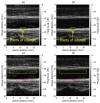
Examples of in vivo measurements. (a) Fundamental B-mode image with ROIs for velocity estimation. (b) Harmonic B-mode image with ROIs for velocity estimation. (c) Fundamental B-mode image with ROIs for contrast measurement. (d) Harmonic B-mode image with ROIs for contrast measurement.
Figure 7
Figure 7
Velocity estimation results in the axial direction without (a) and with (b) frame averaging at a target velocity of −1.0 mm/s. Velocity estimation in the lateral direction without (c) and with (d) frame averaging.
Figure 8
Figure 8
Velocity estimation results in the axial direction without (a) and with (b) frame averaging at a target velocity of −2.0 mm/s. Velocity estimation in the lateral direction without (c) and with (d) frame averaging.
Figure 9
Figure 9
In vivo velocity estimation results. Axial velocity estimates obtained from fundamental (a) and harmonic (b) imaging without frame averaging. Lateral velocity estimates obtained from fundamental (c) and harmonic (d) imaging without frame averaging.
Figure 10
Figure 10
The same as in Figure 9, except with frame averaging applied. Axial velocity estimates obtained from fundamental (a) and harmonic (b) imaging. Lateral velocity estimates obtained from fundamental (c) and harmonic (d) imaging.
Figure 11
Figure 11
Contrasts between vessel lumen and surrounding tissue in fundamental and harmonic imaging.

Similar articles

References

    1. Nair A., Kuban B.D., Tuzcu E.M., Schoenhagen P., Nissen S.E., Vince D.G. Coronary Plaque Classification With Intravascular Ultrasound Radiofrequency Data Analysis. Circulation. 2002;106:2200–2206. doi: 10.1161/01.CIR.0000035654.18341.5E. - DOI - PubMed
    1. Picano E., Paterni M. Ultrasound Tissue Characterization of Vulnerable Atherosclerotic Plaque. Int. J. Mol. Sci. 2015;16:10121–10133. doi: 10.3390/ijms160510121. - DOI - PMC - PubMed
    1. Yamasaki Y., Kodama M., Nishizawa H., Sakamoto K., Matsuhisa M., Kajimoto Y., Kosugi K., Shimizu Y., Kawamori R., Hori M. Carotid intima-media thickness in Japanese type 2 diabetic subjects: Predictors of progression and relationship with incident coronary heart disease. Diab. Care. 2000;23:1310–1315. doi: 10.2337/diacare.23.9.1310. - DOI - PubMed
    1. Kubozono T., Miyata M., Kawasoe S., Ojima S., Yoshifuku S., Miyahara H., Maenohara S., Ohishi M. High Pulse Wave Velocity Has a Strong Impact on Early Carotid Atherosclerosis in a Japanese General Male Population. Circ. J. 2017;81:310–315. doi: 10.1253/circj.CJ-16-0687. - DOI - PubMed
    1. Bots M.L., Hoes A.W., Koudstaal P.J., Hofman A., Grobbee D.E. Common carotid intima-media thickness and risk of stroke and myocardial infarction: The Rotterdam Study. Circulation. 1997;96:1432–1437. doi: 10.1161/01.CIR.96.5.1432. - DOI - PubMed

LinkOut - more resources