. 2025 Apr 13;25(8):2455.
doi: 10.3390/s25082455.

Effect of Tibialis Anterior Neuromuscular Electrical Stimulation-Induced Eccentric Contraction Training on Single-Leg Standing: A Pilot Study

Affiliations

Effect of Tibialis Anterior Neuromuscular Electrical Stimulation-Induced Eccentric Contraction Training on Single-Leg Standing: A Pilot Study

Nayoung Jeong et al. Sensors (Basel). .

Abstract

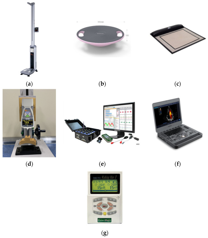
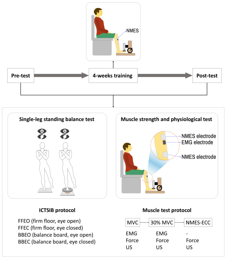
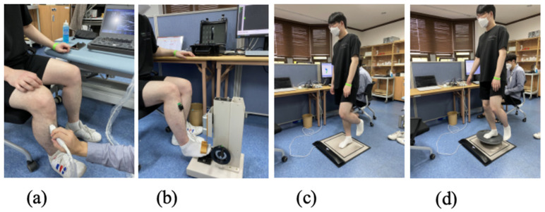
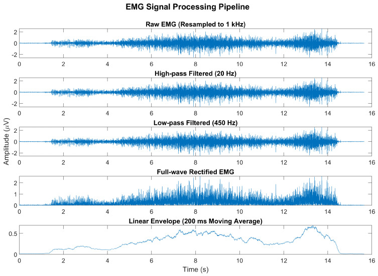
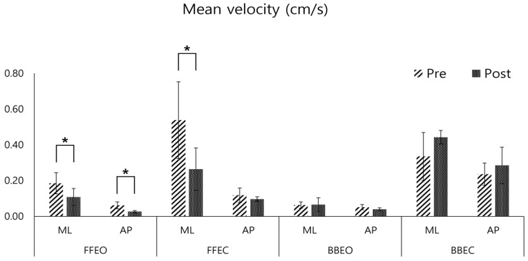
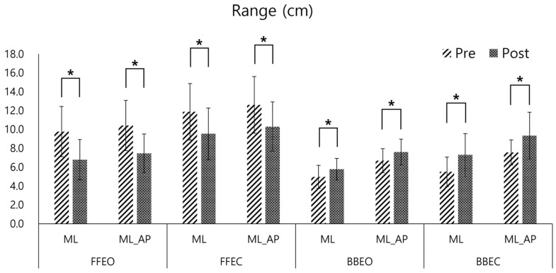
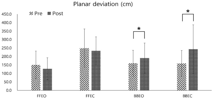
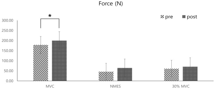
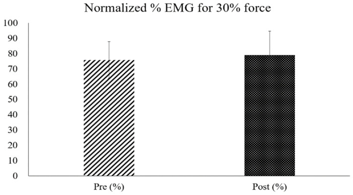
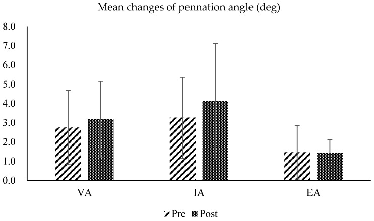
This study explored the impact of a four-week Neuromuscular Electrical Stimulation (NMES)-induced eccentric contraction training on single-leg standing balance and muscle strength in 17 healthy adults. The unique training approach involved active antagonist muscle contraction during NMES. Post-training results revealed significant improvements in balance, with notable reductions in Center of Pressure (CoP) trajectory velocity (mean reduction: 0.07 ± 0.01 cm/s, p < 0.05) and range (mean reduction: 2.98 ± 0.53 cm, p < 0.05) on a firm surface. While increases in dorsiflexion force (mean increase: 21.43 ± 0.79 N, p < 0.05) and muscle activation were observed, these were not statistically significant. Changes in muscle pennation angles were also not significant (mean change: 0.43 ± 0.06 degrees, p > 0.05), underscoring the complexity of muscle adaptation processes. This study highlights NMES's potential in enhancing balance and proprioceptive sensing, suggesting its promising applications in neuromuscular rehabilitation. However, further research is needed to fully understand its impact.

Keywords: center of pressure; dorsiflexion force; electromyography; neuromuscular electrical stimulation; single-leg standing balance; tibialis anterior; ultrasonography.


Conflict of interest statement

The authors declare no conflicts of interest.

Figures

Figure 1
Figure 1
(a) A weight scale (GL-310P, GTECH Co., Ltd., Yangju-si, Republic of Korea); (b) a balance board (Egojin Ltd., Pocheon-si, Republic of Korea); (c) a pressure sensor system (MatScan VersaTek system, Tekscan, Inc., South Boston, MA, USA); (d) a push–pull gauge (ZTA-500N, IMADA Co., Ltd., Toyohashi, Japan); (e) a surface electromyogram (sEMG) sensor system (Trigno EMG sensor, Delsys, Inc., Natick, MA, USA); (f) an ultrasonography system (X5, SonoScape Medical Corp., Shenzhen, China); (g) a neuromuscular electrical stimulation system (EMS1000, Cybermedic Co., Ltd., Iksan-si, Republic of Korea).
Figure 2
Figure 2
Schematic of the experimental protocol including balance and muscle testing before and after 4 weeks of NMES-induced eccentric contraction training.
Figure 3
Figure 3
(a) Ultrasound imaging and measurements; (b) force and EMG measurements; (c) firm floor pressure sensor measurements; (d) balance board pressure sensor measurements.
Figure 4
Figure 4
Representative example of the EMG signal processing pipeline. The raw EMG signal, originally sampled at 2 kHz, was resampled to 1 kHz and processed using a high-pass filter (20 Hz) and a low-pass filter (450 Hz). The signal was then full-wave-rectified and smoothed using a 200 ms moving average to produce a linear envelope of muscle activity.
Figure 5
Figure 5
Pre- and post-comparison of the mean velocity of the center of pressure (COP) in the medio-lateral (ML) and antero-posterior (AP) directions under four conditions: firm floor with eye opened (FFEO), firm floor with eye closed (FFEC), balance board with eye opened (BBEO), balance board with eye closed (BBEC) (* indicates statistical significance).
Figure 6
Figure 6
Pre- and post-comparison of the Center of Pressure (COP) range in the medio-lateral (ML) and Medio-lateral_antero-posterior (ML_AP) directions under four conditions: firm floor with eyes open (FFEO), firm floor with eyes closed (FFEC), balance board with eyes open (BBEO), and balance board with eyes closed (BBEC) (* indicates statistical significance).
Figure 7
Figure 7
Pre- and post-comparison of the center of pressure (COP) planar deviation under four different conditions: firm floor with eyes open (FFEO), firm floor with eyes closed (FFEC), balance board with eyes open (BBEO), and balance board with eyes closed (BBEC) (* indicates statistical significance).
Figure 8
Figure 8
Pre- and post-comparisons of the average dorsiflexion forces of the ankle joint, measured by a push–pull gauge during three different types of dorsiflexion contractions: maximal voluntary contraction (MVC), passive contraction induced by NMES of the tibialis anterior muscle, and contraction at 30% of the MVC strength (* indicates statistical significance).
Figure 9
Figure 9
Comparison of pre- and post-test normalized EMG percentages measured during dorsiflexion at 30% of the MVC strength.
Figure 10
Figure 10
Pre- and post-test comparisons of the pennation angles calculated from ultrasound images of the tibialis anterior muscle, measured under three conditions: maximum voluntary contraction (voluntary activation: VA), passive contraction with NMES alone (involuntary activation; IA), and eccentric contraction during NMES with concurrent contraction of the ankle extensor muscles (eccentric activation; EA).
Figure 11
Figure 11
Example of the pennation angle in ultrasound images of the tibialis anterior muscle ((A) relaxed; (B) voluntary contraction, (C) involuntary contraction, and (D) eccentric contraction).

Similar articles

References

    1. Jones S., Man W.D.C., Gao W., Higginson I.J., Wilcock A., Maddocks M. Neuromuscular electrical stimulation for muscle weakness in adults with advanced disease. Cochrane Database Syst. Rev. 2016;2016:CD009419. doi: 10.1002/14651858.CD009419.pub3. - DOI - PMC - PubMed
    1. Delitto A., Rose S.J., McKowen J.M., Lehman R.C., Thomas J.A., Shively R.A. Electrical stimulation versus voluntary exercise in strengthening thigh musculature after anterior cruciate ligament surgery. Phys. Ther. 1988;68:660–663. doi: 10.1093/ptj/68.5.660. - DOI - PubMed
    1. Bickel C.S., Gregory C.M., Dean J.C. Motor unit recruitment during neuromuscular electrical stimulation: A critical appraisal. Eur. J. Appl. Physiol. 2011;111:2399–2407. doi: 10.1007/s00421-011-2128-4. - DOI - PubMed
    1. Maffiuletti N.A. Physiological and methodological considerations for the use of neuromuscular electrical stimulation. Eur. J. Appl. Physiol. 2010;110:223–234. doi: 10.1007/s00421-010-1502-y. - DOI - PubMed
    1. Enoka R.M. Activation order of motor axons in electrically evoked contractions. Muscle Nerve. 2002;25:763–764. doi: 10.1002/mus.10117. - DOI - PubMed

LinkOut - more resources