. 2025 Oct;48(5):3692-3709.
doi: 10.1007/s10753-025-02292-7. Epub 2025 Apr 26.

A Quinoxaline Derivative as a New Therapeutic Agent for Sepsis through Suppression of TLR4 Signaling Pathways

Affiliations

A Quinoxaline Derivative as a New Therapeutic Agent for Sepsis through Suppression of TLR4 Signaling Pathways

Hamza Hanieh et al. Inflammation. 2025 Oct.

Abstract

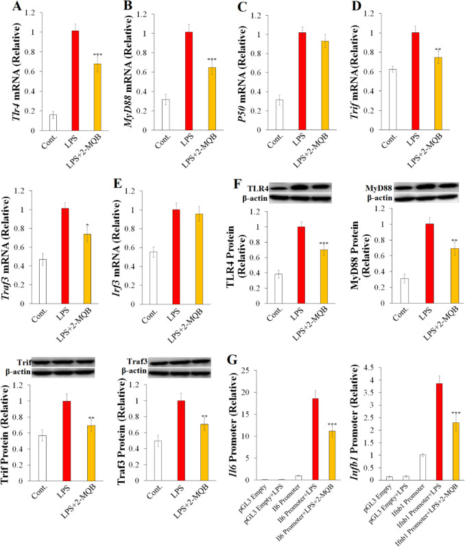
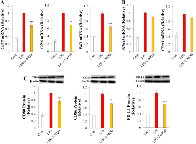
Sepsis is a severe systemic inflammatory syndrome and one of the leading causes of global morbidity and mortality. Preclinical studies have identified several quinoxaline-based compounds with anti-inflammatory properties, but their effects in sepsis have not been investigated. This study aimed to identify a quinoxaline derivative with anti-inflammatory properties in sepsis. Examining the inflammatory response of primary mouse macrophages to Lipopolysaccharides (LPS) revealed that 2-methoxy-N-(3-quinoxalin-2-ylphenyl)benzamide (2-MQB) is a promising molecule. It suppressed the production of several inflammatory cytokines, including Interleukin-1β (IL-1β), IL-6, IL-12p70, Interferon-γ (IFN-γ), IFN-β, and Tumor necrosis factor-α (TNF-α). Importantly, 2-MQB inhibited the transcriptional activities of Toll-like receptor 4 (TLR4) signaling pathways, including Nuclear factor-κB (NF-κB) and Interferon regulatory factor 3 (IRF3). This was accompanied by lower expression of TLR4, Myeloid differentiation primary response 88 (MyD88), TIR Domain-containing adaptor molecule 1 (Trif), and TNF Receptor-associated factor 3 (Traf3). Additionally, 2-MQB selectively reduced the expression of genes encoding CD80, CD86, and Programmed death-ligand 1 (PD-L1). In vivo, 2-MQB improved mice survival, mitigated tissue damage in the spleen, kidney, and lung, and reduced pro-inflammatory cytokine levels in both LPS-induced endotoxin shock and Cecal ligation and puncture (CLP) models. Notably, 2-MQB decreased the numbers of CD4+ and CD8+ T cells in the spleen and inhibited TLR4 signaling pathways in LPS-induced endotoxemia. In conclusion, these results introduce the quinoxaline derivative 2-MQB as a potential therapeutic agent for sepsis by inhibiting TLR4 signaling pathways, paving the way for future clinical applications.

Keywords: Cytokines; Inflammation; Quinoxaline; Sepsis; TLR4 pathways.


Conflict of interest statement

Declarations. Competing Interest: The authors declare no competing interests.

Figures

Fig. 1
Fig. 1
2-MQB suppresses LPS-induced inflammatory response in macrophages. Purified macrophages were cultured for 16 h either without (Cont.) or with LPS (0.2 µg/mL) in the absence or presence of 2-MQB at 10, 20, 30, or 40 µM. (A) the chemical structure of 2-MQB. The concentrations of cytokines in the supernatant, including (B) IL-1β, IL-6, IFN-β, (C) TNF-α, IFN-γ, (D) IL-12p70, (E) IL-8, and (F) IL-10, were determined using ELISA. The data, representing three independent experiments with similar results, are shown as mean ± SD. n = 4 mice per experiment. *P < 0.05, **P < 0.01, and ***P < 0.001 indicate statistical significance compared to LPS alone, one-way ANOVA; horizontal bars indicate statistical comparison between 2-MQB-treated macrophages
Fig. 2
Fig. 2
2-MQB inhibits TLR4 signaling pathways. Purified macrophages were pre-treated with either DMSO or 2-MQB for 30 min, then cultured for 2 h for mRNA collection or 6 h for lysate collection either without (Cont.) or with LPS (0.2 µg/mL) in the absence or presence of 2-MQB at 30 µM. mRNA and proteins levels were determined by qPCR and immunoblotting, respectively. The mRNAs of (A) Tlr4, (B) MyD88, (C) P50, (D) Trif and Traf3, and (E) Irf3 relative to those of LPS alone. (F) Immunoblots and quantification of TLR4, MyD88, Trif, and Traf3 proteins relative to those of LPS alone. (G) Luciferase activities of pGL3 empty or Il6- or Ifnb1-harboring pGL3 transfected in peritoneal macrophages for 12 h, incubated with DMSO (pGL3 Empty) or 2-MQB (30 µM) for 30 min, and then cultured for 6 h without or with LPS (0.2 µg/mL). The data, representing three independent experiments with similar results, are shown as mean ± SD. n = 4 mice per experiment. *P < 0.05, **P < 0.01, and ***P < 0.001 indicate statistical significance compared to LPS alone, one-way ANOVA
Fig. 3
Fig. 3
2-MQB selectively inhibits surface molecules on LPS-stimulated macrophages. Purified macrophages were pre-treated with either DMSO or 2-MQB for 30 min, then cultured for 2 h for mRNA collection or 6 h for lysate collection either without (Cont.) or with LPS (0.2 µg/mL) in the absence or presence of 2-MQB at 30 µM. mRNA and protein levels were determined by qPCR and immunoblotting, respectively. The mRNAs of (A) Cd80, Cd86, and Pdl1, and (B) MhcII and C5ar1 relative to those of LPS alone. (C) Immunoblots and quantification of CD80, CD86, and PD-L1 relative to those of LPS alone. The data, representing three independent experiments with similar results, are shown as mean ± SD. n = 4 mice per experiment. **P < 0.01, and ***P < 0.001 indicate statistical significance compared to LPS alone, one-way ANOVA
Fig. 4
Fig. 4
2-MQB attenuates LPS-induced endotoxin shock. Mice were intraperitoneally injected with vehicle (Cont.) or LPS, without or with 2-MQB. The 2-MQB was injected in mice at 0 and 6 h after LPS injection and observed for 144 h. (A) Weights of the spleen, kidneys, and liver from Control mice and LPS-injected mice (7.5 mg/kg) without or with 2-MQB at 1 or 6 mg/kg on day 6 after LPS injection. (B) Survival (%) of Control mice and LPS-injected mice (20 mg/kg) without or with 2-MQB at 1, 3, or 5 mg/kg. (C) Clinical score of Control mice and LPS-injected mice (20 mg/kg) without or with 2-MQB at 1, 3, or 5 mg/kg. The serum levels of (D) IL-1β, IFN-β, (E) IL-6, TNF-α, IFN-γ, (F) IL-12p70, and (G) IL-10 in blood samples from Control mice and LPS-injected mice (7.5 mg/kg) without or with 2-MQB at 1, 3, or 5 mg/kg were determined using ELISA. The data, representing three independent experiments with similar results, are shown as mean ± SD. (A) n = 4 and (B-G) n = 8 mice per group. *P < 0.05, **P < 0.01, and ***P < 0.001 indicate statistical significance. (A, D-G) one-way ANOVA, compared to LPS treatment alone; (D and E) horizontal bars indicate statistical comparison between 2-MQB treatments. (B) log-rank, and (C) two-way ANOVA; vertical bars indicate statistical comparison, (C) horizontal bar indicates the range of compared data
Fig. 5
Fig. 5
2-MQB mitigates tissue injury in LPS-induced endotoxin shock. Mice were intraperitoneally injected with vehicle (Cont.) or LPS (20 mg/kg), without or with 2-MQB at 1, 3, or 5 mg/kg. The 2-MQB was injected in mice at 0 and 6 h and tissue samples were collected 48 h after LPS injection. (A) Representative histology images of spleen, kidney and lung (200X; scale bar = 100 µm). Serum levels of (B) PCT, (C) CRP, (D) KIM-1 and AST, (E) ALT, and (F) lactate in blood samples from Control mice and LPS-injected mice without or with 2-MQB were determined using ELISA and assay kits. The data, representing three independent experiments with similar results, are shown as mean ± SD. n = 8 mice per group. *P < 0.05, **P < 0.01, and ***P < 0.001 indicate statistical significance compared to LPS treatment alone, (B-F) one-way ANOVA; horizontal bars indicate statistical comparison between 2-MQB treatments
Fig. 6
Fig. 6
2-MQB modulates lymphocytes populations in LPS-induced endotoxin shock. Mice were intraperitoneally injected with LPS (7.5 mg/kg) without or with 2-MQB treatment at 3 or 5 mg/kg at 0 and 6 h after LPS injection. (A) The number of CD4+ and CD8+ T lymphocytes and (B) B lymphocytes per spleen in mice treated with 2-MQB at 3 mg/kg at 0, 6, and 18 h after LPS injection. The number of (C) CD4+ and (D) CD8+ T lymphocytes and (E) B lymphocytes per spleen in mice treated with 2-MQB at 5 mg/kg at 0, 6, and 18 h after LPS injection. The data, representing three independent experiments with similar results, are shown as mean ± SD. (A-E) n = 4 mice per group. *P < 0.05 and **P < 0.01 indicate statistical significance compared to LPS treatment alone at a time point, one-way ANOVA
Fig. 7
Fig. 7
2-MQB inhibits TLR4 signaling in LPS-induced endotoxin shock. Mice were intraperitoneally injected with vehicle (Cont.) or LPS (7.5 mg/kg) without or with 2-MQB treatment at 3 or 5 mg/kg at 0 and 6 h after LPS injection. The mRNA levels were determined by qPCR. The mRNAs of (A) Tlr4, MyD88, Trif, and (B) Traf3 in peritoneal macrophage from Control mice and LPS-injected mice without or with 2-MQB at 3 mg/kg relative to LPS alone. The mRNAs of (C) Tlr4, MyD88, Trif, and Traf3 in peritoneal macrophage from Control mice and LPS-injected mice without or with 2-MQB at 5 mg/kg relative to LPS alone. The data, representing three independent experiments with similar results, are shown as mean ± SD. n = 5 mice per group. **P < 0.01 indicates statistical significance compared to LPS treatment alone, one-way ANOVA
Fig. 8
Fig. 8
2-MQB ameliorates the severity of polymicrobial sepsis. Mice underwent CLP laparotomy and injected with vehicle (Cont.) or LPS, without or with 2-MQB at 3 or 5mg/kg 0 and 6 h after surgery and observed for 144 h. (A) Survival (%) of Control mice and CLP mice without or with 2-MQB. (B) Clinical score of Control mice and CLP mice without or with 2-MQB. The serum levels of (C) IL-1β, IL-6, TNF-α, and IFN-β, and (D) IL-12p70 and IFN-γ were determined using ELISA. (E) Bacterial load (CFU; Colony forming unit) in peritoneal lavage and blood samples obtained at 24 h postsurgery. The data, representing three independent experiments with similar results, are shown as mean ± SD. (A-E) n = 8 mice per group. *P < 0.05, **P < 0.01, and ***P < 0.001 indicate statistical significance. (A) log-rank, (B) two-way ANOVA; vertical bars indicate statistical comparison, (B) horizontal bar indicates the range of compared data. (C-E) one-way ANOVA, compared to LPS treatment alone
Fig. 9
Fig. 9
2-MQB mitigates tissue injury in polymicrobial sepsis. Mice underwent CLP laparotomy and injected with vehicle (Cont.) or 2-MQB at 3 or 5 mg/kg at 0 and 6 h and tissue samples were collected 48 h after surgery. Representative histology images of the spleen, kidney and lung from Control mice and CLP mice without or with 2-MQB (200X; scale bar = 100 µm)

References

    1. Hotchkiss, R.S., L.L. Moldawer, S.M. Opal, K. Reinhart, I.R. Turnbull, and J.-L. Vincent. 2016. Sepsis and septic shock. Nature Reviews Disease Primers 2: 1–21. - PMC - PubMed
    1. Singer, M., C.S. Deutschman, C.W. Seymour, M. Shankar-Hari, D. Annane, M. Bauer, et al. 2016. The third international consensus definitions for sepsis and septic shock (Sepsis-3). Journal of the American Medical Association 315: 801–810. - PMC - PubMed
    1. Rudd, K.E., S.C. Johnson, K.M. Agesa, K.A. Shackelford, D. Tsoi, D.R. Kievlan, et al. 2020. Global, regional, and national sepsis incidence and mortality, 1990–2017: Analysis for the global burden of disease study. The Lancet 395: 200–211. - PMC - PubMed
    1. Cao, M., G. Wang, and J. Xie. 2023. Immune dysregulation in sepsis: Experiences, lessons and perspectives. Cell Death Discovery 9: 465. - PMC - PubMed
    1. Basak, B., and S. Akashi-Takamura. 2024. IRF3 function and immunological gaps in sepsis. Frontiers in Immunology 15: 1336813. - PMC - PubMed