. 2025 Mar 24;30(7):1437.
doi: 10.3390/molecules30071437.

Dipalmitoylphosphatidylcholine Lipid Vesicles for Delivering HMB, NMN, and L-Leucine in Sarcopenia Therapy

Affiliations

Dipalmitoylphosphatidylcholine Lipid Vesicles for Delivering HMB, NMN, and L-Leucine in Sarcopenia Therapy

Alfred Najm et al. Molecules. .

Abstract

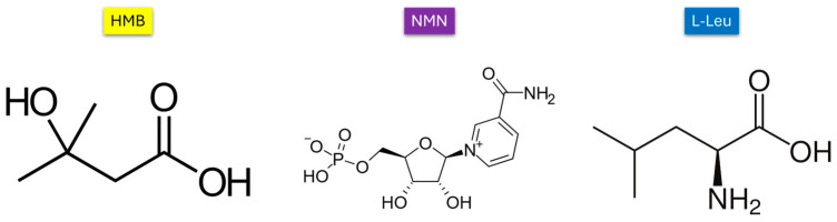
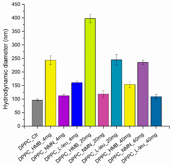
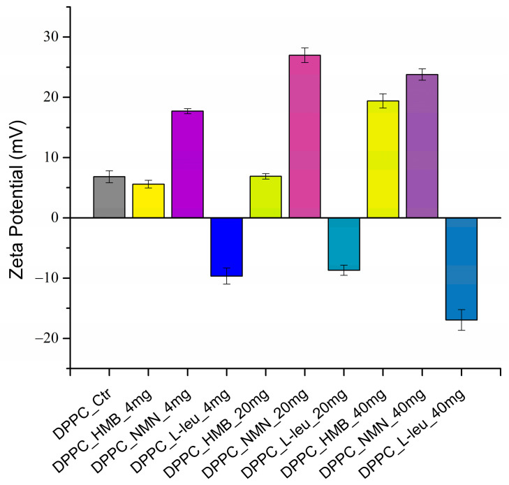
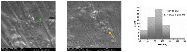
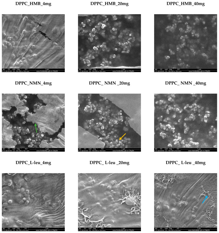
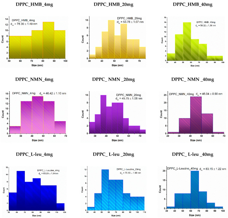
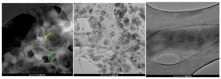
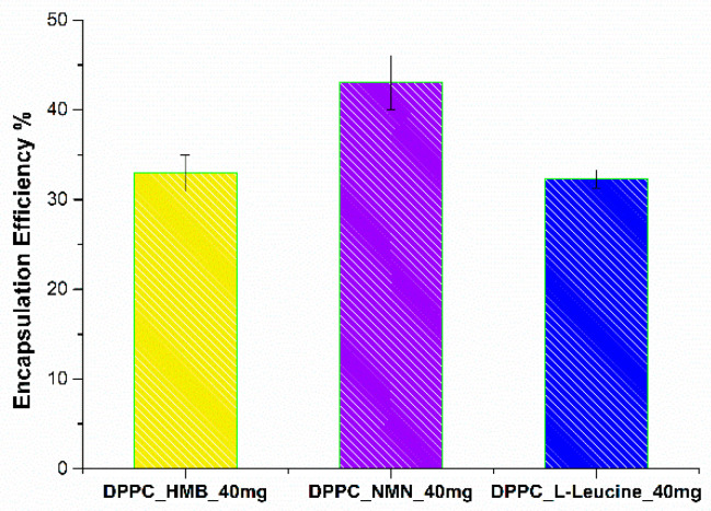
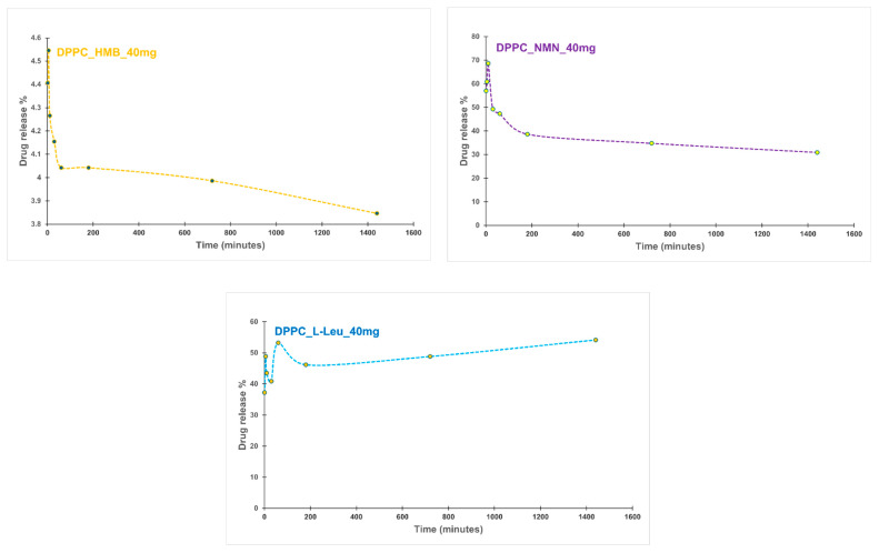
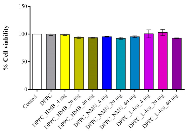
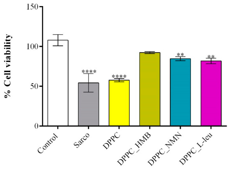
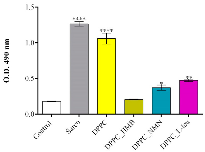
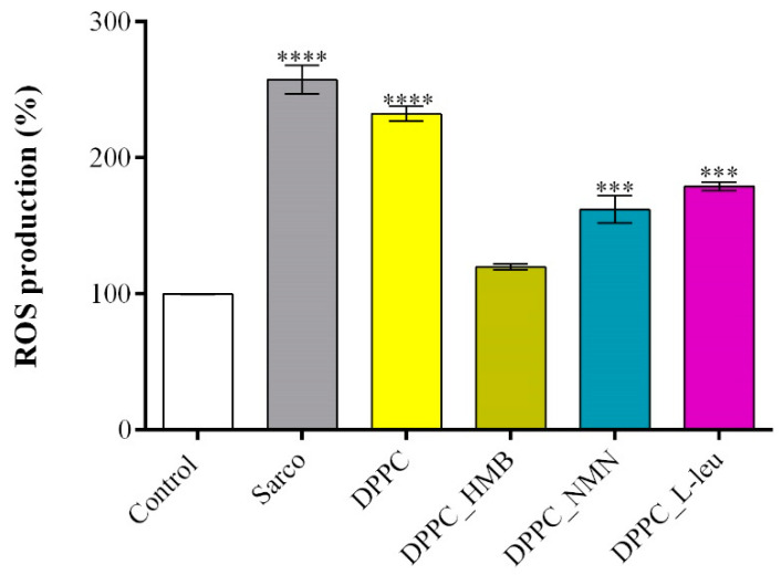
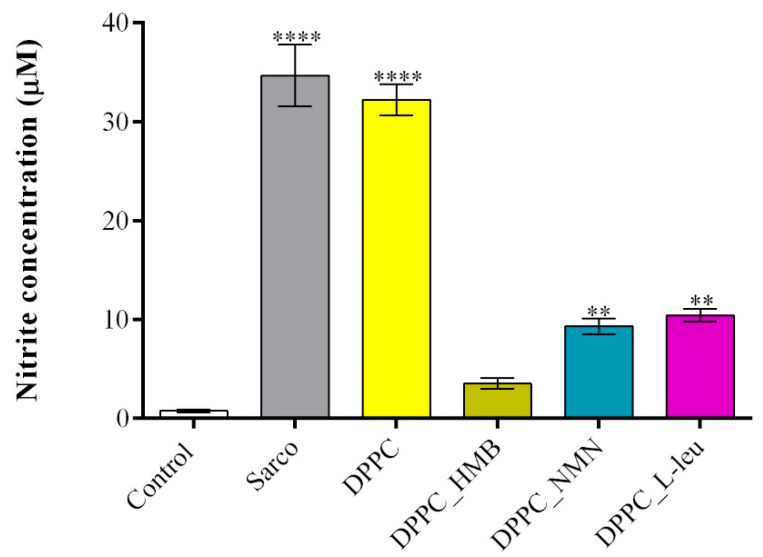
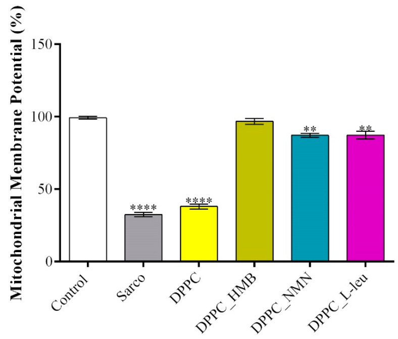
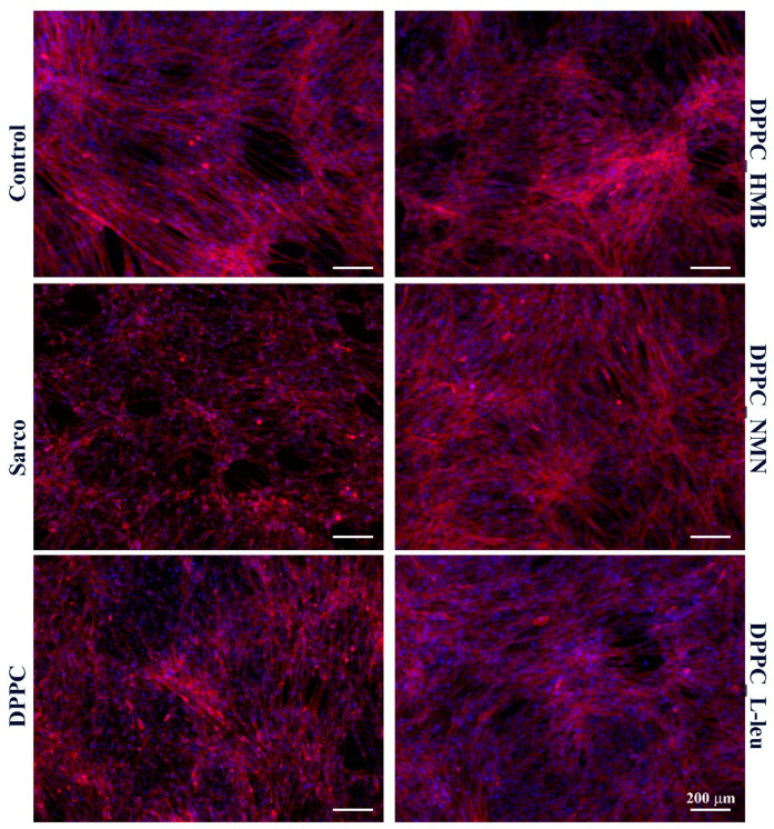
Sarcopenia, characterized by the degeneration of skeletal muscle tissue, has emerged as a significant concern in recent years. This increased awareness stems from advances in research focusing on elderly patients, which have revealed correlations between aging mechanisms and muscle degeneration, beyond the mere fact that tissues age and deteriorate over time. Consequently, the present study aims to address sarcopenia by developing and evaluating DPPC lipid vesicles that encapsulate three distinct drugs: HMB, NMN, and L-Leucine. These drugs are specifically selected for their properties, which facilitate effective interaction with the affected muscle tissue, thereby promoting desired therapeutic effects. Preliminary physicochemical analyses indicate the successful formation of spherical lipid vesicles, characterized by nanometric dimensions and stable membrane integrity. The biological investigations aimed to highlight the potential of DPPC lipid vesicles encapsulating HMB, NMN, and L-Leucine to alleviate sarcopenia-induced cytotoxicity and oxidative stress. Through a comparative evaluation of the three drug formulations, we demonstrate that drug-loaded DPPC vesicles effectively mitigate oxidative damage, preserve mitochondrial function, and maintain cytoskeletal integrity in H2O2-induced C2C12 myotubes, with HMB-loaded vesicles showing the strongest protective effects against muscle degeneration. These findings underscore the therapeutic potential of DPPC-based controlled release systems for sarcopenia treatment and highlight the need for further investigations into their mechanistic role in muscle preservation.

Keywords: DPPC; HMB; L-Leucine; NMN; increased NAD+ levels; lipid vesicles; sarcopenia.


Conflict of interest statement

The authors declare no conflicts of interest.

Figures

Figure 1
Figure 1
Chemical structures of HMB, NMN, and L-Leucine.
Figure 2
Figure 2
Graphic representation of hydrodynamic diameter (nm) as a DLS result of DPPC vesicles dispersed in water.
Figure 3
Figure 3
Graphic representation of zeta potential (mV) as a DLS result of DPPC vesicles dispersed in water.
Figure 4
Figure 4
SEM micrographs and particle size distribution obtained for DPPC_Ctrl (green arrow—lipid matrix, yellow arrow—lipid vesicles).
Figure 5
Figure 5
SEM micrographs obtained for DPPC_HMB, DPPC_NMN, and DPPC_L-Leucine used in three different concentrations, 4 mg, 20 mg, and 40 mg, respectively (green arrow—lipid matrix, yellow arrow—lipid vesicles, blue arrow—fibrillar structures).
Figure 6
Figure 6
Particle size distribution obtained for DPPC_HMB, DPPC_NMN, and DPPC_L-Leucine used in three different concentrations, 4 mg, 20 mg, and 40 mg, respectively.
Figure 7
Figure 7
TEM micrographs obtained for DPPC_HMB_20mg, DPPC_NMN_20mg, and DPPC_L-Leucine_20mg (green arrow—lipid matrix, yellow arrow—lipid vesicles).
Figure 8
Figure 8
Encapsulation efficiency of HMB, NMN, and L-Leucine in DPPC_HMB_40mg, DPPC_NMN_40mg, and DPPC_L-Leucine_40mg.
Figure 9
Figure 9
Drug release profile of DPPC_HMB_40mg, DPPC_NMN_40mg, and DPPC_L-Leucine_40mg.
Figure 10
Figure 10
Graphical representation of C2C12 cell viability as revealed by the MTT assay after 24 h of treatment with DPPC lipid vesicles and drug-loaded formulations at various concentrations. Data are expressed as mean ± standard deviation (SD).
Figure 11
Figure 11
Graphical representation of C2C12 myotube viability after 48 h of treatment with simple DPPC particles and drug-loaded (HMB, NMN, L-Leu) lipid particles. Besides the experimental control, for all conditions, H2O2 was added to induce muscular atrophy in C2C12 myotube cultures. Data are expressed as mean ± standard deviation (SD). Statistical significance was determined using GraphPad Prism; p-values are indicated as follows: p ≤ 0.01 (**) and p ≤ 0.0001 (****) compared to the control group.
Figure 12
Figure 12
Graphical representation of LDH levels released in the culture medium by C2C12 myotubes after 48 h of treatment with simple DPPC particles and drug-loaded (HMB, NMN, L-Leu) lipid particles. Besides the experimental control, for all conditions, H2O2 was added to induce muscular atrophy in C2C12 myotube cultures. Data are expressed as mean ± standard deviation (SD). Statistical significance was determined using GraphPad Prism; p-values are indicated as follows: p ≤ 0.1 (*), p ≤ 0.01 (**) and p ≤ 0.0001 (****) compared to the control group.
Figure 13
Figure 13
Graphical representation of ROS production in C2C12 myotube cell cultures after 48 h of treatment with simple DPPC particles and drug-loaded (HMB, NMN, L-Leu) lipid particles. Besides the experimental control, for all conditions, H2O2 was added to induce muscular atrophy in C2C12 myotubes culture. Data are expressed as mean ± standard deviation (SD). Statistical significance was determined using GraphPad Prism; p-values are indicated as follows: p ≤ 0.001 (***) and p ≤ 0.0001 (****) compared to the control group.
Figure 14
Figure 14
Graphical representation of NO production in C2C12 myotubes cell cultures after 48 h of treatment with simple DPPC particles and drug-loaded (HMB, NMN, L-Leu) lipid particles. Besides the experimental control, for all conditions, H2O2 was added to induce muscular atrophy in C2C12 myotubes culture. Data are expressed as mean ± standard deviation (SD). Statistical significance was determined using GraphPad Prism; p-values are indicated as follows: p ≤ 0.01 (**) and p ≤ 0.0001 (****) compared to the control group.
Figure 15
Figure 15
Graphical representation of MMP of C2C12 myotubes after 48 h of treatment with simple DPPC particles and drug-loaded (HMB, NMN, L-Leu) lipid particles. Besides the experimental control, for all conditions, H2O2 was added to induce muscular atrophy in C2C12 myotubes culture. Data are expressed as mean ± standard deviation (SD). Statistical significance was determined using GraphPad Prism; p-values are indicated as follows: p ≤ 0.01 (**) and p ≤ 0.0001 (****) compared to the control group.
Figure 16
Figure 16
Fluorescence micrographs revealing F-actin filaments (red) and cell nuclei (blue) of C2C12 myotubes after 48 h of treatment with simple DPPC particles and drug-loaded (HMB, NMN, L-Leu) lipid particles. Besides the experimental control, for all conditions, H2O2 was added to induce muscular atrophy in C2C12 myotube cultures.

Similar articles

Cited by

References

    1. von Haehling S., Morley J.E., Anker S.D. From muscle wasting to sarcopenia and myopenia: Update 2012. J. Cachexia Sarcopenia Muscle. 2012;3:213–217. doi: 10.1007/s13539-012-0089-z. - DOI - PMC - PubMed
    1. Tournadre A., Vial G., Capel F., Soubrier M., Boirie Y. Sarcopenia. Jt. Bone Spine. 2019;86:309–314. doi: 10.1016/j.jbspin.2018.08.001. - DOI - PubMed
    1. Keller K. Sarcopenia. Wien. Med. Wochenschr. 2019;169:157–172. doi: 10.1007/s10354-018-0618-2. - DOI - PubMed
    1. Tagliafico A.S., Bignotti B., Torri L., Rossi F. Sarcopenia: How to measure, when and why. La Radiol. Medica. 2022;127:228–237. doi: 10.1007/s11547-022-01450-3. - DOI - PMC - PubMed
    1. Wiedmer P., Jung T., Castro J.P., Pomatto L.C.D., Sun P.Y., Davies K.J.A., Grune T. Sarcopenia—Molecular mechanisms and open questions. Ageing Res. Rev. 2021;65:101200. doi: 10.1016/j.arr.2020.101200. - DOI - PubMed

LinkOut - more resources