Review
. 2025 Mar 26;30(7):1470.
doi: 10.3390/molecules30071470.

Protein O-Fucosyltransferases: Biological Functions and Molecular Mechanisms in Mammals

Affiliations
Review

Protein O-Fucosyltransferases: Biological Functions and Molecular Mechanisms in Mammals

Huilin Hao et al. Molecules. .

Abstract

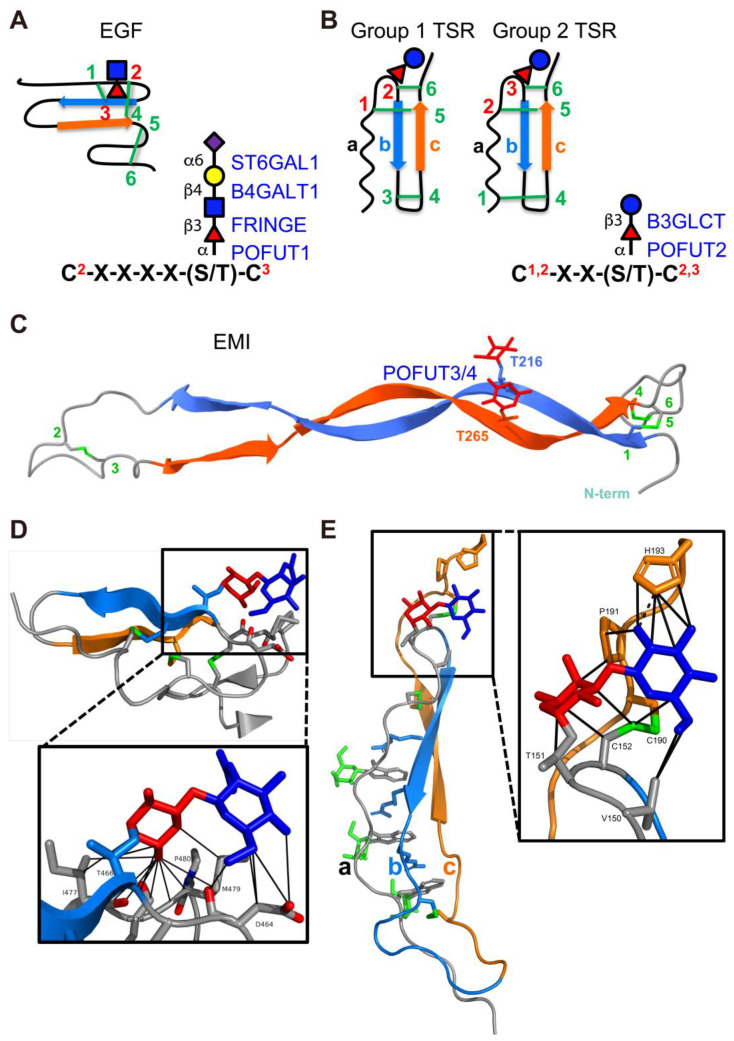
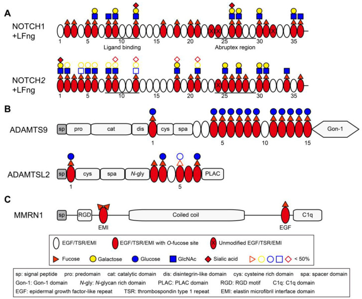
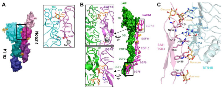
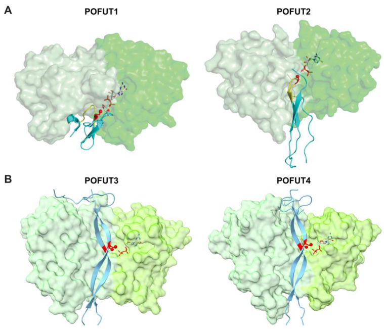
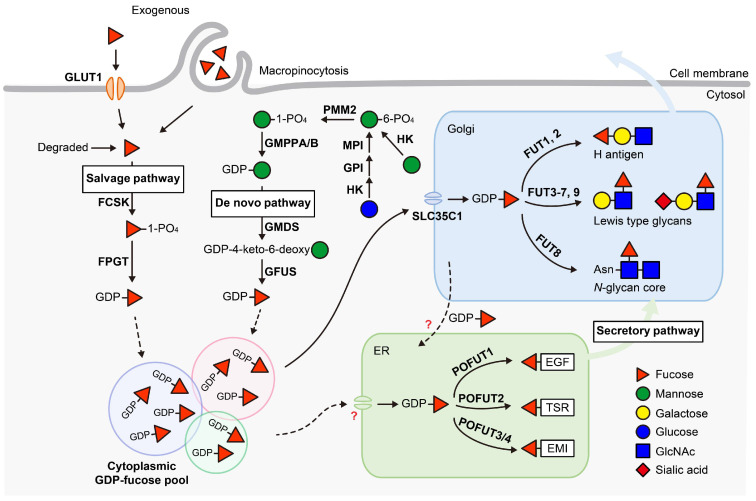
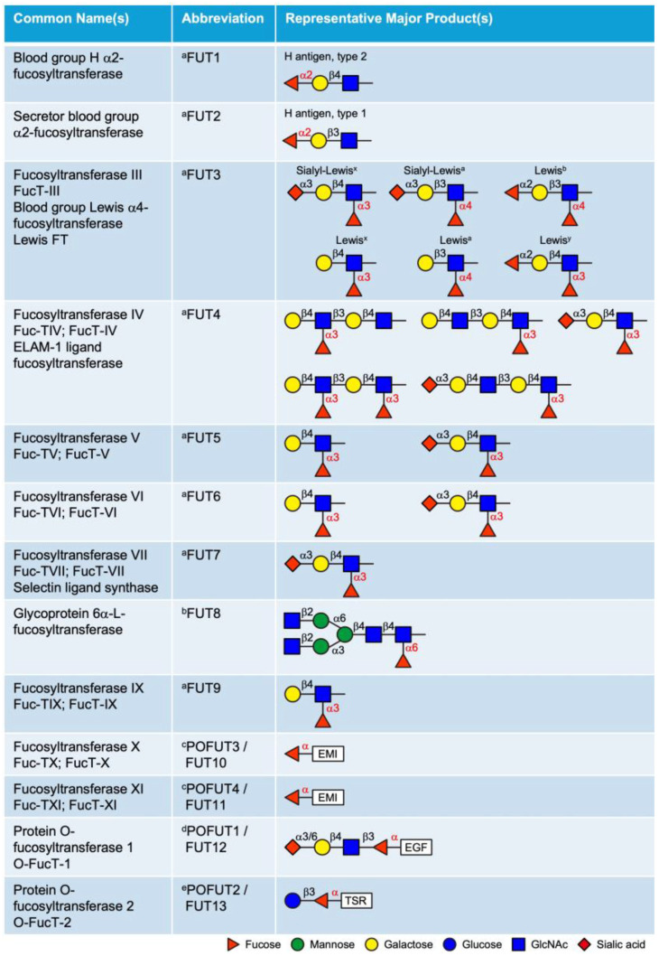
Domain-specific O-fucosylation is an unusual type of glycosylation, where the fucose is directly attached to the serine or threonine residues in specific protein domains via an O-linkage. O-fucosylated proteins play critical roles in a wide variety of biological events and hold important therapeutic values, with the most studied being the Notch receptors and ADAMTS proteins. O-fucose glycans modulate the function of the proteins they modify and are closely associated with various diseases including cancer. In mammals, alongside the well-documented protein O-fucosyltransferase (POFUT) 1-mediated O-fucosylation of epidermal growth factor-like (EGF) repeats and POFUT2-mediated O-fucosylation of thrombospondin type 1 repeats (TSRs), a new type of O-fucosylation was recently identified on elastin microfibril interface (EMI) domains, mediated by POFUT3 and POFUT4 (formerly FUT10 and FUT11). In this review, we present an overview of our current knowledge of O-fucosylation, integrating the latest findings and with a particular focus on its biological functions and molecular mechanisms.

Keywords: EGF; EMI; FUT10; FUT11; Notch; O-fucosylation; POFUT1; POFUT2; POFUT3; POFUT4; TSR.


Conflict of interest statement

The authors declare no conflicts of interest.

Figures

Figure 3
Figure 3
EGF repeats, TSRs, and EMI domains are modified by O-fucose glycans. (A) (left) Disulfide bonding pattern of an EGF repeat. Cysteines are numbered, disulfide bonds are in green, β-strands are indicated as blue and orange arrows. O-fucose site is marked with fucose (red triangle), GlcNAc (blue square), Galactose (yellow circle), and Neu5Ac (purple diamond). (right) Consensus sequence for POFUT1 modification. C2 and C3 are the second and third conserved cysteine in the EGF repeat. X indicates any amino acid. Enzymes responsible for modification are in blue with linkages indicated. (B) (left) Two distinct disulfide bonding patterns for TSRs. Cysteines are numbered, disulfide bonds are in green, β-strands are indicated as blue and orange arrows. O-fucose site is marked with fucose (red triangle) and glucose (blue circle). (right) Consensus sequence for POFUT2 modification. The C’s can be C1 and C2 or C2 and C3, depending on whether the TSR is Group 1 or Group 2. Enzymes responsible for modification are in blue with linkages indicated. (C) AlphaFold2 predicted structure of the EMI domain from MMRN1 showing the disulfide bonding pattern. Cysteines are numbered, disulfide bonds are in green, β-strands are indicated as blue and orange arrows. The two O-fucose sites are indicated and marked with fucose residues in red. POFUT3 and POFUT4 are responsible for modification and indicated in blue. (D) Structure of NOTCH1 EGF12 modified with a GlcNAcβ1-3Fucose disaccharide (from PDB ID 4D0E). β-strands colored as in (A). Fucose in red, GlcNAc in blue, disulfide bonds in green, oxygen atoms highlighted in red. Box shows zoomed in region highlighting interactions of the disaccharide with underlying amino acids identified by MolProbity [79] (van der Waals, solid lines). Structures rendered in PyMOL (version 2.2.2). (E) Structure of Properdin TSR2 modified with Glucoseβ1-3Fucose disaccharide (from PDB ID 6RUS). The three strands (a, b, and c) of the TSRs are colored as in (B). Fucose in red, glucose in blue, mannose in green, disulfide bonds in green. Box shows zoomed in region highlighting interactions of the disaccharide with underlying amino acids identified by MolProbity [79] (H-bonds, dashed lines; van der Waals, solid lines). Structures rendered in PyMOL (version 3.0.4). Panels A, B and D are modified with permission from Ref. [44].
Figure 4
Figure 4
Domain maps of representative EGF/TSR/EMI-containing proteins showing distribution of O-fucosylation. (A) Domain maps of mouse NOTCH1 EGF1–36 and NOTCH2 EGF1–36. The majority of EGF repeats are modified with O-fucose glycans with more than half elongated by Lfng (modified from Ref. [66] with permission). (B) Domain maps of human ADAMTS9 and mouse ADAMTSL2. The majority of TSRs in ADAMTS9 and ADAMTSL2 are modified with Glucoseβ1-3Fucose disaccharide by POFUT2 and B3GLCT (modified from Refs. [59,230] with permission) [213]. (C) Domain map of human MMRN1 showing the EMI domain at protein N-terminal and can be modified with two fucose residues.
Figure 5
Figure 5
O-fucose glycans form direct contacts with binding partners. (A) Co-crystal structure of NOTCH1 EGF11–13 (magenta/purple) and DLL4 N-EGF1 (blue/green) (modified with permission from Ref. [72]). Zoomed panel shows direct interaction between fucose residue on NOTCH1 EGF12 with residues in DLL4. (B) Co-crystal structure of NOTCH1 EGF8–12 (magenta) and JAG1 N-EGF3 (green) (modified with permission from Ref. [73]). Zoomed panels show direct interactions between fucose residues on EGF8 and EGF12 with residues in JAG1. (C) Co-crystal structure of RTN4R (cyan) and BAI1 TSR3 (pink) (modified with permission from Ref. [235]). O-fucose forms direct interactions with residues in RTN4R.
Figure 6
Figure 6
The cleft in each POFUT shows a high degree of complementarity with its respective substrate domains. (A) Crystal structures of POFUT1 with EGF domain (left panel, overlay of PDB IDs 5KXH and 5KY3) and POFUT2 with TSR (right panel, PDB ID 5FOE). (B) AlphaFold2-multimer predicted structure of human POFUT3 (FUT10) (left panel, residues 81–479) and human POFUT4 (FUT11) (right panel, residues 73–492) with human MMRN1 EMI domain. POFUT1, POFUT2, POFUT3 and POFUT4 enzymes are displayed in surface representations with A domains in light green and B domains in darker green. Folded domains (EGF repeats for POFUT1; TSR for POFUT2; EMI for POFUT3 and POFUT4) are in cyan cartoon with respective consensus sequences of POFUT1 and POFUT2 in yellow. Acceptor amino acid is shown in red sticks, with the acceptor oxygen as a red sphere. Donor nucleotides, white sticks. Panel A is modified with permission from Ref. [332].
Figure 1
Figure 1
GDP-fucose biosynthesis and transport. GDP-fucose is synthesized in the cytosol via two distinct pathways. The de novo pathway converts mannose or glucose into GDP-fucose, while the salvage pathway utilizes exogenous fucose or fucose salvaged from degraded glycoconjugates. The enzymes responsible for each step are listed and emphasized in bold. GDP-fucose derived from different origins is stored in distinct pools in cytosol and selectively transported into the Golgi by the GDP-fucose transporter SLC35C1 or into the ER by an as-yet-unknown transporter. The transport of GDP-fucose between the Golgi and ER GDP-fucose pools has been observed, most likely by retrograde transport. Once inside the Golgi or ER, GDP-fucose is transferred to specific substrates by 13 fucosyltransferases (FUTs) in mammals. In the Golgi, nine FUTs (FUT1-9) transfer GDP-fucose to glycan substrates. In the ER, four POFUTs (POFUT1-4) transfer GDP-fucose to proteins containing specific domain structures. These modified proteins are then either secreted or expressed on the cell surface. Dashed arrows indicate trafficking pathways with unknown mechanisms. “?” indicates unknown transporters. HK, hexokinase; GPI, glucose-6-phosphate isomerase; MPI, mannose-6-phosphate isomerase; PMM2, phosphomannomutase 2; GMPPA/B, GDP-mannose pyrophosphorylase A/B; GMDS, GDP-mannose 4,6-dehydratase; GFUS, GDP-L-fucose synthase; GLUT1, glucose transporter type 1; FCSK, L-fucose kinase; FPGT, fucose-1-phosphate guanylytransferase; EGF, epidermal growth factor-like repeat; TSR, thrombospondin type 1 repeat; EMI, elastin microfibril interface domain; FUT, fucosyltransferase; POFUT, protein O-fucosyltransferase.
Figure 2
Figure 2
List of 13 fucosyltransferases (FUTs) in mammals and their representative products. The linkage of the fucose added by each enzyme is labeled in red. a These enzymes add fucose to oligosaccharide chains on glycolipids, N-glycans, mucin O-glycans or glycoRNAs. b This enzyme adds fucose specifically to the innermost GlcNAc of the chitobiose unit in N-glycans. c This modification is observed only in EMI domains; see Figure 3C. d This modification is observed only in EGF repeats with the consensus sequence C2XXXX[S/T]C3; see Figure 3A. e This modification is observed only in TSRs with the consensus sequence C1−2XX[S/T]C2−3; see Figure 3B. EGF, epidermal growth factor-like repeat; TSR, thrombospondin type 1 repeat; EMI, elastin microfibril interface domain. Updated from Schneider et al., 2017 [24].

Similar articles

Cited by

References

    1. Ginsburg V. Formation of guanosine diphosphate L-fucose from guanosine diphosphate D-mannose. J. Biol. Chem. 1960;235:2196–2201. - PubMed
    1. Tonetti M., Sturla L., Bisso A., Benatti U., De Flora A. Synthesis of GDP-L-fucose by the human FX protein. J. Biol. Chem. 1996;271:27274–27279. - PubMed
    1. Coffey J.W., Miller O.N., Sellinger O.Z. The metabolism of L-fucose in the rat. J. Biol. Chem. 1964;239:4011–4017. - PubMed
    1. Kaufman R.L., Ginsburg V. The metabolism of L-fucose by HeLa cells. Exp. Cell Res. 1968;50:127–132. - PubMed
    1. Ishihara H., Heath E.C. The metabolism of L-fucose. IV. The biosynthesis of guanosine diphosphate L-fucose in porcine liver. J. Biol. Chem. 1968;243:1110–1115. - PubMed

LinkOut - more resources