. 2025 Jun;301(6):108515.
doi: 10.1016/j.jbc.2025.108515. Epub 2025 Apr 24.

The late-stage steps of Burkholderia cenocepacia protein O-linked glycan biosynthesis are conditionally essential

Affiliations

The late-stage steps of Burkholderia cenocepacia protein O-linked glycan biosynthesis are conditionally essential

Leila Jebeli et al. J Biol Chem. 2025 Jun.

Abstract

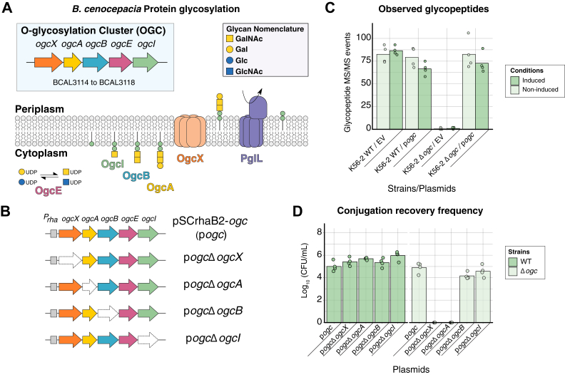
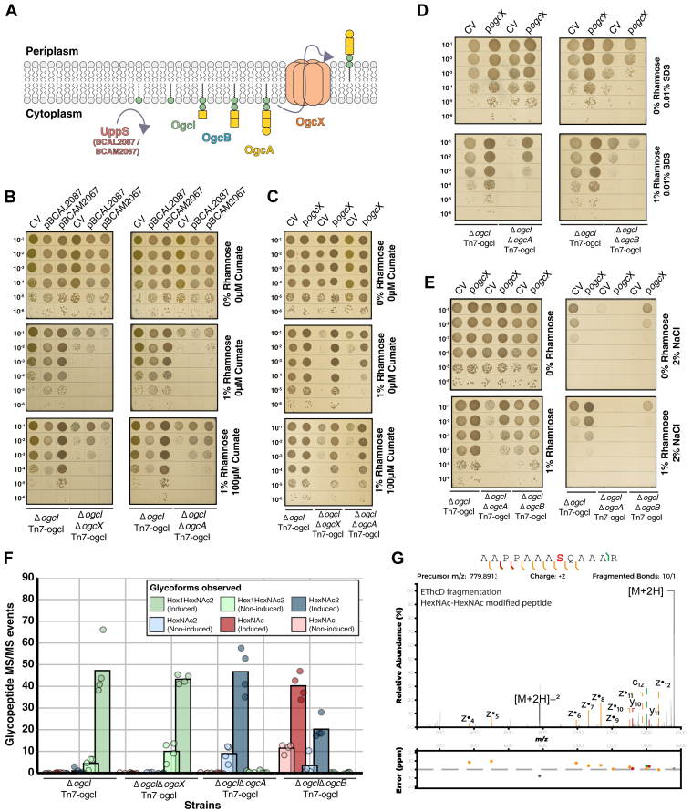
Periplasmic O-linked protein glycosylation is a highly conserved process observed across the Burkholderia genus. Within Burkholderia, protein glycosylation requires the five-gene cluster known as the O-glycosylation cluster (OGC, ogcXABEI), which facilitates the construction of the O-linked trisaccharide attached to periplasmic proteins. Previous studies have reported conflicting results regarding the essentiality of ogcA, predicted to be responsible for the addition of the final carbohydrate of the O-linked trisaccharide, and ogcX, the putative O-linked glycan flippase. Within this work, we aimed to dissect the impact of the loss of ogcA and ogcX on Burkholderia cenocepacia viability. We demonstrate that the loss of either ogcA or ogcX is detrimental if glycosylation is initiated, leading to marked phenotypic effects. Proteomic analysis supports that the loss of ogcA/ogcX both blocks glycosylation and drives pleotropic effects in the membrane proteome, resulting in the loss of membrane integrity. Consistent with this, strains lacking ogcA and ogcX exhibit increased sensitivity to membrane stressors, including antibiotics, and demonstrate marked changes in membrane permeability. These effects are consistent with the fouling of the undecaprenyl pool due to dead-end O-linked glycan intermediates, and consistent with this, we show that modulation of the undecaprenyl pool through the overexpression of undecaprenyl pyrophosphate synthase (UppS) or the OGC flippase (OgcX) restores viability, while expression of early-stage OGC biosynthesis genes (ogcI and ogcB) reduces B. cenocepacia viability. These findings demonstrate that disrupting O-linked glycan biosynthesis or transport appears to dramatically impact B. cenocepacia viability, supporting the assignment of ogcA and ogcX as conditionally essential.

Keywords: Burkholderia; Burkholderia cenocepacia; glycoproteomics; glycosylation; post-translational modifications; proteomics.


Conflict of interest statement

Conflict of interest The authors declare that they have no conflicts of interest with the contents of this article.

Figures

Figure 1
Figure 1
Loss of ogcX and ogcA impacts viability of B. cenocepacia Δogc.A, diagram of the O-glycosylation biosynthesis pathway in Burkholderia species and the genetic organization of the ogc cluster (BCAL3114–BCAL3118 within B. cenocepacia J2315) (47). O-glycosylation biosynthesis involves the stepwise construction of a conserved trisaccharide composed of Gal-GalNAc2 by the glycosyltransferases, OgcI, OgcB and OgcA, with the nucleotide-sugar donors UDP-GalNAc and UDP-Gal generated by the epimerase OgcE. The resulting trisaccharide is then thought to be translocated across the inner membrane by the putative flippase, OgcX. Once in the periplasm, the glycans can be transferred to proteins by the oligosaccharyltransferase, PglL. Monosaccharides have been annotated according to the Symbol Nomenclature for Glycans (SNFG) (119). B, graphic representation of pogc and variants lacking ogcX, ogcA, ogcB and ogcI. C, quantification of glycopeptide and protein levels encoded by the ogc cluster identified from whole cell proteomic analysis of B. cenocepacia WT and Δogc containing either the empty vector pSCrhaB2 (EV) or pSCrhaB2-ogc (pogc) with and without induction with 1% rhamnose (n = 4). D, conjugation of pogc derivatives into B. cenocepacia K56-2 WT and Δogc reveals differences in recovery rates of pogcΔogcX and pogcΔogcA within B. cenocepacia Δogc (n = 4).
Figure 2
Figure 2
Loss of ogcX or ogcA leads to reduced growth when glycosylation is initiated.A, graphic representation of strains ΔogcX, ΔogcA, ΔogcB and ΔogcAB generated within the chromosomal inducible background B. cenocepacia ΔogcI Tn7-ogcI. B, spot plate assays assessing growth of strains with and without 1% rhamnose induction. Induction results in reduced colony size of ΔogcX and ΔogcA strains to 10% of the size observed in B. cenocepacia ΔogcI Tn7-ogcI (n = 4). C, quantification of glycopeptides identified from whole-cell proteomic analysis of strains. Glycopeptides were assessed in the absence (light green) or presence (dark green) of rhamnose induction (n = 4). Induction of ogcI expression by 1% rhamnose led to the restoration of glycosylation only in B. cenocepacia ΔogcI Tn7-ogcI with no glycosylation observed in the ΔogcX, ΔogcB, and ΔogcAB strains.
Figure 3
Figure 3
Complementation of ΔogcA and ΔogcX restores growth and glycosylation.A, spot plate assays of B. cenocepacia ΔogcI Tn7-ogcI and B. cenocepacia ΔogcIΔogcA Tn7-ogcI containing pSCrhaB2 (EV), pSCrhaB2-ogcA-Met1 (pogcAMet1), or pSCrhaB2-ogcA-Met2 (pogcAMet2) with and without 1% rhamnose induction revealing complementation of ΔogcA with ogcA variants restores viability and colony size. B and C, quantification of glycopeptides and OgcA levels from whole-cell proteomic analysis of B. cenocepacia ΔogcI Tn7-ogcI and B. cenocepacia ΔogcIΔogcA Tn7-ogcI strains carrying EV, ogcAMet1, or ogcAMet2. Induction with 1% rhamnose results in the detection of OgcA within B. cenocepacia ΔogcIΔogcA Tn7-ogcI and the restoration of glycosylation (n = 4, ΔogcI ΔogcA Tn7-ogcI ogcA-Met1 induced group n = 3). D, spot plate assays of B. cenocepacia ΔogcI Tn7-ogcI and B. cenocepacia ΔogcIΔogcX Tn7-ogcI carrying the control vector pcumate-sfGFP (CV) and pcumate-ogcX-his (pogcX) with and without 1% rhamnose induction revealing complemented ΔogcX restores viability and colony size. E and F, quantification of glycopeptides and OgcX levels from whole-cell proteomic analysis of B. cenocepacia ΔogcI Tn7-ogcI and B. cenocepacia ΔogcIΔogcX Tn7-ogcI strains carrying sfGFP or ogcX plasmids. Induction results in the detection of OgcX within B. cenocepacia ΔogcIΔogcX Tn7-ogcI and the restoration of glycosylation (n = 4). Imputed values correspond to missing quantitation events resulting from the low abundance or absence of OgcX or OgcA within biological replicates.
Figure 4
Figure 4
Proteomic analysis of strains in response to glycosylation initiation. DIA proteomic analysis of ΔogcI Tn7-ogcI, ΔogcIΔogcA Tn7-ogcI, ΔogcIΔogcB Tn7-ogcI, and ΔogcIΔogcX Tn7-ogcI strains with and without rhamnose induction (n = 4). A, principal component analysis (PCA) of ΔogcI Tn7-ogcI strains under induced and non-induced conditions. Biological groups are observed to form distinct clusters, with ΔogcIΔogcA Tn7-ogcI and ΔogcIΔogcX Tn7-ogcI strains separating along PC1 upon induction. B and C, upset plot and Venn diagram of proteomic alterations, defined as >two-fold-change and −log10(p-value) > 2 observed across strains, reveal that induction results in hundreds of protein alterations within ΔogcIΔogcA Tn7-ogcI and ΔogcIΔogcX Tn7-ogcI strains compared to ΔogcIΔogcB Tn7-ogcI or ΔogcI Tn7-ogcI. D, enrichment analysis of GO terms associated with the altered proteins in ΔogcIΔogcX Tn7-ogcI strain reveals an over-representation of proteomic changes in membrane-associated protein classes. E, 2D scatter plots comparing proteome changes observed within ΔogcIΔogcA Tn7-ogcI and ΔogcIΔogcX Tn7-ogcI in response to glycosylation initiation. Proteins associated with membrane GO-terms are color-coded dark blue with the opacity corresponding to the average p-values observed across both comparisons. F, zoomed in 2D scatter plots of proteins observed to increase upon induction within ΔogcIΔogcA Tn7-ogcI and ΔogcIΔogcX Tn7-ogcI with membrane proteins of note highlighted.
Figure 5
Figure 5
Loss of ogcA and ogcX impacts the membrane integrity of B. cenocepacia.A, spot plate assays of strains in response to membrane (0.01% SDS) and osmotic (2% NaCl) stressors with and without 1% rhamnose induction (n = 4). The deletion of ogcX and ogcA increases sensitivity to 0.01% SDS when glycosylation is initiated, while both mutants appear sensitive to osmotic stress (2% NaCl) regardless of induction. B and C, spot plate assays of complemented ΔogcogcA Tn7-ogcI and ΔogcogcX Tn7-ogcI strains in response to membrane (0.01% SDS) and osmotic (2% NaCl) stress with and without 1% rhamnose induction demonstrating the restoration of growth within complemented strains (n = 4). The empty vector plasmid pSCrhaB2 (EV) and control vector pCumate-sfGFP (CV) correspond to negative control plasmids for pogcAMet1/pogcAMet2 and pogcX, respectively. D and E, hoechst 33,342 and NPN uptake assays of strains reveal loss of ogcA and ogcX leads to increased fluorescence upon induction, supporting enhanced membrane permeability (n = 6 for Hoechst 33,342, n = 5 for NPN). The fluorescence intensities have been normalised against bacterial viability counts (Fig. S9).
Figure 6
Figure 6
Loss of ogcA and ogcX sensitises B. cenocepacia to antimicrobials. Spot plate assays of B. cenocepacia strains, including ΔogcI Tn7-ogcI, ΔogcIΔogcX Tn7-ogcI, ΔogcIΔogcA Tn7-ogcI, and ΔogcIΔogcB Tn7-ogcI on CAMHA containing various antibiotics, with or without induction with 1% rhamnose. Rhamnose-induced glycosylation resulted in increased susceptibility to tetracycline, rifampicin, trimethoprim and ceftazidime in strains lacking ogcA and ogcX. Data representative of four biological replicates per strain per antimicrobial.
Figure 7
Figure 7
Overexpression of UppS or OgcX modulates ogc associated defects in B. cenocepacia.A, proposed enzymes responsible for the de novo synthesis of the undecaprenyl pool and steps within the ogc biosynthesis. Within B. cenocepacia, two putative undecaprenyl pyrophosphate synthases (UppS) are assigned corresponding to BCAL2087 and BCAM2067, while OgcI is responsible for ogc initiation and OgcB the addition of the second GalNAc (47). The functions of OgcA, as the glycotransferase responsible for the addition of the final Gal and OgcX, as the flippase responsible for glycan translocation have only been inferred to date. B, spot plate assays of the B. cenocepacia ΔogcI Tn7-ogcI, ΔogcIΔogcX Tn7-ogcI and ΔogcIΔogcA Tn7-ogcI containing the control vector pcumate-sfGFP (CV), pcumate-BCAL2087 or pcumate-BCAM2067 reveal the initiation of glycosylation reduces viability while induction of putative UppS partially restores the defect observed within ΔogcA and ΔogcX strains (n = 4). C, spot plate assays of the B. cenocepacia ΔogcI Tn7-ogcI, ΔogcIΔogcX Tn7-ogcI and ΔogcIΔogcA Tn7-ogcI containing the control vector (CV) and pcumate-ogcX (pogcX) demonstrating the complementation of ΔogcIΔogcA Tn7-ogcI with pogcX restores growth (n = 4). D and E, spot plate assays of the B. cenocepacia ΔogcI Tn7-ogcI, ΔogcIΔogcA Tn7-ogcI and ΔogcIΔogcB Tn7-ogcI containing the control vector (CV) and pogcX in response to membrane (0.01% SDS) and osmotic (2% NaCl) stresses with glycosylation initiation demonstrating that pogcX restores resistance to stress in ΔogcIΔogcA Tn7-ogcI yet does not suppress the defect observed in ΔogcIΔogcB Tn7-ogcI (n = 4). F, quantification of glycopeptides from whole-cell proteomic analysis of B. cenocepacia Tn7-ogcI strains carrying pogcX. Induction results in the detection of glycosylation with varying glycoforms observed across strains (n = 4). G, EThcD fragmentation of the HexNAc2 modified peptide 397AAPPAAASQAAAR409 (BCAL1674 Uniprot: B4E8U6) confirming the localization of the HexNAc2 glycosylation event residue S404.
Figure 8
Figure 8
Modulation of early steps in ogc biosynthesis impacts B. cenocepacia viability. Spot plate assays of B. cenocepacia K56-2 (WT) containing the expression vectors pSCrhaB2 (EV), pSCrhaB2-ogc (pogc), pSCrhaB2-ogcI (pogcI), pSCrhaB2-ogcA-Met1 (pogcAMet1), pSCrhaB2-ogcB (pogcB), pSCrhaB2-ogcAB-Met1 (pogcABMet1). Induction of ogcI and ogcB results in reduced viability (n = 4).

Similar articles

References

    1. Touz E.T., Mengin-Lecreulx D. Undecaprenyl phosphate synthesis. EcoSal Plus. 2008;3 doi: 10.1128/ecosalplus.4.7.1.7. - DOI - PubMed
    1. Workman S.D., Strynadka N.C.J. A slippery scaffold: synthesis and recycling of the bacterial cell wall carrier lipid. J. Mol. Biol. 2020;432:4964–4982. - PubMed
    1. Hong Y., Cunneen M.M., Reeves P.R. The Wzx translocases for Salmonella enterica O-antigen processing have unexpected serotype specificity. Mol. Microbiol. 2012;84:620–630. - PubMed
    1. Liu M.A., Stent T.L., Hong Y., Reeves P.R. Inefficient translocation of a truncated O unit by a Salmonella Wzx affects both O-antigen production and cell growth. FEMS Microbiol. Lett. 2015;362 - PubMed
    1. Barreteau H., Magnet S., El Ghachi M., Touzé T., Arthur M., Mengin-Lecreulx D., et al. Quantitative high-performance liquid chromatography analysis of the pool levels of undecaprenyl phosphate and its derivatives in bacterial membranes. J. Chromatogr. B Analyt Technol. Biomed. Life Sci. 2009;877:213–220. - PubMed

MeSH terms

LinkOut - more resources