Review
. 2025 Jun;301(6):108548.
doi: 10.1016/j.jbc.2025.108548. Epub 2025 Apr 24.

Phase separation as a key mechanism in plant development, environmental adaptation, and abiotic stress response

Affiliations
Review

Phase separation as a key mechanism in plant development, environmental adaptation, and abiotic stress response

Karina C Pougy et al. J Biol Chem. 2025 Jun.

Abstract

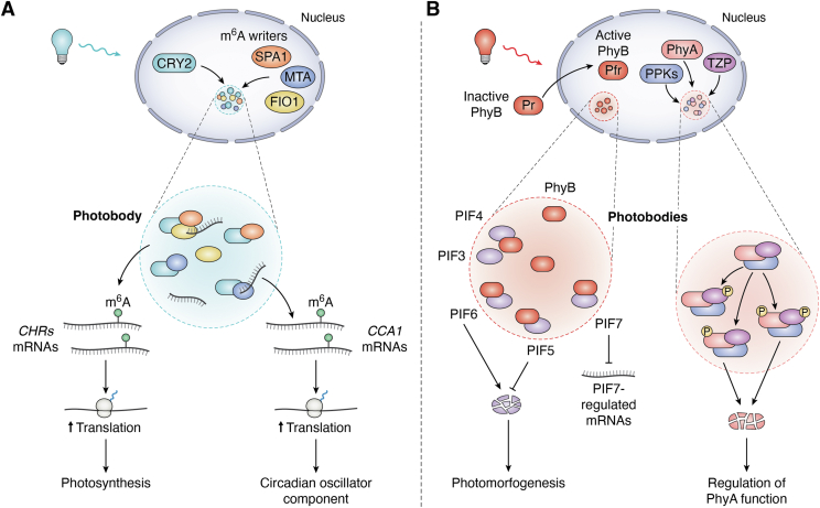
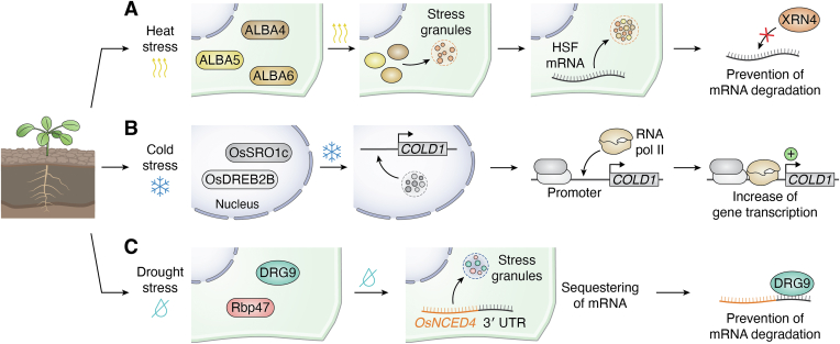
In memoriam of Professor Anderson de Sá Pinheiro, principal investigator at the Laboratory of Molecular Biology (LabMol) at the Institute of Chemistry, Federal University of Rio de Janeiro (UFRJ). Prof. Pinheiro passed away prematurely at the age of 44, on March 1, 2025. Prof. Pinheiro was a distinguished figure in the fields of biochemistry and structural biology in Brazil. He earned his bachelor's degree in Pharmacy in 2000, his master's degree in 2003, and his Ph.D. in 2007, all in Biological Chemistry at UFRJ. He continued his academic journey with postdoctoral research at Brown University in the United States (2007-2011). Upon returning to Brazil, he became an Associate Professor in the Department of Biochemistry at UFRJ (2011-2025). He led the Laboratory of Molecular Biochemistry (LaBMol), focusing on the study of RNA-binding proteins related to cancer and neurodegenerative disorders, as well as plant responses to abiotic stress. His research followed two main fronts-analyzing protein structures and dynamics using solution NMR spectroscopy and investigating the relationship between structural features and liquid-liquid phase separation, along with its role in protein function. Beyond research, Prof. Anderson was deeply committed to education, mentoring numerous students and contributing to various academic committees. During his brief but impactful career, he made significant contributions to the structural biology community, serving as President of the Brazilian Association of Nuclear Magnetic Resonance Users (AUREMN) and as Scientific Director of the Brazilian Biophysical Society (SBBf). This review marks Professor Pinheiro's 50th published article. His untimely passing is a profound loss to the scientific community, but his legacy endures through his scientific contributions and the many lives he has touched. Liquid-liquid phase separation is a fundamental biophysical process in which biopolymers, such as proteins, nucleic acids, and their complexes, spontaneously demix into distinct coexisting phases. This phenomenon drives the formation of membraneless organelles-cellular subcompartments without a lipid bilayer that perform specialized functions. In plants, phase-separated biomolecular condensates play pivotal roles in regulating gene expression, from genome organization to transcriptional and post-transcriptional processes. In addition, phase separation governs plant-specific traits, such as flowering and photosynthesis. As sessile organisms, plants have evolved to leverage phase separation for rapid sensing and response to environmental fluctuations and stress conditions. Recent studies highlight the critical role of phase separation in plant adaptation, particularly in response to abiotic stress. This review compiles the latest research on biomolecular condensates in plant biology, providing examples of their diverse functions in development, environmental adaptation, and stress responses. We propose that phase separation represents a conserved and dynamic mechanism enabling plants to adapt efficiently to ever-changing environmental conditions. Deciphering the molecular mechanisms underlying phase separation in plant stress responses opens new avenues for biotechnological strategies aimed at engineering stress-resistant crops. These advancements have significant implications for agriculture, particularly in addressing crop productivity in the face of climate change.

Keywords: condensate; growth; phase; plant; separation; stress.


Conflict of interest statement

Conflict of interest The authors declare that they have no conflicts of interest with the contents of this article.

Figures

None
This tribute to the late Prof. Anderson Pinheiro was previously allowed by JBC with the agreement of the associate editor Dr. Karin Musier-Forsyth.
Figure 1
Figure 1
Representative biomolecular condensates in plants. Schematic representation of a plant cell highlighting major organelles alongside examples of biomolecular condensates. Cytoplasmic and nuclear condensates are depicted as clusters of color-coded circles, each labeled with the name of the protein undergoing phase separation, an icon representing the associated biological process or external stimulus, as indicated in the figure legend, and the relevant citation. Flowering: FCA (pink) co-condenses with FLL2 (pink) to regulate flowering (flower icon) via the autonomous pathway. In response to cold (snowflake icon), FRIGIDA (FRI) (light blue) undergoes phase separation to regulate flowering through the vernalization pathway. Carbon fixation: EPYC1 (dark purple) co-condenses with RuBisCO (dark purple) to form the pyrenoid within chloroplasts, increasing carbon fixation (CO2 icon). Light signaling: In response to blue light (sun icon), CRY2 (light cyan) co-condenses with MTA (light cyan) to promote m6A RNA methylation. In response to red light (sun icon) or low temperatures (snowflake icon), PhyB forms nuclear photobodies through LLPS, facilitating photomorphogenesis. Heat stress: In response to heat (curved arrows icon), ELF3 undergoes nuclear phase separation to regulate flowering (flower icon) and hypocotyl elongation. In addition, the RNA-binding proteins GRP7 (dark blue) and AGO1 (yellow) undergo cytoplasmic phase separation under heat stress, facilitating heat stress adaptation. Cold stress: In response to cold (snowflake icon), OsSRO1c (light gray) co-condenses with the transcription factor OsDREB2B (dark gray) in the nucleus, regulating the cold stress response in rice. Drought stress: Under drought conditions (crossed drop icon), DRG9 (dark cyan) and the m6A reader ECT8 (orange) undergo cytoplasmic phase separation to enhance the drought stress response.
Figure 2
Figure 2
Phase separation as a mechanism for chromatin organization. The Agenet domain-containing protein, ADCP1 (blue), specifically recognizes the silenced chromatin mark H3K9me2 (pink sphere), undergoing phase separation. In addition, the high mobility group protein, HMGA (purple), co-condenses with ADCP1, promoting the formation of heterochromatin foci. The methyltransferase MtSUVR2 (dark orange) undergoes phase separation to catalyze H3K9me2 (pink sphere) deposition. Furthermore, MtSUVR2 condensates recruit the recombinase MtRAD51 (light orange), facilitating DNA double-strand break repair. Finally, histone H1 (green) undergoes phase separation through its C-terminal intrinsically disordered tail, contributing to chromatin compaction.
Figure 3
Figure 3
Phase separation and RNA processing.A, miRNA maturation in phase-separated Dicing bodies (D-bodies). The disordered zinc finger protein SERRATE (SE) (dark green) forms condensates that recruit DICER-LIKE 1 (DCL1) (medium green), (HYPONASTIC LEAVES 1) HYL1 (light green), and pri-miRNAs. HYL1 and DCL1 collaborate to process pri-miRNA into pre-miRNA, which is subsequently converted into the mature miRNA/∗ duplex. B, siRNA biogenesis in phase-separated siRNA bodies. SUPPRESSOR OF GENE SILENCING 3 (SGS3) (dark orange) forms condensates that recruit various components, including RNA-DEPENDENT RNA POLYMERASE 6 (RDR6) (light purple), DICER-LIKE 2 (DCL2) (light orange), DICER-LIKE 4 (DCL4) (pink), and ssRNAs. RDR6 converts ssRNA into dsRNA, which is then processed by DCL2 and DCL4 into the final easiRNA.
Figure 4
Figure 4
Phase separation regulation of flowering time in the autonomous, vernalization, and photoperiod pathways. Schematic representation illustrating the role of phase separation in the regulation of FLC and FT transcription across different flowering pathways in plants. A, autonomous pathway. FCA (dark violet) and FLL2 (light violet) co-condense in the nucleus, forming a molecular scaffold that recruits additional factors, including FPA, FY, CPSF30, CPSF100, and FIL1. This condensate promotes the proximal polyadenylation of the FLC antisense transcript COOLAIR. The 3′-proximally polyadenylated COOLAIR associates with the FLC locus, leading to its transcriptional repression. The downregulation of FLC facilitates the transition to flowering in spring. B, vernalization pathway. Left panel: During warm seasons, FRIGIDA (FRI) (blue) is diffusely distributed in the nucleus, where it recruits proteins such as FR1, FS1, SVF24, and FSX, which facilitate the deposition of transcriptionally active epigenetic marks, including H3K36me3 and H3K4me1 (pink spheres), at the FLC locus. These marks activate FLC transcription, inhibiting flowering. Right panel: During winter, FRI (blue) forms condensates that sequester transcription factors (light red and green) together with a distally polyadenylated form of COOLAIR, blocking RNA polymerase activity at the FLC locus. This repression of FLC transcription enables flowering in spring. C, photoperiod pathway. Upon light stimulation, CONSTANS (CO) (light orange) and the nuclear factors NF-YB2 (yellow) and NF-YC9 (dark orange) form condensates that promote CO binding to the FT promoter region. This interaction enhances FT transcription, driving flowering.
Figure 5
Figure 5
Light-induced phase separation of cryptochromes and phytochromes into photobodies. Schematic representation of the mechanism by which light stimuli trigger the phase transition of cryptochrome 2 (CRY2) and phytochromes A and B (PhyA and PhyB) into photobodies. A, CRY2-mediated photobody formation. Upon blue light absorption, CRY2 (dark cyan) interacts with m6A writers, including MTA (blue) and FIO1 (yellow), undergoing phase separation to form photobodies that sequester mRNAs. CRY2 (dark cyan)-MTA (blue) condensates deposit m6A methylation onto CCA1 mRNA, increasing its translation and regulating the circadian rhythm. Similarly, CRY2 (dark cyan)-FIO1 (yellow)-SPA1 (orange) condensates deposit m6A methylation onto CHR mRNAs, facilitating their translation and contributing to photosynthesis. B, phytochrome-mediated photobody formation. In its inactive Pr state, PhyB (dark red) resides in the cytoplasm. Absorption of red light converts PhyB (dark red) to its active Pfr state, triggering its relocation to the nucleus, where it undergoes phase separation to form photobodies. PhyB condensates recruit PIF transcription factors (light violet), which promote skotomorphogenesis, although with distinct effects. The interaction of photoactivated PhyB with PIF6 leads to PIF6 degradation, thereby promoting photomorphogenesis. Conversely, sequestration of PIF5 by PhyB photobodies protects it from degradation, whereas sequestration of PIF7 inhibits its chromatin binding and the transcription of PIF7 target genes. In addition, upon far-red light absorption, PhyA (light red) interacts with TZP (purple) to form condensates that recruit PPKs (blue). These kinases phosphorylate both PhyA and TZP. PhyA phosphorylation targets it for degradation, fine-tuning its regulatory functions.
Figure 6
Figure 6
Phase separation mechanisms involved in the abiotic stress response in plants. Schematic representation illustrating examples of the molecular mechanisms that utilize LLPS to mediate environmental stress responses in plants. A, heat stress (top row). Upon exposure to heat stress (curved arrows icon), ALBA proteins—ALBA4 (brown), ALBA5 (yellow), and ALBA6 (orange)—undergo phase separation, relocating to stress granules (SGs). This redistribution enables ALBA proteins to sequester heat shock transcription factor (HSF) mRNAs, protecting them from XRN4-mediated degradation and promoting heat tolerance. B, cold stress (middle row). During cold stress (snowflake icon), OsSRO1c (dark gray) co-condenses with the transcription factor OsDREB2B (light gray) in the nucleus. This interaction facilitates the binding of OsDREB2B to the COLD1 gene promoter, increasing its transcription and contributing to cold tolerance. C, drought stress (bottom row). Under drought conditions (crossed drop icon), DRG9 (dark cyan) undergoes phase separation and translocates to SGs, where it colocalizes with Rbp47 (red). This relocation allows DRG9 to sequester OsNCED4 mRNA within SGs, protecting it from degradation and enhancing drought tolerance.

Similar articles

References

    1. Banani S.F., Lee H.O., Hyman A.A., Rosen M.K. Biomolecular condensates: organizers of cellular biochemistry. Nat. Rev. Mol. Cell Biol. 2017;18:285–298. - PMC - PubMed
    1. Shin Y., Brangwynne C.P. Liquid phase condensation in cell physiology and disease. Science. 2017;357 - PubMed
    1. Boeynaems S., Alberti S., Fawzi N.L., Mittag T., Polymenidou M., Rousseau F., et al. Protein phase separation: a new phase in cell biology. Trends Cell. Biol. 2018;28:420–435. - PMC - PubMed
    1. Gomes E., Shorter J. The molecular language of membraneless organelles. J. Biol. Chem. 2019;294:7115–7127. - PMC - PubMed
    1. Hofmann S., Kedersha N., Anderson P., Ivanov P. Molecular mechanisms of stress granule assembly and disassembly. Biochim. Biophys. Acta Mol. Cell Res. 2021;1868 - PMC - PubMed

LinkOut - more resources