. 2025 Apr 26;11(1):95.
doi: 10.1038/s41531-025-00965-6.

Subthalamic stimulation evokes hyperdirect high beta interruption and cortical high gamma entrainment in Parkinson's disease

Affiliations

Subthalamic stimulation evokes hyperdirect high beta interruption and cortical high gamma entrainment in Parkinson's disease

Ádám József Berki et al. NPJ Parkinsons Dis. .

Abstract

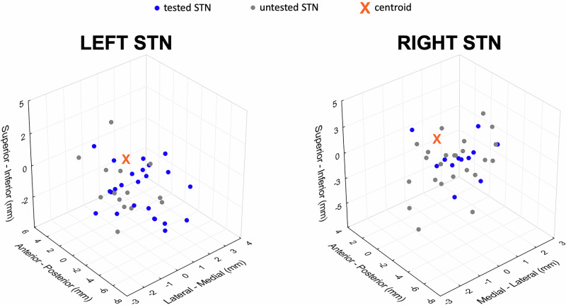
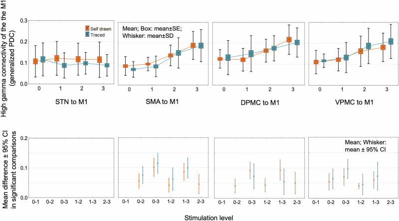
Compound network dynamics in beta and gamma bands determine the severity of bradykinesia in Parkinson's disease. We explored its subthalamic stimulation related changes parallel with improvement of complex hand movements. Thirty eight patients with Parkinson's disease treated with bilateral stimulation accomplished voluntary and traced spiral drawing with their more affected hand on a digital tablet. A 64 channel electroencephalography was recorded, low and high beta and gamma power was computed in subthalamic and motor cortical sources at four stimulation levels. Subthalamic cortical effective connectivity was calculated, and subnetwork models were created. Beta power decreased, and gamma power increased in sources ipsilateral to stimulation with increasing stimulation intensity. Networks comprising the primary motor cortex played a dominant role in predicting the improvement of voluntary drawing speed. Subthalamic stimulation diminished the hyperdirect high beta information processing and promoted the cortico cortical interactions of the primary motor cortex in the high gamma band.


Conflict of interest statement

Competing interests: M.M. is the Deputy Editor-in-chief of this journal. Other authors have no competing interest to declare.

Figures

Fig. 1
Fig. 1. Active contact locations relative to the center point of the dorsolateral STN.
The active contact locations are presented in the MNI space; the x-axis represents the medial-lateral, the y-axis anterior-posterior, and the z-axis superior-inferior plane. Tested contact locations in each STN contralateral to the movement are presented with blue dots. Gray dots depict contact locations of untested STN (ipsilateral to the movement) with stable stimulation. The center point of the dorsolateral STN is the point of reference, represented by an orange cross. MNI Montreal Neurological Institute, STN subthalamic nucleus.
Fig. 2
Fig. 2. Average velocity and entropy of velocity of spiral drawing in the four levels of stimulation.
a Average tangential velocity increased in both self-paced and traced spiral drawing tasks. b Entropy of velocity decreased when switching the stimulation on but did not follow further intensity increase in each task. Repeated-measures ANOVA revealed a significant main effect of stimulation level. Significant post hoc comparisons are presented in the bottom row (p < 0.05).
Fig. 3
Fig. 3. Absolute beta and gamma power changes in the primary motor cortex.
In the primary motor cortex, subthalamic stimulation decreased absolute low and high beta band power stepwise in the tested hemisphere (contralateral to the movement). At the same time, low and high gamma activity was raised with increasing stimulation intensity. Power differences in significant post hoc comparisons of stimulation level effect are presented in the middle row. Beta and gamma power remained unchanged in the untested hemisphere, with rising stimulation intensity. Bottom row: in the not-tested hemisphere (ipsilateral to the movement), beta and gamma power remained unchanged with elevating stimulation intensity. M1 primary motor cortex.
Fig. 4
Fig. 4. Absolute beta and gamma power changes in the subthalamic nucleus.
Stimulation-induced power changes in the low beta, high beta, low gamma, and high gamma bands in the STN are similar to the changes observed in the primary motor cortex (Fig. 3). STN subthalamic nucleus.
Fig. 5
Fig. 5. Stimulation effects on task-related hyperdirect high-beta effective connectivity in PD patients.
Effective connectivity between the motor cortical areas and STN decreased in the high beta band with increasing stimulation intensity. Significant comparisons of stimulation level effects are presented in the bottom row. The figure visualizes the direction of coherence from motor cortical areas toward the subthalamic nucleus. However, this effect was bidirectional; the opposite directional effective connectivity is demonstrated in Supplementary Fig. 7. DPMC dorsal premotor cortex, M1 primary motor cortex, SMA supplementary motor cortex, STN subthalamic nucleus, PD Parkinson’s disease, VPMC ventral premotor cortex.
Fig. 6
Fig. 6. Stimulation effects on task-related hyperdirect high-beta effective connectivity in PD patients according to the tested hemisphere.
The comparison of the tested left and tested right hemispheres revealed a similar stimulation effect on connectivity values with the merged analysis (Fig. 5): effective connectivity between the motor cortical areas and STN decreased in the high beta band with increasing stimulation intensity. Effective connectivity in patients with tested left hemisphere did not differ from those with tested right hemisphere in any stimulation level and pair of brain regions (p > 0.05). The figure presents the direction of coherence from motor cortical areas toward the subthalamic nucleus. However, this effect was bidirectional; the opposite directional effective connectivity is demonstrated in Supplementary Fig. 8. DPMC dorsal premotor cortex, M1 primary motor cortex, SMA supplementary motor cortex, STN subthalamic nucleus, PD Parkinson’s disease, VPMC ventral premotor cortex.
Fig. 7
Fig. 7. STN stimulation increases the high-gamma effective connectivity between the premotor cortical areas, and the M1.
Ramping subthalamic stimulation increased the effective connectivity between the premotor cortical areas and M1 in the high gamma frequency band; however, the high gamma STN-M1 effective connectivity was unaffected. Significant post hoc comparisons of the stimulation level effect are presented in the lower row. The effect was bidirectional; effective connectivity from the direction of M1 to other structures is presented in Supplementary Fig. 9. DPMC dorsal premotor cortex, M1 primary motor cortex, SMA supplementary motor cortex, STN subthalamic nucleus, PD Parkinson’s disease, VPMC ventral premotor cortex.
Fig. 8
Fig. 8. Study protocol.
EEG electroencephalography, STIM OFF stimulation OFF.
Fig. 9
Fig. 9. Signal processing pipeline.
a Based on individual T1 MRI sequences, we reconstructed a lead-field matrix using the finite-element method. b High-density EEG acquisition during traced and self-paced spiral drawing at four different stimulation levels. c Estimation of coherent neural activity and source extraction from the selected regions. CSF cerebrospinal fluid, DLPFC dorsolateral prefrontal cortex, DPMC dorsal premotor cortex, EEG electroencephalography, LFM lead-field matrix, MRI magnetic resonance imaging, M1 primary motor cortex, SMA supplementary motor cortex, STN subthalamic nucleus, VC visual cortex, VPMC ventral premotor cortex.

Similar articles

References

    1. Oswal, A. et al. Neural signatures of hyperdirect pathway activity in Parkinson’s disease. Nat. Commun.12, 5185 (2021). - PMC - PubMed
    1. He, S. et al. Beta-triggered adaptive deep brain stimulation during reaching movement in Parkinson’s disease. Brain146, 5015–5030 (2023). - PMC - PubMed
    1. Piña-Fuentes, D. et al. Acute effects of adaptive Deep Brain Stimulation in Parkinson’s disease. Brain Stimul.13, 1507–1516 (2020). - PMC - PubMed
    1. Oehrn, C. R. et al. Chronic adaptive deep brain stimulation versus conventional stimulation in Parkinson’s disease: a blinded randomized feasibility trial. Nat. Med.10.1038/s41591-024-03196-z (2024). - PMC - PubMed
    1. Feldmann, L. K. et al. Toward therapeutic electrophysiology: beta-band suppression as a biomarker in chronic local field potential recordings. NPJ Parkinsons Dis.8, 44 (2022). - PMC - PubMed

LinkOut - more resources