. 2025 Apr 26;44(5):103.
doi: 10.1007/s00299-025-03485-y.

Recombinant production of spider silk protein in Physcomitrella photobioreactors

Affiliations

Recombinant production of spider silk protein in Physcomitrella photobioreactors

Maryam Ramezaniaghdam et al. Plant Cell Rep. .

Abstract

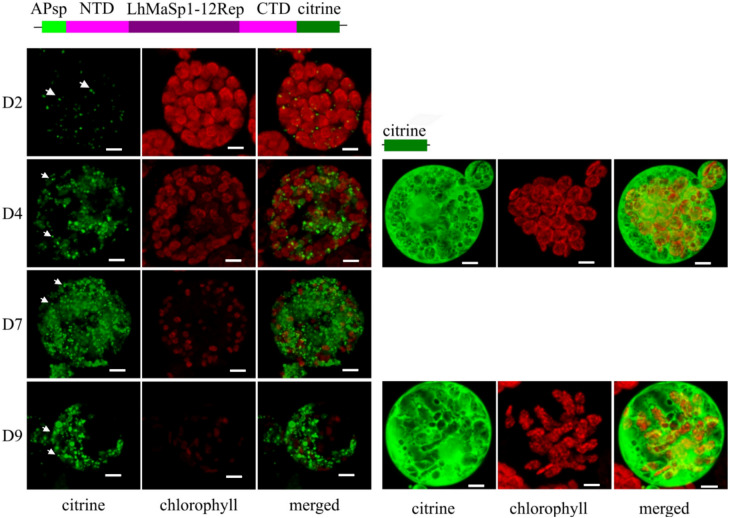
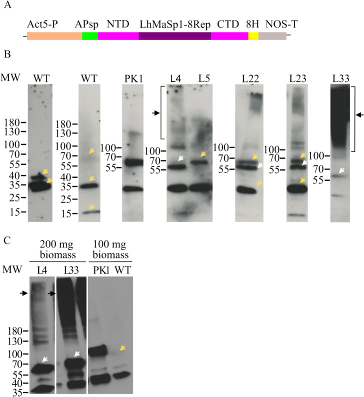
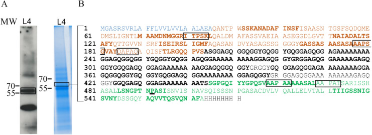
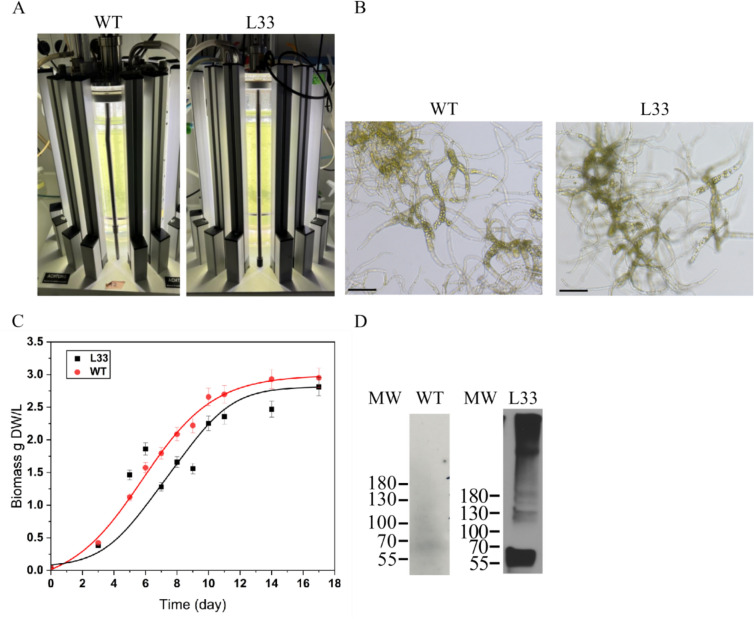
We report the successful moss-produced recombinant spider silk key protein component containing both the N- and the C-terminal domain. Spider dragline silk stands out as a remarkable biomaterial, representing one of nature's toughest fibres. Its strength rivals that of many synthetic fibres used commercially, rendering it applicable across various industrial and medical domains. However, its widespread utilisation requires cost-effective mass production. Biotechnology presents a promising avenue for achieving this goal, particularly through the production of recombinant dragline silk proteins in transgenic plant systems. This study aimed to assess the feasibility of producing one key protein component of dragline silk, MaSp1, from the western black widow spider, Latrodectus hesperus, the protein LhMaSp1, in the moss Physcomitrella (Physcomitrium patens). Here, we present the successful recombinant production of spider silk protein containing both the N- and C-terminal domains of LhMaSp1 in moss cells. The production of recombinant LhMaSp1 protein in Physcomitrella was performed in shake flasks and in five-litre photobioreactors and the correct synthesis of LhMaSp1 was proven via mass spectrometry. We estimate that the yield of recombinant spider silk protein in Physcomitrella bioreactors is above 0.82 mg/g fresh weight.

Keywords: Bioreactor; Bryotechnology; Dragline silk protein; Plant-made protein; Smart materials; Spidroins.


Conflict of interest statement

Declarations. Conflict of interest: All authors declare no conflict of interest.

Figures

Fig. 1
Fig. 1
Confocal microscopy images showing the production of NTD-LhMaSp1-12Rep-CTD-citrine fusion protein in Physcomitrella protoplasts. Schematic representation of the fusion protein is at the top. The images were recorded 2 (D2), 4 (D4), 7 (D7), and 9 days (D9) after protoplast transfection and show the successful production of NTD-LhMaSp1-12Rep-CTD-citrine in Physcomitrella (left panel). Protein aggregates are observed from NTD-LhMaSp1-12Rep-CTD fused to citrine (white arrows). The right panel shows the citrine expression control on days 4 (D4) and 9 (D9) after protoplast transfection. All images are 3D-rendered Z-stack images. NTD: N-terminal domain. CTD: C-terminal domain. Bars = 5 µm
Fig. 2
Fig. 2
Confocal microscopy images showing the localisation of NTD-LhMaSp1-12Rep-CTD fused to citrine in the secretory pathway of Physcomitrella protoplasts. Schematic representation of the fusion proteins is on top. The mCerulean-KDEL construct was used as a positive control for ER localisation. A Chlorophyll autofluorescence. B, C mCerulean and citrine channels, respectively. The arrows in Z-stack images point to the fluorescence signal of the ramified network and arrows in single slices point to the fluorescence signal of the nuclear envelope. A dense concentration of mCerulean-KDEL and NTD-LhMaSp1-12Rep-CTD-citrine signals is present in a region encircling the nucleus, suggesting its association with the rough ER. Contour drawings are provided to highlight this area. D Citrine and mCerulean signals do not overlap but demonstrate considerable coverage across similar areas (contour drawings). E Merged channels. The upper images display 3D-rendered Z-stack images, while the lower ones depict individual slices of single cells. Grey-scale channels are provided for better visibility. The images were recorded 5 days after protoplast transfection. Bars = 5 µm
Fig. 3
Fig. 3
Western blot analysis showing successful production of recombinant NTD-LhMaSp1-8Rep-CTD-8Htag protein in stable Physcomitrella lines. A Schematic representation of the expression cassette NTD-LhMaSp1-8Rep-CTD-8Htag. The plasmid contains the CDS of aspartic protease signal peptide (APsp), eight codon-optimized repetitive poly-alanine blocks of the L. hesperus MaSp1 protein core region, the full sequence of N- and C-terminal domains (NTD and CTD), 8 × Histidine-tag (8H), under the control of the Physcomitrella actin5 promoter and the NOS terminator. B Immunodetection of NTD-LhMaSp1-8Rep-CTD-8Htag was performed via Western blot using an anti-His-tag antibody (18,184, Thermo Fischer Scientific). Some of the best-producing lines are presented here. A signal slightly above 55 kDa was detected (white arrows). Almost all transgenic lines show high molecular mass signals higher than 180 kDa (black arrows). C Western blot of the two best NTD-LhMaSp1-8Rep-CTD-8Htag producing lines L4 and L33 with the elution fraction from the His SpinTrap column shows single bands at ~ 56 kDa (white arrows) and high molecular mass signals (black arrows). Unspecific signals also present in the WT are marked with yellow arrows. WT: wild type, L: line. PK1: positive control; transgenic moss line producing the 58 kDa His-tagged protein MFHR1. Gradient 4–15% SDS gels were used. Western blot under reducing conditions. MW: PageRuler Prestained Protein Ladder (Thermo Fisher Scientific)
Fig. 4
Fig. 4
Mass spectrometric analysis of NTD-LhMaSp1-8Rep-CTD-8Htag. A Total protein of protonema material of L4 was recovered after acetone precipitation. The protein band at ~ 56 kDa which is highlighted in the rectangle on the Coomassie-stained SDS-PAGE and corresponds to the band on the Western blot marked with a rectangle, was utilized for mass spectrometry. Anti-His-tag antibody (18,184, Thermo Fischer Scientific) was used for detection. B NTD-LhMaSp1-8Rep-CTD-8Htag peptides identified by mass spectrometry are shown in bold. Blue: aspartic protease signal peptide (APsp). Orange: N-terminal domain. Black: repetitive poly-alanine and glycine blocks. Green: C-terminal domain. Potential sites for hydroxylation of proline are shown in rectangles. Identified hydroxyproline is underlined. Peptides for MS analysis were generated with chymotrypsin. Western blot under reducing conditions. MW: PageRuler Prestained Protein Ladder (Thermo Fisher Scientific)
Fig. 5
Fig. 5
Cultivation of Physcomitrella WT and NTD-LhMaSp1-8Rep-CTD-8Htag producing line (L33) in photobioreactors. A Five-litre photobioreactor set-up of WT and NTD-LhMaSp1-8Rep-CTD-8Htag-producing line L33 was illuminated with LED lamps. After 2 days, the lights increased from 160 to 350 µmol/m2s. B Morphology of protonema tissue from WT and L33 at day 5 of cultivation. Bars = 150 µm. C Biomass accumulation of WT and L33 in the photobioreactor over a period of 17 days. Data represent mean ± standard deviation (SD) from three biological replicates. WT (red points) and L33 (black points) showed exponential growth until day 8, after which the growth rate began to slow. Solid lines represent fitted curves for WT (red) and L33 (black). Statistical analysis showed no significant difference between the two lines (t-test, p > 0.05). Biomass density in both lines reached almost 3 g DW/L on day 17. D 100 mg biomass of both lines at day 8 was harvested for anti-His Western blot analysis. The result shows the production of NTD-LhMaSp1-8Rep-CTD-8Htag in both forms of monomers and soluble multimers. Anti-His-tag antibody (18,184, Thermo Fisher Scientific) was used. Western blot under reducing conditions. MW: PageRuler Prestained Protein Ladder (Thermo Fisher Scientific)

Similar articles

References

    1. Altman GH, Diaz F, Jakuba C, Calabro T, Horan RL, Chen J, Lu H, Richmond J, Kaplan DL (2003) Silk-based biomaterials. Biomaterials 24:401–416. 10.1016/S0142-9612(02)00353-8 - PubMed
    1. Andresen PA, Kaasen I, Styrvold OB, Boulnois GJ, Strøm AR (1988) Molecular cloning, physical mapping and expression of the bet genes governing the osmoregulatory choline-glycine betaine pathway of Escherichia coli. J Gen Microbiol 134:1737–1746. 10.1099/00221287-134-6-1737 - PubMed
    1. Anton AM, Heidebrecht A, Mahmood N, Beiner M, Scheibel T, Kremer F (2017) Foundation of the outstanding toughness in biomimetic and natural spider silk. Biomacromol 18:3954–3962. 10.1021/acs.biomac.7b00990 - PubMed
    1. Askarieh G, Hedhammar M, Nordling K, Saenz A, Casals C, Rising A, Johansson J, Knight SD (2010) Self-assembly of spider silk proteins is controlled by a pH-sensitive relay. Nature 465:236–238. 10.1038/nature08962 - PubMed
    1. Ayoub NA, Garb JE, Tinghitella RM, Collin MA, Hayashi CY (2007) Blueprint for a high-performance biomaterial: full-length spider dragline silk genes. PLoS ONE 2:1–13. 10.1371/journal.pone.0000514 - PMC - PubMed

LinkOut - more resources