. 2025 Apr 26;15(1):14666.
doi: 10.1038/s41598-025-98939-4.

Profiling the cell-specific small non-coding RNA transcriptome of the human placenta

Affiliations

Profiling the cell-specific small non-coding RNA transcriptome of the human placenta

Nikita Telkar et al. Sci Rep. .

Abstract

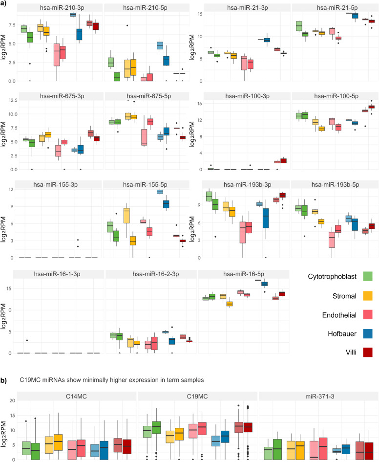
The human placenta is the composite of multiple cell types, each which contributes uniquely to placental function. Small non-coding RNAs (sncRNAs) are regulators of gene expression and can be cell-specific. The sncRNA transcriptome of individual placental cell types has not yet been investigated due to difficulties in their procurement and isolation. Using a custom sequencing method, we explored the expression of seven sncRNA species (miRNA, piRNA, rRNA, scaRNA, snRNA, snoRNA, tRNA) from whole chorionic villi and four major sample-matched FACS-sorted cell type (cytotrophoblast, stromal, endothelial, Hofbauer) samples from 9 first trimester and 17 term placentas. After normalization for technical variables, samples clustered primarily by cell type lineage. No sncRNAs were uniquely expressed by cell type, however, mean expression differed by cell type for 115 sncRNAs. Known placentally-expressed sncRNAs showed differing expression by cell type and trimester. Expression of few sncRNAs varied by sex. Lastly, sample-matched sncRNA expression and DNA methylation correlation was not significant, although high correlation (> R2 ± 0.6) was observed for some sncRNA-CpG pairs. This study represents the first exploration of the sncRNA transcriptome of bulk placental villi and placental cell types, informing about the expression and regulatory patterns underlying human placental development.

Keywords: Chorionic villi; MiRNA; Placenta; Small non-coding RNA; Trophoblast.


Conflict of interest statement

Declarations. Competing interests: The authors declare no competing interests. Ethics approval and consent to participate: Written informed consent was obtained from all participants including a tissue banking consent. This study was approved by The University of British Columbia and BC Women’s and Children’s Hospital research ethics board in Vancouver, BC, Canada (certificate number: H16-02280).

Figures

Fig. 1
Fig. 1
Expression of known placentally-expressed miRNAs. All reads are per million. (a) The expression of 7 miRNAs known to be associated with placental/pregnancy disorders, where the lighter coloured boxplots on the left are first trimester samples, and the darker colour boxplots are term samples. The thick horizontal line in the middle of each boxplot represents the median of the data (Quartile 2). The bottom box represents the first quartile (Q1) i.e., 25% of the data falls below this value, and the top box represents the third quartile (Q3), i.e., 75% of the data falls below this value. The total box represents the interquartile range (IQR), which is the middle 50% of the data (from Q1 to Q3). The vertical line extending from the middle of the boxplot represents the total spread of the data, with outliers as independent points. (b) Combined expression of the 89 C14MC, 59 C19MC, and 8 miR-371-3 cluster miRNAs in first trimester and term samples in the different cell types and matched whole villi.
Fig. 2
Fig. 2
Cell type-associated sncRNA expression. (a) Clustering heatmap (Z-scores of log2-transformed normalized expression counts) of the 115 cell type-associated sncRNAs. (b) Expression of two examples each of cell type-associated sncRNAs detected per cell type. n represents the number of sncRNAs designated as cell type-associated for that specific cell type. The thick horizontal line in the middle of each boxplot represents the median of the data (Quartile 2). The bottom box represents the first quartile (Q1) i.e., 25% of the data falls below this value, and the top box represents the third quartile (Q3), i.e., 75% of the data falls below this value. The total box represents the interquartile range (IQR), which is the middle 50% of the data (from Q1 to Q3). The vertical line extending from the middle of the boxplot represents the total spread of the data, with outliers as independent points. (c) Chromosomal location of each of the 115 cell type-associated sncRNAs.
Fig. 3
Fig. 3
Pathways predicted for the 10 cytotrophoblast cell-associated miRNAs. Gene sets for which all 10 cytotrophoblast cell-associated miRNAs were significant at a BH p-value <= 0.05.
Fig. 4
Fig. 4
SncRNAs showing variable expression by sex in term sorted-cell placental samples. (a) Autosomal sncRNAs which showed differing expression by sex in each cell type. The lighter coloured boxplots on the left are XX samples and the darker coloured boxplots are XY samples. The thick horizontal line in the middle of each boxplot represents the median of the data (Quartile 2). The bottom box represents the first quartile (Q1) i.e., 25% of the data falls below this value, and the top box represents the third quartile (Q3), i.e., 75% of the data falls below this value. The total box represents the interquartile range (IQR), which is the middle 50% of the data (from Q1 to Q3). The vertical line extending from the middle of the boxplot represents the total spread of the data, with outliers as independent points. (b) X-linked sncRNAs with differing expression by sex in cytotrophoblast sorted-cell samples.

Update of

Similar articles

References

    1. Mercuri, N. D. & Cox, B. J. The need for more research into reproductive health and disease. eLife11, e75061 (2022). - PMC - PubMed
    1. Turco, M. Y. & Moffett, A. Development of the human placenta. Dev. Camb. Engl.146, dev163428 (2019). - PubMed
    1. Wang, Y. & Zhao, S. Cell types of the placenta. In Vascular Biology of the Placenta (Morgan & Claypool Life Sciences, 2010). - PubMed
    1. Martin et al. Human placenta and trophoblast development: key molecular mechanisms and model systems. Cell. Mol. Life Sci.76, 3479–3496 (2019). - PMC - PubMed
    1. Reyes, L. & Golos, T. G. Hofbauer cells: their role in healthy and complicated pregnancy. Front. Immunol.9, 2628 (2018). - PMC - PubMed

Substances

LinkOut - more resources