. 2025 Apr 26;82(1):177.
doi: 10.1007/s00018-025-05702-z.

Calciprotein particle-activated endothelial cells aggravate smooth muscle cell calcification via paracrine signalling

Collaborators, Affiliations

Calciprotein particle-activated endothelial cells aggravate smooth muscle cell calcification via paracrine signalling

Lian Feenstra et al. Cell Mol Life Sci. .

Abstract

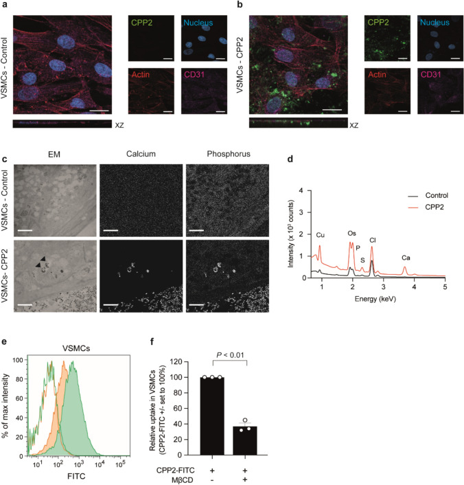
Background: Vascular calcification is highly prevalent in Chronic Kidney Disease (CKD) and is associated with markedly increased cardiovascular risk. High serum phosphate in CKD increases calcification propensity via generation of circulating calciprotein particles (CPP2), crystalline nanoaggregates composed of calcium, phosphate, and serum proteins. CPP2 induce vascular calcification in vascular smooth muscle cells (VSMCs) in vitro. In vivo, endothelial cells, rather than VSMCs are primarily exposed to CPP2, yet understanding the influence of endothelial cells on vascular calcification is limited.

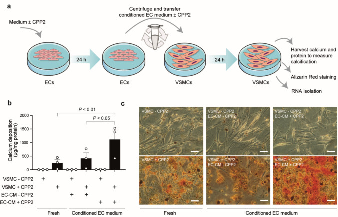
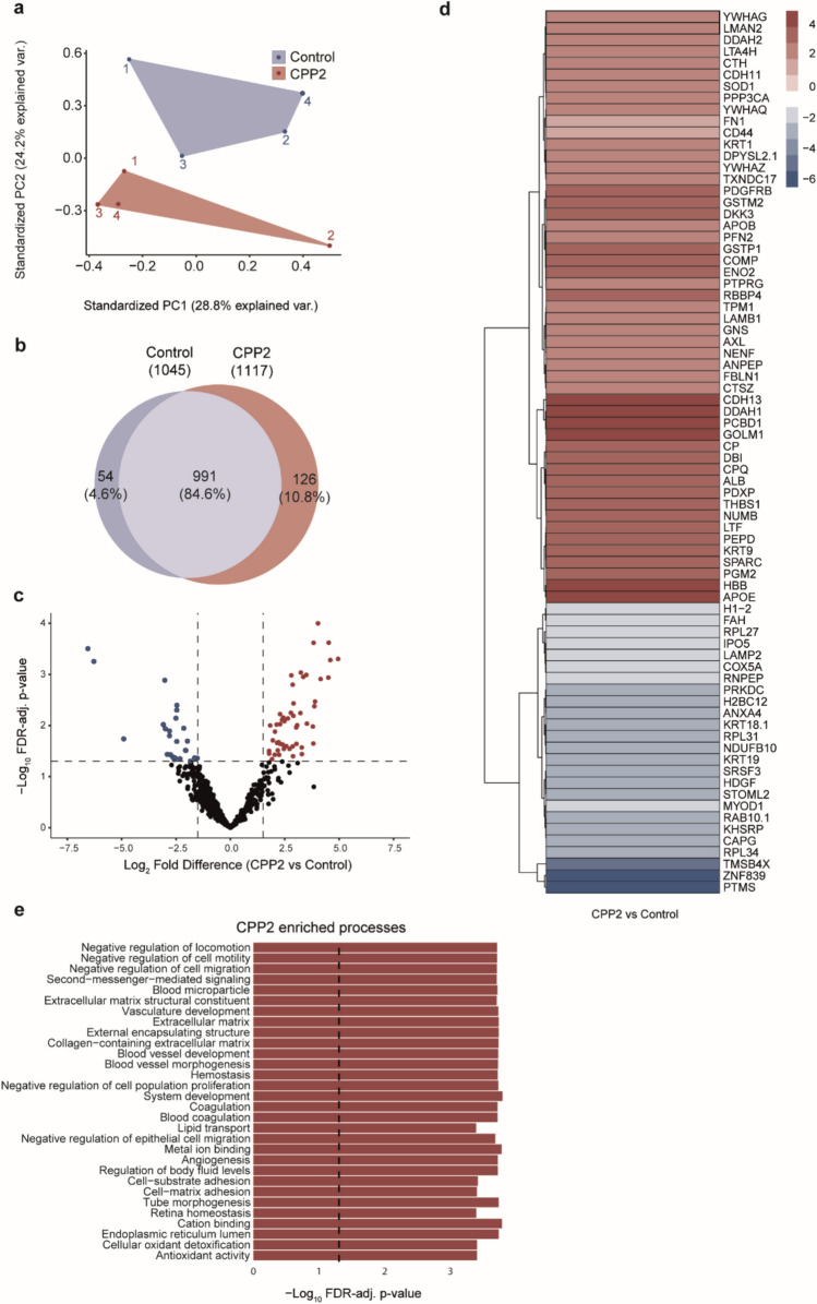
Methods: We investigated calcification-promoting signalling by endothelial cells on VSMCs. Effects of CPP2 exposure to endothelial cells on CPP2 uptake, endothelial cell activation, and endothelial cell-derived secretome were studied. Effects of the secretome on VSMC calcification were investigated. Using NanoString nCounter analysis the effects of CPP2-activated endothelial cell-conditioned medium on VSMCs gene expression were mapped.

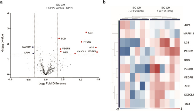
Results: Endothelial cells internalise CPP2 and elevate ICAM-1, E-selectin, and VCAM-1-mRNA expression, indicating endothelial activation. VSMCs cultured in conditioned medium from CPP2-activated endothelial cells demonstrated enhanced calcification, suggesting that CPP2-activated endothelial cells release pro-calcifying soluble factors. Mass spectrometry was utilized to identify 1171 proteins in the CPP2-activated endothelial cells' secretome. Among these, 76 proteins were differentially expressed compared to control endothelial cells' secretome, including proteins related to blood vessel development, extracellular matrix remodelling, and oxidative stress-related processes. Finally, endothelial cell-derived paracrine factors present in conditioned medium enhanced mRNA-expression of calcification-related factors in VSMCs.

Conclusions: CPP2-activated endothelial cells promote VSMC calcification via paracrine signalling. In response to these paracrine factors, VSMCs increase the expression of pro-calcification genes.

Keywords: Calciprotein particles; Chronic kidney disease; Endothelial cell activation; Paracrine signalling; Vascular calcification.


Conflict of interest statement

Declarations. Conflict of interest: G. Krenning is Chief Scientific Officer of Sulfateq B.V. (Groningen, the Netherlands), a company that develops small molecule therapeutics. Sulfateq B.V. has no small molecule in development for anti-circulating calciprotein particle (CPP2) therapy at present and had no influence on the content of this paper. E.R. Smith is a stockholder and former scientific advisor of Calciscon AG and has also received honoraria from CSL Vifor. Funding from Astellas received by R.A. Pol had no influence on the content of the paper. Astellas was not involved in the design of the study, collection of data, analysis and interpretation of data, management, manuscript preparation or the decision to submit the paper for publication. All other authors report no conflict of interest. Ethics approval: The study was conducted in compliance with the principle of the Declaration of Helsinki. All patients gave written informed consent. The protocol was reviewed and approved by the Institutional Review Board of the UMCG (number 2014–077).

Figures

Fig. 1
Fig. 1
Correlations of soluble VCAM-1 (sVCAM-1) and CPP counts in healthy kidney donor and CKD patients. a sVCAM-1 levels (pg/ml) measured in plasma of healthy kidney donors (n = 17) and CKD patients (n = 34). Data presented as mean ± SEM and tested with Mann–Whitney U test. b Correlation of sVCAM-1 levels and serum CPP1 counts (104 particles/ml). c Correlation of sVCAM-1 levels and serum CPP2 counts (104 particles/ml) (two-tailed nonparametric Spearman correlation). The correlation coefficient is indicated with the symbol r. P < 0.05 was considered statistically significant
Fig. 2
Fig. 2
Endothelial uptake op CPP2 in vitro. a, b Confocal images of ECs treated without (a) or with (b) FITC-labelled CPP2 (green), 25 µg calcium per ml CPP2 solution (= 25 µg/ml). XZ planes demonstrate internalisation of CPP2. Nucleus = blue, actin skeleton = red and CD31 = pink. Scale bars represent 20 μm. c Electron microscopical analysis of ECs showing CPP2 uptake. CPP2-containing vesicles are marked with an arrowhead. Insert in left bottom panel shows a magnification of a CPP2-containing vesicle. Calcium and phosphate content of the particles is visualised with EDX. Scale bars represent 2 μm. d EDX spectra showing copper (Cu), osmium (Os), phosphate (P), sulphur (S), chloride (Cl) and calcium (Ca) for unstimulated ECs (control, black line) and the CPP2 stimulated ECs (red line) in ECs. e Flow cytometric analysis demonstrated the uptake of FITC-labelled CPP2 (green) and the inhibition thereof in the presence of 2.5 mM methyl-β-cyclodextrin (MβCD; orange). Lines indicate incubation at 4 °C (extracellular binding), filled histograms indicate incubation at 37 °C (endocytosis + extracellular binding). f Quantification of CPP2 endocytosis in the absence or presence of endocytosis inhibitor MβCD. Data is presented as mean ± SEM of n = 3 individual experiments (each consisting of 3 biological replicates). One sample t-test was performed against a hypothetical value of 100% and P < 0.05 was considered statistically significant
Fig. 3
Fig. 3
Uptake of CPP2 by VSMCs in vitro. Confocal images of VSMCs (a, b) treated without (a) or with (b) FITC-labelled CPP2 (green). a, b XZ planes show internalization of CPP2 (25 µg/ml) by VSMCs. Nucleus = blue, actin skeleton = red and CD31 = pink (negative in VSMCs). Scale bars represent 20 μm. c Electron microscopical analysis of VSMCs showing CPP2 uptake. CPP2-containing vesicles are marked with an arrowhead. Scale bars represent 2 μm. d EDX spectra showing copper (Cu), osmium (Os), phosphate (P), sulfur (S), chloride (Cl) and calcium (Ca) for unstimulated VSMCs (control, black line) and the CPP2 stimulated VSMCs (red line). e, f Flow cytometric analysis quantified the uptake of FITC-labelled CPP2. Endocytosis of CPP2 (green) was inhibited by 2.5 mM methyl-β-cyclodextrin (MβCD; orange) in VSMCs. Lines indicate incubation at 4 °C (extracellular binding), filled histograms indicate incubation at 37 °C (endocytosis + extracellular binding). Data is presented as mean ± SEM of n = 3 individual experiments (each consisting of 3 biological replicates). One sample t-test against a hypothetical value of 100%, P < 0.05 considered statistically significant
Fig. 4
Fig. 4
Conditioned medium of activated ECs increased calcification in VSMCs. a Schematic overview of conditioned EC medium model. Briefly, ECs were incubated for 24 h with or without 25 µg/ml CPP2. After 24 h the medium from ECs was collected (“EC-CM + CPP2” and “EC-CM – CPP2”) and centrifuged to remove remaining CPP2. Next, the conditioned medium was added to VSMCs in the absence (“VSMC – CPP2”) or presence (“VSMC + CPP2”) of 25 µg/ml CPP2. VSMCs were incubated for 24 h after which calcium deposition was measured and RNA was isolated. b VSMCs calcium deposition measured after culture in fresh VSMC medium with or without 25 µg/ml CPP2 (“VSMC + CPP2” and “VSMC – CPP2”, respectively) only, or after culture in EC conditioned medium (“EC-CM – CPP2” and “EC-CM + CPP2”) for 24 h. EC conditioned medium (EC-CM) was derived from ECs exposed to CPP2 or not, as schematically depicted in panel (a). Medium from CPP2-activated ECs (“EC-CM + CPP2” induced higher calcification levels than medium from unstimulated ECs (“EC-CM – CPP2”). Data is presented as mean ± SEM of n = 3 individual experiments (each consisting of 3 biological replicates). One way ANOVA followed by Sidák specific multiple comparisons test and P < 0.05 was considered statistically significant. c Representative Alizarin Red staining visualises calcium depositions in VSMCs under various conditions. Scale bars represent 100 μm
Fig. 5
Fig. 5
VSMCs exposed to CPP2-activated ECs conditioned medium display differential gene expression patterns. a Volcano plot of NanoString nCounter gene expression analysis data indicating significantly upregulated (red dots) and downregulated (blue dots) genes in VSMCs that were cultured in conditioned medium from HUVECs exposed to 25 µg/ml CPP2 (“EC-CM + CPP2”) vs. VSMCs cultured in conditioned medium from HUVECs not exposed to CPP2 (“EC-CM – CPP2”) (fold change ≤ – 1.25 or ≥ 1.25 and P < 0.05), b Heatmap displaying clustering of the “EC-CM -CPP2” and “EC-CM + CPP2” conditioned medium exposed VSMC samples based on the significantly differentially expressed genes
Fig. 6
Fig. 6
Secretome analysis of conditioned ECs medium. a Principal component analysis (PCA) showing the first two components that explain the variance between CPP2-exposed and non-exposed ECs samples. b Venn diagram showing overlapping (n = 991 proteins) and differentially secreted proteins (n = 180 proteins) in non-exposed (control) and CPP2-exposed ECs. c Volcano plot revealing 76 significantly differentially expressed proteins in medium from CPP2-activated versus unstimulated ECs with cut-off values of FDR-adjusted P-value < 0.05 and Log2 fold change > 1.5. Significantly increased and decreased proteins are indicated in red and blue, respectively. d Heat-map indicating Log2 fold change protein expression of the 76 identified proteins. e Gene set enrichment analysis (GSEA) of Gene Ontology (GO) terms using ClusterProfiler including the differentially expressed (increased) proteins as shown in panel d. Displayed are the top-30 enriched processes based on the CPP2 secretome

References

    1. Noce A, Canale MP, Capria A et al (2015) Coronary artery calcifications predict long term cardiovascular events in non diabetic Caucasian hemodialysis patients. Aging 7:269–279. 10.18632/aging.100740 - PMC - PubMed
    1. Cano-Megías M, Guisado-Vasco P, Bouarich H et al (2019) Coronary calcification as a predictor of cardiovascular mortality in advanced chronic kidney disease: a prospective long-term follow-up study. BMC Nephrol 20:188. 10.1186/s12882-019-1367-1 - PMC - PubMed
    1. Schwarz U, Buzello M, Ritz E et al (2000) Morphology of coronary atherosclerotic lesions in patients with end-stage renal failure. Nephrol Dial Transplant 15:218–223. 10.1093/ndt/15.2.218 - PubMed
    1. Zhang Y-X, Tang R-N, Wang L-T, Liu B-C (2021) Role of crosstalk between endothelial cells and smooth muscle cells in vascular calcification in chronic kidney disease. Cell Prolif 54:e12980. 10.1111/cpr.12980 - PMC - PubMed
    1. Eddington H, Hoefield R, Sinha S et al (2010) Serum phosphate and mortality in patients with chronic kidney disease. Clin J Am Soc Nephrol 5:2251–2257. 10.2215/CJN.00810110 - PMC - PubMed

MeSH terms

Substances

LinkOut - more resources